
Spatial Profiling with SignalStar® Multiplex IHC
SignalStar Multiplex IHC assay kits from CST provide customizable, flexible, and highly-validated antibody panels for your spatial biology research.
Amplify multiple targets simultaneously in FFPE tissue with high sensitivity and specificity—and generate results on up to 8 targets in just 2 days.
What is SignalStar Multiplex IHC?
SignalStar Multiplex IHC technology uses antibodies, oligonucleotides, and fluorophores to interrogate cellular presence, location, function, and target co-expression patterns. SignalStar assays enable the detection of multiple phenotypic and functional markers while maintaining spatial context and tissue architecture. These insights are essential in spatial biology for understanding how cells organize and interact to influence the tissue microenvironment to drive disease progression and therapeutic responses.
How Multiplex IHC Enables Spatial Profiling
Multiplex IHC (mIHC) is a versatile and powerful tool for spatial biology that can be used to answer a wide range of research questions. Whether you need to understand the native state of targets and cells, maximize data acquisition, understand cellular function, or detect proteins because RNA information just isn't enough—mIHC is the ideal solution.
Understand biology and function in the native state
Multiplex IHC enables simultaneous detection of multiple phenotypic and functional protein targets, including protein modifications such as phosphorylation, within a single tissue sample, maintaining the original spatial and cellular context of targets and providing insights into the underlying function of cells and tissues.
Maximize data acquisition
Multiplex IHC enables you to maximize the amount of data acquired from an FFPE sample, reducing the need for additional tissue samples.
Deeper post-translational insight
While RNA analysis can provide information about gene expression, it can't capture post-translational protein modifications such as phosphorylation, glycosylation, and acetylation. Multiplex IHC allows for the detection of multiple protein modifications simultaneously, providing a more comprehensive understanding of the underlying biology.
How it Works
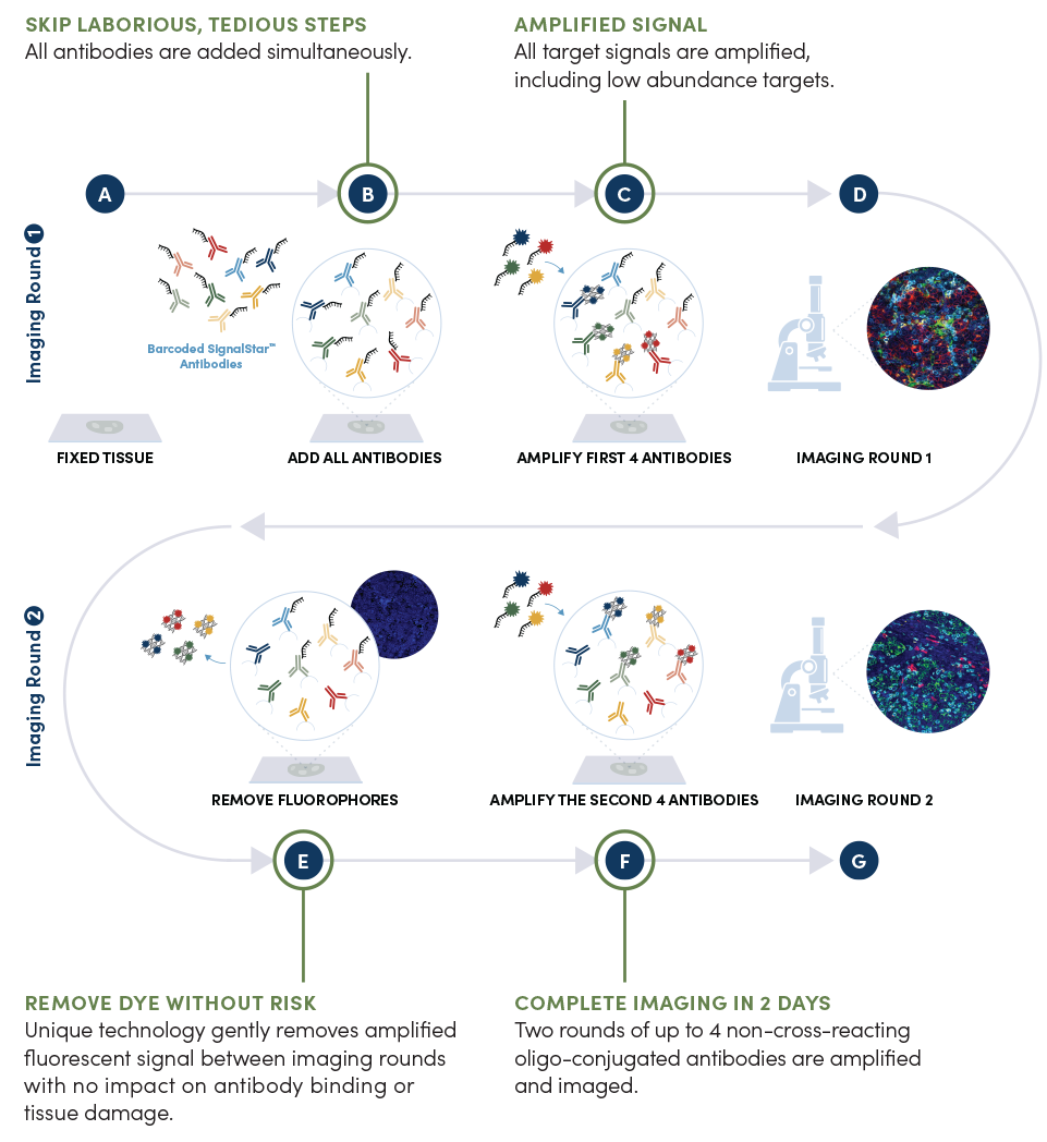
The SignalStar mIHC assay allows for the simultaneous labeling of up to 8 targets in FFPE tissue. Deparaffinized and rehydrated FFPE tissue sections undergo antigen retrieval (A), and all antibodies in your plex size of choice (3-8 maximum unique oligo-conjugated antibodies) are added in a cocktail, in one primary incubation step (B). A network of complementary oligos with fluorescent dyes (channels: 488, 594, 647, 750) amplify the signal of up to 4 oligo-conjugated antibodies in the first round of imaging by building oligo-fluorophore constructs attached to the antibody (C-D). If the plex size is greater than 4, the first round of oligonucleotides and fluorophores are gently removed (E), and a second round of amplification is performed to visualize up to 4 additional oligo-conjugated antibodies (F). The two images are then aligned and fused computationally with either proprietary or open-source software to generate the 8-plex image (G).
Tissue Preparation
- Collection and fixation of the tissue is crucial as it directly affects sample integrity and experimental results. Tissue collection, preservation, and fixation procedures vary depending on the sample or the marker of interest.
- Cross-linking fixatives, like formalin, are common for preparing samples for multiplex IHC as they preserve the structural integrity and architecture of the tissue.
Antigen Retrieval
- Fixatives used to preserve the structural integrity of the tissue sample may mask the epitope your antibody is designed to recognize.
- Several methods exist for revealing epitopes masked by fixation, including proteolytic-induced antigen retrieval or heat-induced epitope retrieval (HIER).
Antibody Incubation
Choose from a growing menu of IHC antibodies validated to work in any combination with our universal manual and automated protocols. If your target isn’t available as a SignalStar conjugate, you can select a primary antibody and add a SignalStar® Secondary Antibody to use unconjugated, IHC-validated antibodies instead. These fully validated protocols allow you to design and modify your panels easily.
- All antibodies are added simultaneously, eliminating antibody cycling and any possibility of epitope degradation or masking during multiple imaging rounds.
- A short post-stain fixation with 10% neutral buffered formalin guarantees antibodies remain on the slide during rounds of amplification and imaging.
Oligo Amplification
- Oligos amplify the fluorescent signal, enabling the detection of a wide range of targets, with a dynamic range that outshines tissue autofluorescence.
- Complementary oligos specific to each antibody are added for the first 4 oligo-conjugated antibodies, followed by additional oligos with fluorophores to exponentially amplify the signal.
- Fluorophores are spectrally distinct to lessen the need for spectral unmixing.
- Gentle removal of the oligos containing the fluorophores leaves the antibodies intact, enabling a second round of amplification and imaging.
- Pair any antibody with almost any fluorophore for your specific experiments.
What You Can Do With SignalStar Multiplex IHC
Generate Results in Just 2 Days
Timelines for panel optimization and validation for other multiplex technologies used for spatial profiling can take weeks to months. SignalStar technology minimizes that time with optimized, ready-to-go panels. Select targets from a growing menu of IHC-validated antibodies for use in FFPE tissues.
SignalStar assays amplify the fluorescent signal of up to 8 proteins in a single tissue. These highly-sensitive assays give you the signal you need the first time, so you can confidently analyze limited, precious FFPE tissue samples.
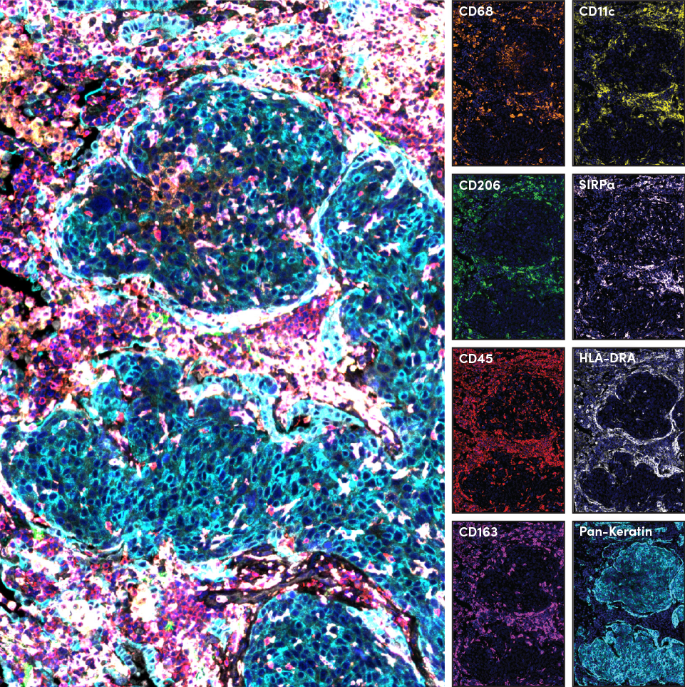
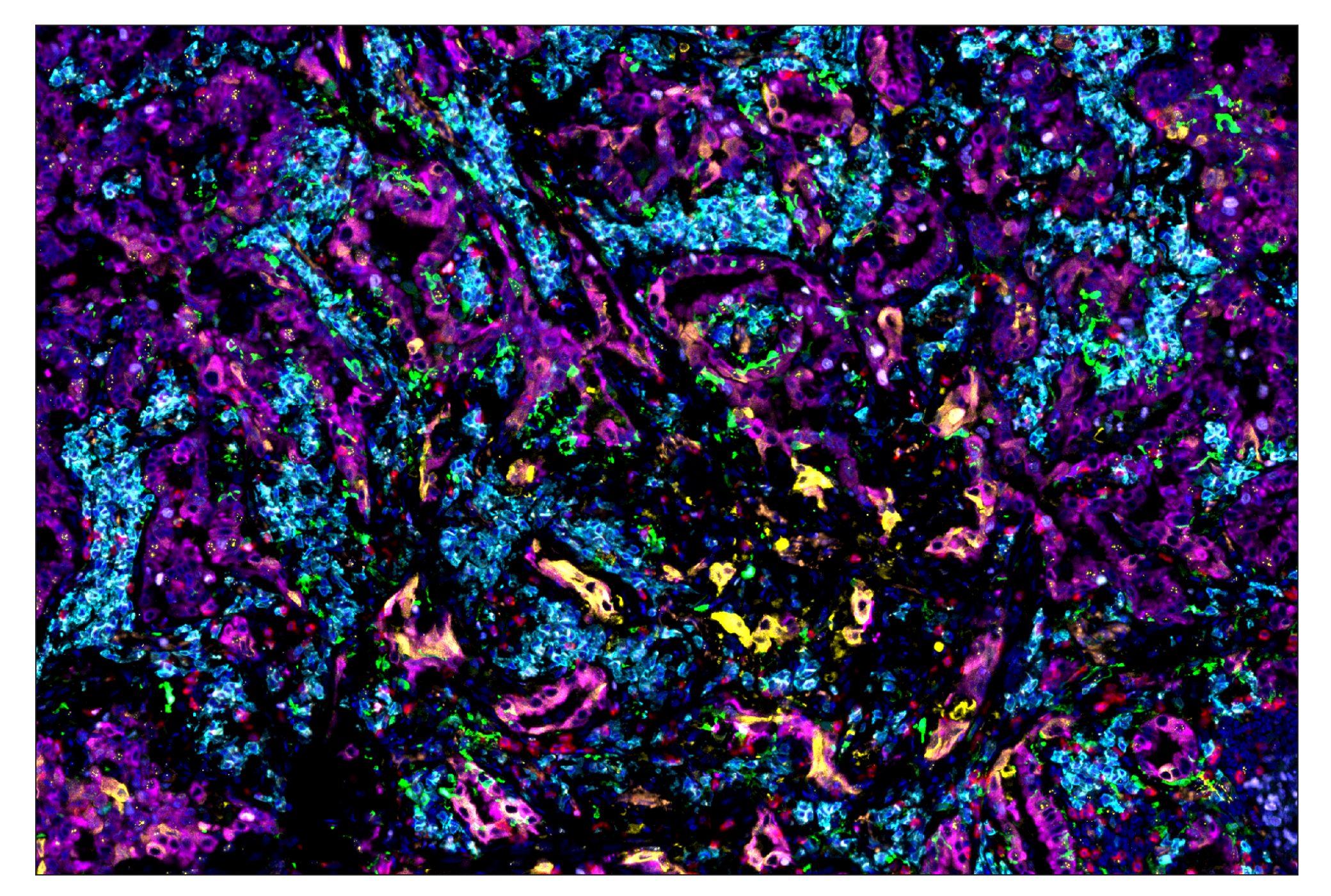
SignalStar immunohistochemical analysis of paraffin-embedded human squamous cell lung carcinoma using Pan-Keratin (C11) & CO-0003-488 SignalStar® Oligo-Antibody Pair #63566 (cyan), CD68 (D4B9C) & CO-0007-594 SignalStar® Oligo-Antibody Pair #77318 (orange), SIRPα/SHPS1 (D6I3M) & CO-0034-647 SignalStar® Oligo-Antibody Pair #80150 (pink), CD163 (D6U1J) & CO-0022-750 SignalStar® Oligo-Antibody Pair #71043 (magenta), CD206/MRC1 (E2L9N) & CO-0035-488 SignalStar® Oligo-Antibody Pair #99626 (green), CD11c (D3V1E) & CO-0017-594 SignalStar® Oligo-Antibody Pair #85384 (yellow), CD45 (Intracellular Domain) (D9M8I) & CO-0013-647 SignalStar® Oligo-Antibody Pair #32740 (red), and HLA-DRA (E9R2Q) & CO-0023-750 SignalStar® Oligo-Antibody Pair #58446 (white). All fluorophores have been assigned a pseudocolor, as indicated. Multiplex staining was performed on the BOND RX autostainer by Leica Biosystems.
Design Panels Faster
Our online, easy-to-use SignalStar Multiplex IHC Panel Builder does the design work so you don’t have to. Protocols are validated and only need minimal optimization—it works out of the box for most experiments.
SignalStar technology stains consistently regardless of the panel you design. Protocols are validated and optimized, and demonstrate reproducibility across different antibody combinations, experiments, and imaging instruments.
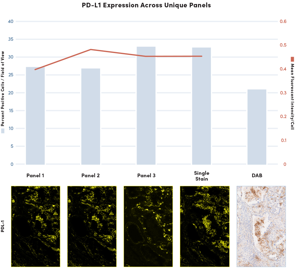
SignalStar multiplex immunohistochemical analysis of paraffin-embedded human medullary thyroid carcinoma within a tissue microarray. Panel 1: PD-L1 (E1L3N®) & CO-0005-647 SignalStar® Oligo-Antibody Pair #52085 (yellow), PD-1 (Intracellular Domain) (D4W2J) & CO-0008-594 SignalStar® Oligo-Antibody Pair #35347 (not shown), CD68 (D4B9C) & CO-0007-488 SignalStar® Oligo-Antibody Pair #73071 (not shown), and CD3ε (D7A6E) & CO-0001-750 SignalStar® Oligo-Antibody Pair #51754 (not shown). Panel 2: PD-L1 (E1L3N®) & CO-0005-647 SignalStar® Oligo-Antibody Pair #52085 (yellow), PD-1 (Intracellular Domain) (D4W2J) & CO-0008-594 SignalStar® Oligo-Antibody Pair #35347 (not shown), CD68 (D4B9C) & CO-0007-488 SignalStar® Oligo-Antibody Pair #73071 (not shown), and CD8 (D8A8Y) & CO-0004-750 SignalStar® Oligo-Antibody Pair #62750 (not shown). Panel 3: PD-L1 (E1L3N®) & CO-0005-647 SignalStar® Oligo-Antibody Pair #52085 (yellow), PD-1 (Intracellular Domain) (D4W2J) & CO-0008-594 SignalStar® Oligo-Antibody Pair #35347 (not shown), Ki-67 (8D5) & CO-0014-488 SignalStar® Oligo-Antibody Pair #89034 (not shown), and CD8ɑ (D8A8Y) & CO-0004-750 SignalStar® Oligo-Antibody Pair #62750 (not shown). All fluorophores were assigned a pseudocolor, as indicated. Multiplex staining was performed on the BOND RX autostainer by Leica Biosystems. Percent positive PD-L1 cells were quantified in matched regions of interest from serial sections for three unique SignalStar 4-plex panels compared to single-plex SignalStar staining using PD-L1 (E1L3N®) & CO-0005-647 SignalStar® Oligo Antibody Pair #52085 (yellow) and chromogenic staining using PD-L1 (E1L3N®) XP® Rabbit mAb #13684.
Redesign Panels Easily
Many solutions used for spatial biology lack the flexibility to evolve and adapt to changing research needs. SignalStar panels give you the flexibility to easily change targets and redesign panels as your needs change.
Amplify Your Signal
Unlike many other solutions, SignalStar mIHC panels offer amplification, which lets you see more than before and detect targets with low expression levels.
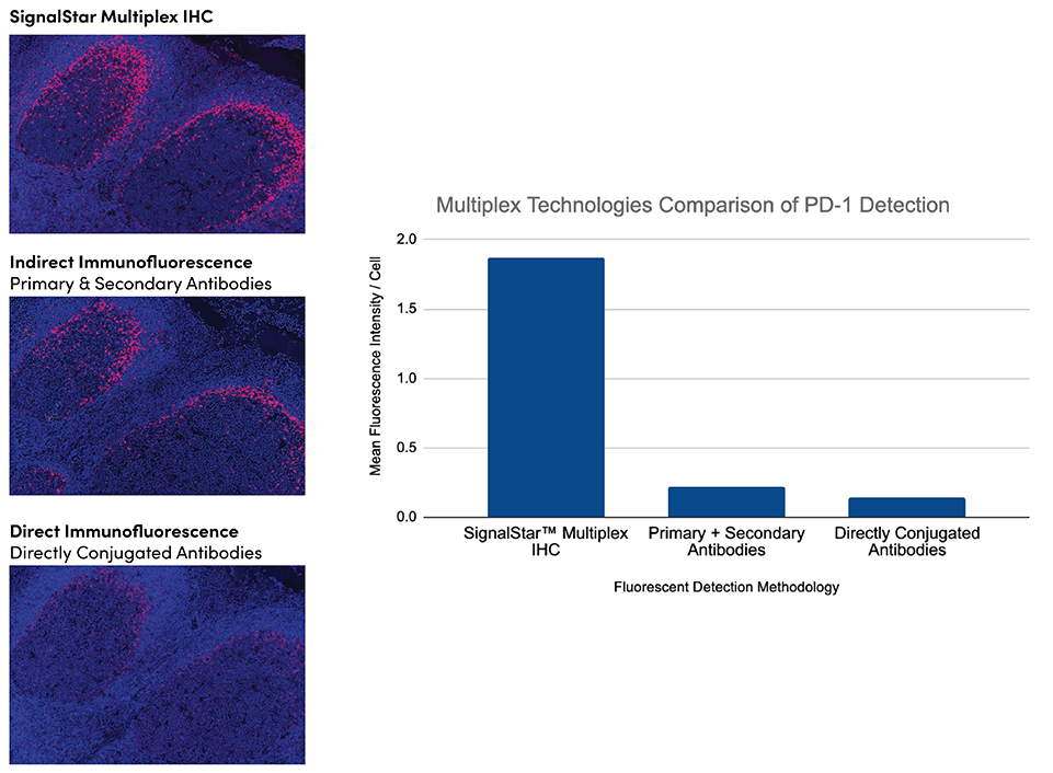
SignalStar multiplex immunohistochemical analysis of paraffin-embedded human tonsil using PD-1 (Intracellular Domain) (D4W2J) & CO-0008-647 SignalStar™ Oligo-Antibody Pair #56837 (red) and ProLong Gold Antifade Reagent with DAPI #8961 (blue), compared to PD-1 (Intracellular Domain) (D4W2J) XP® Rabbit mAb #86163 detected with anti-rabbit IgG (H+L), F(ab')2 Fragment (Alexa Fluor® 647 Conjugate) #4414, and PD-1 (Intracellular Domain) (D4W2J) XP® Rabbit mAb (Alexa Fluor® 647 Conjugate). All fluorophores have been assigned a pseudocolor, as indicated. Multiplex staining was performed on the BOND RX autostainer by Leica Biosystems. Mean fluorescence intensity of PD-1 positive cells was quantified in matched regions of interest from serial tissue sections for the SignalStar assay compared to the indirect and direct fluorescent methodologies.
Reproducible Every Time
CST validates antibodies in the SignalStar assay and guarantees them to work as expected. Every SignalStar antibody is validated against the chromogenic gold standard, and is consistently reproducible across replicates.
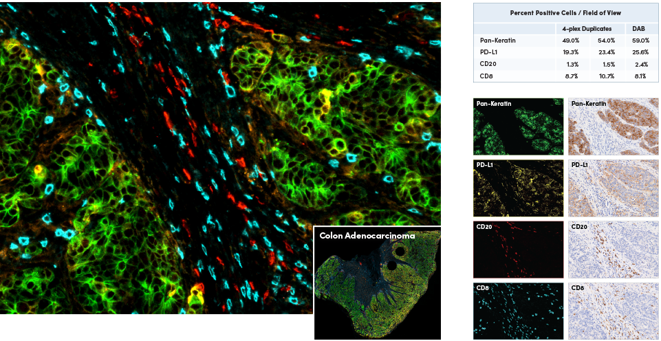
SignalStar multiplex immunohistochemical analysis of paraffin-embedded human colorectal adenocarcinoma using Pan-Keratin (C11) & CO-0003-488 SignalStar® Oligo-Antibody Pair #63566 (green), PD-L1 (E1L3N®) & CO-0005-594 SignalStar® Oligo-Antibody Pair #28249 (yellow), CD20 (E7B7T) & CO-0011-647 SignalStar® Oligo-Antibody Pair #36775 (red), and CD8a (D8A8Y) & CO-0004-750 SignalStar® Oligo-Antibody Pair #62750 (cyan). Representative individual and 4-plex images are included. All fluorophores were assigned a pseudocolor, as indicated. Multiplex staining was performed on the BOND RX autostainer by Leica Biosystems. Percent positive cells were quantified in matches regions of interest from serial sections for the SignalStar assay (n=2) compared to the chromogenic.
Use Your Existing Instrumentation
Stable fluorescent signals with distinct emission peaks make SignalStar mIHC panels compatible with your existing fluorescence imagers.
Image your slides using a microscope compatible with channels 488, 594, 647, and 750 nm to process 3-8 plex assays. With a 6-hour protocol run time, it's possible to stain and image a 4-plex assay in 1 day or an 8-plex in 2 days. The automated protocol enables overnight staining, greatly reducing hands-on time.
Considerations when designing your own Multiplex IHC Panel
Eliminate Antibody Cycling
Add all your antibodies at once without having to worry about epitope loss or masking.
Results You Can Trust
Every antibody used in a SignalStar panel has been independently validated for use in its intended application and guaranteed to perform as expected just like all CST® antibodies—to provide you with reliable, reproducible results, the first time and every time.
Cell Signaling Technology, CST, and SignalStar are trademarks of Cell Signaling Technology, Inc. Cy is a registered trademark of GE Healthcare. All other trademarks are the property of their respective owners. Visit cellsignal.com/legal/trademark-information for more information.
U.S. Patent No. 10,781,477, foreign equivalents, and child patents deriving therefrom.