| REACTIVITY | H |
Product Information
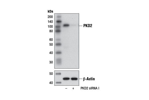
CST recommends transfection with 100 nM SignalSilence® PKD2 siRNA I 48 to 72 hours prior to cell lysis. For transfection procedure, follow protocol provided by the transfection reagent manufacturer. Please feel free to contact CST with any questions on use.
Each vial contains the equivalent of 100 transfections, which corresponds to a final siRNA concentration of 100 nM per transfection in a 24-well plate with a total volume of 300 μl per well.
Oligonucleotide synthesis is monitored base by base through trityl analysis to ensure appropriate coupling efficiency. The oligo is subsequently purified by affinity-solid phase extraction. The annealed RNA duplex is further analyzed by mass spectrometry to verify the exact composition of the duplex. Each lot is compared to the previous lot by mass spectrometry to ensure maximum lot-to-lot consistency.
Protein kinase D2 (PKD2) is one of three members of the protein kinase D family, including PKD1/PKCμ and PKD3/PKCν, that belong to the calcium/calmodulin superfamily of serine/threonine protein kinases (1,2). PKDs contain a conserved, carboxy-terminal catalytic domain, an amino-terminal regulatory region hallmarked by a PH domain that coordinates subcellular localization, and two zinc-finger/C1 lipid-binding domains that mediate activation of the enzyme in response to diacylglycerol (DAG) or phorbol ester (2,3). In addition to lipid-mediated activation, PKD catalytic activity can also be stimulated via phosphorylation of critical serine residues within the activation loop of the enzyme (4-8). Novel PKCs, such as PKCη and PKCε, have been shown to phosphorylate PKD1 at Ser744 and Ser748 (Ser706 and Ser710 in human PKD2), resulting in alleviation of autoinhibition of the enzyme mediated by PH domain interactions with the catalytic domain (5). Phosphorylation and activation of PKD isoforms has also been described for other upstream kinases. For example, casein kinase 2 (CK2) has been shown to phosphorylate PKD2 at Ser244, which promotes nuclear accumulation of PKD2, phosphorylation of HDAC7, and expression of Nur77 (9). Although only a handfull of PKD2 effectors have been identified, PKD2 has been implicated in regulating an array of cellular events, including cell survival, development, growth, migration, and transformation (10-14). PKD2-mediated phosphorylation of at least one known substrate, phosphatidylinositol 4-kinase type IIIβ (PI4KIIIβ), also implicates PKD2 in the formation and regulation of exocytotic transport vesicles from the trans Golgi network (15).
Explore pathways related to this product.
STRING - Known and Predicted Protein-Protein Interactions.
Except as otherwise expressly agreed in a writing signed by a legally authorized representative of CST, the following terms apply to Products provided by CST, its affiliates or its distributors. Any Customer's terms and conditions that are in addition to, or different from, those contained herein, unless separately accepted in writing by a legally authorized representative of CST, are rejected and are of no force or effect.
Products are labeled with For Research Use Only or a similar labeling statement and have not been approved, cleared, or licensed by the FDA or other regulatory foreign or domestic entity, for any purpose. Customer shall not use any Product for any diagnostic or therapeutic purpose, or otherwise in any manner that conflicts with its labeling statement. Products sold or licensed by CST are provided for Customer as the end-user and solely for research and development uses. Any use of Product for diagnostic, prophylactic or therapeutic purposes, or any purchase of Product for resale (alone or as a component) or other commercial purpose, requires a separate license from CST. Customer shall (a) not sell, license, loan, donate or otherwise transfer or make available any Product to any third party, whether alone or in combination with other materials, or use the Products to manufacture any commercial products, (b) not copy, modify, reverse engineer, decompile, disassemble or otherwise attempt to discover the underlying structure or technology of the Products, or use the Products for the purpose of developing any products or services that would compete with CST products or services, (c) not alter or remove from the Products any trademarks, trade names, logos, patent or copyright notices or markings, (d) use the Products solely in accordance with CST Product Terms of Sale and any applicable documentation, and (e) comply with any license, terms of service or similar agreement with respect to any third party products or services used by Customer in connection with the Products.