| # | Product Name | Applications | Reactivity | |
|---|---|---|---|---|
| 3108 | Phospho-SMAD2 (Ser465/467) (138D4) Rabbit mAb |
|
H M R Mi |
| REACTIVITY | H M R |
| SENSITIVITY | Endogenous |
| MW (kDa) | 60 |
| SOURCE | Rabbit |
Product Information
| Application | Dilution |
|---|---|
| Western Blotting | 1:1000 |
For western blots, incubate membrane with diluted primary antibody in 5% w/v nonfat dry milk, 1X TBS, 0.1% Tween® 20 at 4°C with gentle shaking, overnight.
NOTE: Please refer to primary antibody product webpage for recommended antibody dilution.
NOTE: Prepare solutions with reverse osmosis deionized (RODI) or equivalent grade water.
Load 20 µl onto SDS-PAGE gel (10 cm x 10 cm).
NOTE: Loading of prestained molecular weight markers (#59329, 10 µl/lane) to verify electrotransfer and biotinylated protein ladder (#7727, 10 µl/lane) to determine molecular weights are recommended.
NOTE: Volumes are for 10 cm x 10 cm (100 cm2) of membrane; for different sized membranes, adjust volumes accordingly.
* Avoid repeated exposure to skin.
posted June 2005
revised June 2020
Protocol Id: 263
Human, Mouse, Rat
Chicken, Xenopus, Zebrafish
Polyclonal antibodies are produced by immunizing animals with a synthetic phosphopeptide corresponding to residues surrounding Ser465/467 of human Smad2. Antibodies are purified by protein A and peptide affinity chromatography.
Members of the SMAD family of signal transduction molecules are components of a critical intracellular pathway that transmit TGF-β signals from the cell surface into the nucleus. Three distinct classes of SMADs have been defined: the receptor-regulated SMADs (R-SMADs), which include SMAD1, 2, 3, 5, and 9; the common-mediator SMAD (co-SMAD), SMAD4; and the antagonistic or inhibitory SMADs (I-SMADs), SMAD6 and 7 (1-5). Activated type I receptors associate with specific R-SMADs and phosphorylate them on a conserved carboxy-terminal SSXS motif. The phosphorylated R-SMADs dissociate from the receptor and form a heteromeric complex with SMAD4, initiating translocation of the heteromeric SMAD complex to the nucleus. Once in the nucleus, SMADs recruit a variety of DNA binding proteins that function to regulate transcriptional activity (6-8).
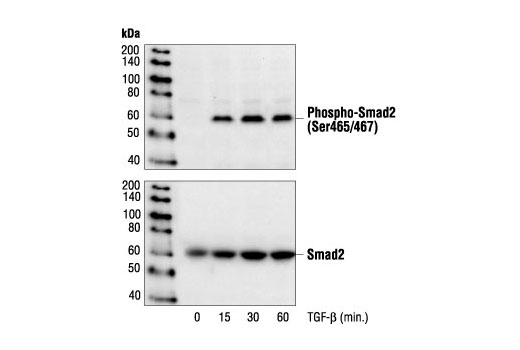
Following stimulation by TGF-beta, Smad2 and Smad3 become phosphorylated at their carboxyl termini by the receptor kinase (serines 465 and 467 on Smad2; serines 423 and 425 on Smad3) by TbetaR-I (9-11). Following phosphorylation, Smad2 and Smad3 form a heteromeric complex with the co-smad family member Smad4. These complexes are translocated to the nucleus where they bind DNA and regulate gene transcription.
Explore pathways related to this product.
STRING - Known and Predicted Protein-Protein Interactions.
Except as otherwise expressly agreed in a writing signed by a legally authorized representative of CST, the following terms apply to Products provided by CST, its affiliates or its distributors. Any Customer's terms and conditions that are in addition to, or different from, those contained herein, unless separately accepted in writing by a legally authorized representative of CST, are rejected and are of no force or effect.
Products are labeled with For Research Use Only or a similar labeling statement and have not been approved, cleared, or licensed by the FDA or other regulatory foreign or domestic entity, for any purpose. Customer shall not use any Product for any diagnostic or therapeutic purpose, or otherwise in any manner that conflicts with its labeling statement. Products sold or licensed by CST are provided for Customer as the end-user and solely for research and development uses. Any use of Product for diagnostic, prophylactic or therapeutic purposes, or any purchase of Product for resale (alone or as a component) or other commercial purpose, requires a separate license from CST. Customer shall (a) not sell, license, loan, donate or otherwise transfer or make available any Product to any third party, whether alone or in combination with other materials, or use the Products to manufacture any commercial products, (b) not copy, modify, reverse engineer, decompile, disassemble or otherwise attempt to discover the underlying structure or technology of the Products, or use the Products for the purpose of developing any products or services that would compete with CST products or services, (c) not alter or remove from the Products any trademarks, trade names, logos, patent or copyright notices or markings, (d) use the Products solely in accordance with CST Product Terms of Sale and any applicable documentation, and (e) comply with any license, terms of service or similar agreement with respect to any third party products or services used by Customer in connection with the Products.