| REACTIVITY | H |
| SENSITIVITY | Endogenous |
| MW (kDa) | 82 |
| SOURCE | Rabbit |
Product Information
| Application | Dilution |
|---|---|
| Western Blotting | 1:1000 |
For western blots, incubate membrane with diluted primary antibody in 5% w/v BSA, 1X TBS, 0.1% Tween® 20 at 4°C with gentle shaking, overnight.
NOTE: Please refer to primary antibody product webpage for recommended antibody dilution.
From sample preparation to detection, the reagents you need for your Western Blot are now in one convenient kit: #12957 Western Blotting Application Solutions Kit
NOTE: Prepare solutions with reverse osmosis deionized (RODI) or equivalent grade water.
Load 20 µl onto SDS-PAGE gel (10 cm x 10 cm).
NOTE: Loading of prestained molecular weight markers (#59329, 10 µl/lane) to verify electrotransfer and biotinylated protein ladder (#7727, 10 µl/lane) to determine molecular weights are recommended.
NOTE: Volumes are for 10 cm x 10 cm (100 cm2) of membrane; for different sized membranes, adjust volumes accordingly.
* Avoid repeated exposure to skin.
posted June 2005
revised June 2020
Protocol Id: 10
Human
Polyclonal antibodies are produced by immunizing animals with a synthetic peptide corresponding to residues surrounding Gly267 of human A20/TNFAIP3. Antibodies were purified by protein A and peptide affinity chromatography.
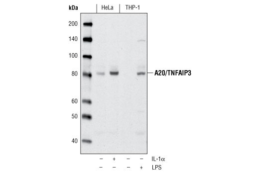
A20, also referred to as TNF-α-induced protein 3 (TNFAIP3), is cytokine-inducible protein that functions to inhibit apoptosis and activate NF-κB (1,2). It was first identified as a TNF-α inducible primary response gene in human umbilical vein endothelial cells, and encodes a 790-amino acid protein containing seven Cys2/Cys2-zinc finger motifs (3). Constitutive expression of A20 is observed in lymphoid tissues (4), but it is transiently expressed in a variety of cell types in response to inflammatory signals such as TNF-α (3,5), IL-1 (3,5), phorbol esters (6), and LPS (7). Expression of A20 can confer resistance to apoptosis and NF-κB activation triggered by these signals, probably through interference with TRAF (TNF receptor associated factor) family members (8,9), and interaction with the NF-κB inhibiting protein ABIN (10). Studies also show that A20 contains site-specific ubiquitin modifying activity that can contribute to its biological functions (11,12). The amino-terminus of A20 contains de-ubiquitinating (DUB) activity for Lys63 branches, such as those found in TRAF6 and RIP, while the carboxyl-terminus contains ubiquitin ligase (E3) activity for Lys48 branches of the same substrates and leads to their degradation (12).
Explore pathways related to this product.
STRING - Known and Predicted Protein-Protein Interactions.
Except as otherwise expressly agreed in a writing signed by a legally authorized representative of CST, the following terms apply to Products provided by CST, its affiliates or its distributors. Any Customer's terms and conditions that are in addition to, or different from, those contained herein, unless separately accepted in writing by a legally authorized representative of CST, are rejected and are of no force or effect.
Products are labeled with For Research Use Only or a similar labeling statement and have not been approved, cleared, or licensed by the FDA or other regulatory foreign or domestic entity, for any purpose. Customer shall not use any Product for any diagnostic or therapeutic purpose, or otherwise in any manner that conflicts with its labeling statement. Products sold or licensed by CST are provided for Customer as the end-user and solely for research and development uses. Any use of Product for diagnostic, prophylactic or therapeutic purposes, or any purchase of Product for resale (alone or as a component) or other commercial purpose, requires a separate license from CST. Customer shall (a) not sell, license, loan, donate or otherwise transfer or make available any Product to any third party, whether alone or in combination with other materials, or use the Products to manufacture any commercial products, (b) not copy, modify, reverse engineer, decompile, disassemble or otherwise attempt to discover the underlying structure or technology of the Products, or use the Products for the purpose of developing any products or services that would compete with CST products or services, (c) not alter or remove from the Products any trademarks, trade names, logos, patent or copyright notices or markings, (d) use the Products solely in accordance with CST Product Terms of Sale and any applicable documentation, and (e) comply with any license, terms of service or similar agreement with respect to any third party products or services used by Customer in connection with the Products.