| Cat. # | Size | Qty. | Price |
|---|---|---|---|
| 67184S | 500 µl (100 tests) |
|
| REACTIVITY | H |
| SENSITIVITY | Endogenous |
| MW (kDa) | |
| Source/Isotype | Mouse IgG1 kappa |
Product Information
| Application | Dilution |
|---|---|
| Flow Cytometry (Fixed/Permeabilized) | 1:20 |
| Flow Cytometry (Live) | 1:20 |
This antibody has been validated for Flow Cytometry using the FoxP3/Transcription Factor Fixation/Permeabilization Kit #43481. Please refer to the protocol provided with the kit for detailed instructions.
Protocol Id: 1804
NOTE: Prepare solutions with reverse osmosis deionized (RODI) or equivalent grade water.
NOTE: When including fluorescent cellular dyes in your experiment (including viability dyes, DNA dyes, etc.), please refer to the dye product page for the recommended protocol. Visit www.cellsignal.com for a full listing of cellular dyes validated for use in flow cytometry.
NOTE: Count cells using a hemocytometer or alternative method.
NOTE: If using whole blood, lyse red blood cells and wash by centrifugation prior to immunostaining.
NOTE: Human Fc receptors cross-react with rabbit IgG. When cells of interest express high levels of Fc receptor protein (for example, macrophage/monocyte lineages), pre-incubate live cells with human Fc block prior to immunostaining with rabbit antibodies.
NOTE: Optimal centrifugation conditions will vary depending upon cell type and reagent volume. Generally, 150-300g for 1-5 minutes will be sufficient to pellet the cells.
posted June 2017
revised January 2022
Protocol Id: 1504
Human
This monoclonal antibody was purified from tissue culture supernatant via affinity chromatography. The purified antibody was conjugated under optimal conditions, with unreacted dye removed from the preparation.
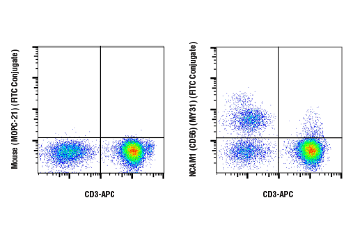
NCAM (neural cell adhesion molecule, CD56) is an adhesion glycoprotein with five extracellular immunoglobulin-like domains followed by two fibronectin type III repeats. Structural diversity is introduced by alternative splicing resulting in different cytoplasmic domains (1). NCAM mediates neuronal attachment, neurite extension, and cell-cell interactions through homo and heterophilic interactions. PSA (polysialic acid) post-translationally modifies NCAM and increases the metastatic potential of small cell lung carcinoma, Wilms' tumor, neuroblastoma, and rhabdomyosarcoma (2). CD56 is commonly used along with CD3 and CD16 to identify human natural killer (NK) cells (mouse NK cells do not express CD56) (3). Human NK cells are CD3-CD56+. The large subset with high CD16 expression are mature cytotoxic NK cells, while those with low CD16 expression are immature precursors and cytokine producers (4,5).
Except as otherwise expressly agreed in a writing signed by a legally authorized representative of CST, the following terms apply to Products provided by CST, its affiliates or its distributors. Any Customer's terms and conditions that are in addition to, or different from, those contained herein, unless separately accepted in writing by a legally authorized representative of CST, are rejected and are of no force or effect.
Products are labeled with For Research Use Only or a similar labeling statement and have not been approved, cleared, or licensed by the FDA or other regulatory foreign or domestic entity, for any purpose. Customer shall not use any Product for any diagnostic or therapeutic purpose, or otherwise in any manner that conflicts with its labeling statement. Products sold or licensed by CST are provided for Customer as the end-user and solely for research and development uses. Any use of Product for diagnostic, prophylactic or therapeutic purposes, or any purchase of Product for resale (alone or as a component) or other commercial purpose, requires a separate license from CST. Customer shall (a) not sell, license, loan, donate or otherwise transfer or make available any Product to any third party, whether alone or in combination with other materials, or use the Products to manufacture any commercial products, (b) not copy, modify, reverse engineer, decompile, disassemble or otherwise attempt to discover the underlying structure or technology of the Products, or use the Products for the purpose of developing any products or services that would compete with CST products or services, (c) not alter or remove from the Products any trademarks, trade names, logos, patent or copyright notices or markings, (d) use the Products solely in accordance with CST Product Terms of Sale and any applicable documentation, and (e) comply with any license, terms of service or similar agreement with respect to any third party products or services used by Customer in connection with the Products.