| Cat. # | Size | Qty. | Price |
|---|---|---|---|
| 9257S | 100 µl (50 tests) |
|
| REACTIVITY | H M R Hm Sc |
| SENSITIVITY | Endogenous |
| MW (kDa) | |
| Source/Isotype | Mouse IgG1 |
Product Information
| Application | Dilution |
|---|---|
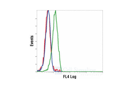
| Flow Cytometry (Fixed/Permeabilized) | 1:50 |
All reagents required for this protocol may be efficiently purchased together in our Intracellular Flow Cytometry Kit (Methanol) #13593, or individually using the catalog numbers listed below.
NOTE: Prepare solutions with reverse osmosis deionized (RODI) or equivalent grade water.
NOTE: When including fluorescent cellular dyes in your experiment (including viability dyes, DNA dyes, etc.), please refer to the dye product page for the recommended protocol. Visit www.cellsignal.com for a full listing of cellular dyes validated for use in flow cytometry.
NOTE: Adherent cells or tissue should be dissociated and in single-cell suspension prior to fixation.
NOTE: Optimal centrifugation conditions will vary depending upon cell type and reagent volume. Generally, 150-300g for 1-5 minutes will be sufficient to pellet the cells.
NOTE: If using whole blood, lyse red blood cells and wash by centrifugation prior to fixation.
NOTE: Antibodies targeting CD markers or other extracellular proteins may be added prior to fixation if the epitope is disrupted by formaldehyde and/or methanol. The antibodies will remain bound to the target of interest during the fixation and permeabilization process. However, note that some fluorophores (including PE and APC) are damaged by methanol and thus should not be added prior to permeabilization. Conduct a small-scale experiment if you are unsure.
NOTE: Count cells using a hemocytometer or alternative method.
posted July 2009
revised June 2020
Protocol Id: 407
Human, Mouse, Rat, Hamster, S. cerevisiae
Monoclonal antibody is produced by immunizing animals with a synthetic phosphopeptide corresponding to residues around Thr183/Tyr185 of human SAPK/JNK. The antibody was conjugated to Alexa Fluor® 647 under optimum conditions with an F/P ratio of 2-6. This antibody was conjugated to Alexa Fluor® 647 under optimal conditions with an F/P ratio of 2-6. The Alexa Fluor® 647 dye is maximally excited by red light (e.g. 633 nm He-Ne laser). Antibody conjugates of the Alexa Fluor® 647 dye produce bright far-red-fluorescence emission, with a peak at 665 nm.
The stress-activated protein kinase/Jun-amino-terminal kinase SAPK/JNK is potently and preferentially activated by a variety of environmental stresses, including UV and gamma radiation, ceramides, inflammatory cytokines, and in some instances, growth factors and GPCR agonists (1-6). As with the other MAPKs, the core signaling unit is composed of a MAPKKK, typically MEKK1-MEKK4, or by one of the mixed lineage kinases (MLKs), which phosphorylate and activate MKK4/7. Upon activation, MKKs phosphorylate and activate the SAPK/JNK kinase (2). Stress signals are delivered to this cascade by small GTPases of the Rho family (Rac, Rho, cdc42) (3). Both Rac1 and cdc42 mediate the stimulation of MEKKs and MLKs (3). Alternatively, MKK4/7 can be activated in a GTPase-independent mechanism via stimulation of a germinal center kinase (GCK) family member (4). There are three SAPK/JNK genes each of which undergoes alternative splicing, resulting in numerous isoforms (3). SAPK/JNK, when active as a dimer, can translocate to the nucleus and regulate transcription through its effects on c-Jun, ATF-2, and other transcription factors (3,5).
Explore pathways related to this product.
STRING - Known and Predicted Protein-Protein Interactions.
Except as otherwise expressly agreed in a writing signed by a legally authorized representative of CST, the following terms apply to Products provided by CST, its affiliates or its distributors. Any Customer's terms and conditions that are in addition to, or different from, those contained herein, unless separately accepted in writing by a legally authorized representative of CST, are rejected and are of no force or effect.
Products are labeled with For Research Use Only or a similar labeling statement and have not been approved, cleared, or licensed by the FDA or other regulatory foreign or domestic entity, for any purpose. Customer shall not use any Product for any diagnostic or therapeutic purpose, or otherwise in any manner that conflicts with its labeling statement. Products sold or licensed by CST are provided for Customer as the end-user and solely for research and development uses. Any use of Product for diagnostic, prophylactic or therapeutic purposes, or any purchase of Product for resale (alone or as a component) or other commercial purpose, requires a separate license from CST. Customer shall (a) not sell, license, loan, donate or otherwise transfer or make available any Product to any third party, whether alone or in combination with other materials, or use the Products to manufacture any commercial products, (b) not copy, modify, reverse engineer, decompile, disassemble or otherwise attempt to discover the underlying structure or technology of the Products, or use the Products for the purpose of developing any products or services that would compete with CST products or services, (c) not alter or remove from the Products any trademarks, trade names, logos, patent or copyright notices or markings, (d) use the Products solely in accordance with CST Product Terms of Sale and any applicable documentation, and (e) comply with any license, terms of service or similar agreement with respect to any third party products or services used by Customer in connection with the Products.