View Featured Offers >>
9708
Phospho-Histone H3 (Ser10) Antibody (Alexa Fluor® 488 Conjugate)
Antibody Conjugates
Polyclonal Antibody

Phospho-Histone H3 (Ser10) Antibody (Alexa Fluor® 488 Conjugate) #9708

Citations (48)

We recommend the following alternatives

# Product Name Applications Reactivity
  • Immunofluorescence
  • Flow Cytometry
H M R Mk Z XP
Filter:
  1. IF
  2. F
Immunocytochemical staining of untreated NIH/3T3 cells, using Phospho-Histone H3 (Ser10) Antibody (Alexa Fluor® 488 conjugate) (green) and DAPI (blue).
Flow cytometric analysis of THP1 cells treated with paclitaxel, using Phospho-Histone H3 (Ser10) Antibody (Alexa Fluor® 488 conjugate) versus propidium iodide (DNA content). The box indicates phospho-histone H3 positive cells.

Supporting Data

REACTIVITY H M R Mk Dm
SENSITIVITY Endogenous
MW (kDa)
SOURCE Rabbit

Application Key:

  • WB-Western Blot
  • IP-Immunoprecipitation
  • IHC-Immunohistochemistry
  • ChIP-Chromatin Immunoprecipitation
  • C&R-CUT&RUN
  • C&T-CUT&Tag
  • DB-Dot Blot
  • eCLIP-eCLIP
  • IF-Immunofluorescence
  • F-Flow Cytometry

Species Cross-Reactivity Key:

  • H-Human
  • M-Mouse
  • R-Rat
  • Hm-Hamster
  • Mk-Monkey
  • Vir-Virus
  • Mi-Mink
  • C-Chicken
  • Dm-D. melanogaster
  • X-Xenopus
  • Z-Zebrafish
  • B-Bovine
  • Dg-Dog
  • Pg-Pig
  • Sc-S. cerevisiae
  • Ce-C. elegans
  • Hr-Horse
  • GP-Guinea Pig
  • Rab-Rabbit
  • All-All Species Expected

Product Description

This Cell Signaling Technology antibody is conjugated to Alexa Fluor® 488 fluorescent dye and tested in-house for direct flow cytometry and immunofluorescent analysis in human and mouse cells. The antibody is expected to exhibit the same species cross-reactivity as the unconjugated Phospho-Histone H3 (Ser10) Antibody #9701.

Product Usage Information

Application Dilution
Immunofluorescence (Immunocytochemistry) 1:50
Flow Cytometry (Fixed/Permeabilized) 1:50

Storage

Supplied in PBS (pH 7.2), less than 0.1% sodium azide and 2 mg/ml BSA. Store at 4°C. Do not aliquot the antibody. Protect from light. Do not freeze.

Protocol

PRINT

View >Collapse >

Immunofluorescence (Immunocytochemistry)

A. Solutions and Reagents

NOTE: Prepare solutions with reverse osmosis deionized (RODI) or equivalent grade water.

  1. 20X Phosphate Buffered Saline (PBS): (9808) To prepare 1L 1X PBS: add 50 ml 20X PBS to 950 ml dH2O, mix. Adjust pH to 8.0.
  2. Formaldehyde: 16%, methanol free, Polysciences, Inc. (cat# 18814), use fresh and store opened vials at 4°C in dark, dilute in 1X PBS for use.
  3. Blocking Buffer (1X PBS / 5% normal goat serum (#5425) / 0.3% Triton™ X-100): To prepare 10 ml: add 0.5 ml normal goat serum and 0.5 ml 20X PBS to 9.0 ml dH2O, mix. While stirring, add 30 µl Triton™ X-100.
  4. Antibody Dilution Buffer: (1X PBS / 1% BSA / 0.3% Triton™ X-100): To prepare 10 ml, add 30 µl Triton™ X-100 to 10 ml 1X PBS. Mix well then add 0.1 g BSA (#9998), mix.
  5. Prolong® Gold AntiFade Reagent (#9071), Prolong® Gold AntiFade Reagent with DAPI (#8961).

B. Specimen Preparation - Cultured Cell Lines (IF-IC)

NOTE: Cells should be grown, treated, fixed and stained directly in multi-well plates, chamber slides or on coverslips.

  1. Aspirate liquid, then cover cells to a depth of 2–3 mm with 4% formaldehyde diluted in 1X PBS.

    NOTE: Formaldehyde is toxic, use only in a fume hood.

  2. Allow cells to fix for 15 min at room temperature.
  3. Aspirate fixative, rinse three times in 1X PBS for 5 min each.
  4. Proceed with Immunostaining (Section C).

C. Immunostaining

NOTE: All subsequent incubations should be carried out at room temperature unless otherwise noted in a humid light-tight box or covered dish/plate to prevent drying and fluorochrome fading.

  1. Block specimen in Blocking Buffer for 60 min.
  2. While blocking, prepare primary antibody by diluting as indicated on product webpage in Antibody Dilution Buffer.
  3. Aspirate blocking solution, apply diluted primary antibody.
  4. Incubate overnight at 4°C.
  5. Rinse three times in 1X PBS for 5 min each.
  6. Coverslip slides with Prolong® Gold Antifade Reagent (#9071) or Prolong® Gold Antifade Reagent with DAPI (#8961).
  7. For best results, allow mountant to cure overnight at room temperature. For long-term storage, store slides flat at 4°C protected from light.

posted November 2006

revised November 2013

Protocol Id: 182

Flow Cytometry, Methanol Permeabilization Protocol for Directly Conjugated Antibodies

A. Solutions and Reagents

All reagents required for this protocol may be efficiently purchased together in our Intracellular Flow Cytometry Kit (Methanol) #13593, or individually using the catalog numbers listed below.

NOTE: Prepare solutions with reverse osmosis deionized (RODI) or equivalent grade water.

  1. 1X Phosphate Buffered Saline (PBS): To prepare 1 L 1X PBS: add 100 ml 10X PBS (#12528) to 900 ml water mix.
  2. 4% Formaldehyde, Methanol-Free (#47746)
  3. 100% Methanol (#13604): Chill before use
  4. Antibody Dilution Buffer: Purchase ready-to-use Flow Cytometry Antibody Dilution Buffer (#13616), or prepare a 0.5% BSA PBS buffer by dissolving 0.5 g Bovine Serum Albumin (BSA) (#9998) in 100 ml 1X PBS. Store at 4°C.

NOTE: When including fluorescent cellular dyes in your experiment (including viability dyes, DNA dyes, etc.), please refer to the dye product page for the recommended protocol. Visit www.cellsignal.com for a full listing of cellular dyes validated for use in flow cytometry.

B. Fixation

NOTE: Adherent cells or tissue should be dissociated and in single-cell suspension prior to fixation.

NOTE: Optimal centrifugation conditions will vary depending upon cell type and reagent volume. Generally, 150-300g for 1-5 minutes will be sufficient to pellet the cells.

NOTE: If using whole blood, lyse red blood cells and wash by centrifugation prior to fixation.

NOTE: Antibodies targeting CD markers or other extracellular proteins may be added prior to fixation if the epitope is disrupted by formaldehyde and/or methanol. The antibodies will remain bound to the target of interest during the fixation and permeabilization process. However, note that some fluorophores (including PE and APC) are damaged by methanol and thus should not be added prior to permeabilization. Conduct a small-scale experiment if you are unsure.

  1. Pellet cells by centrifugation and remove supernatant.
  2. Resuspend cells in approximately 100 µl 4% formaldehyde per 1 million cells. Mix well to dissociate pellet and prevent cross-linking of individual cells.
  3. Fix for 15 min at room temperature (20-25°C).
  4. Wash by centrifugation with excess 1X PBS. Discard supernatant in appropriate waste container. Resuspend cells in 0.5-1 ml 1X PBS. Proceed to Permeabilization step.
    1. Alternatively, cells may be stored overnight at 4°C in 1X PBS.

C. Permeabilization

  1. Permeabilize cells by adding ice-cold 100% methanol slowly to pre-chilled cells, while gently vortexing, to a final concentration of 90% methanol.
  2. Permeabilize for a minimum of 10 min on ice.
  3. Proceed with immunostaining (Section D) or store cells at -20°C in 90% methanol.

D. Immunostaining

NOTE: Count cells using a hemocytometer or alternative method.

  1. Aliquot desired number of cells into tubes or wells. (Generally, 5x10⁵ to 1x10⁶ cells per assay.)
  2. Wash cells by centrifugation in excess 1X PBS to remove methanol. Discard supernatant in appropriate waste container. Repeat if necessary.
  3. Resuspend cells in 100 µl of diluted primary antibody, prepared in Antibody Dilution Buffer at a recommended dilution or as determined via titration.
  4. Incubate for 1 hr at room temperature. Protect from light.
  5. Wash by centrifugation in Antibody Dilution Buffer or 1X PBS. Discard supernatant. Repeat.
  6. Resuspend cells in 200-500 µl of 1X PBS and analyze on flow cytometer.

posted July 2009

revised June 2020

Protocol Id: 407

Specificity / Sensitivity

Phospho-Histone H3 (Ser10) Antibody (Alexa Fluor® 488 Conjugate) detects endogenous levels of histone H3 only when phosphorylated at serine 10. The antibody does not cross-react with other phosphorylated histones or with acetylated histones.

Species Reactivity:

Human, Mouse, Rat, Monkey, D. melanogaster

Species predicted to react based on 100% sequence homology:

Xenopus

Source / Purification

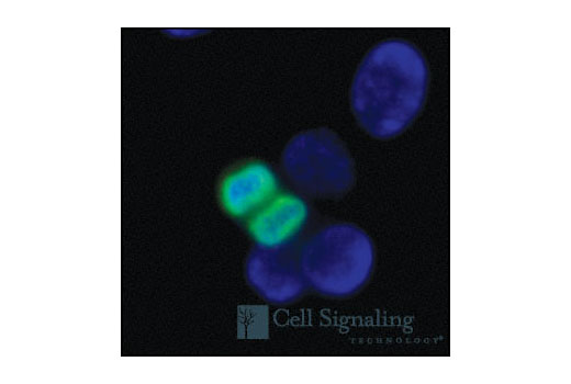
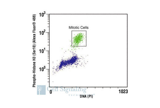
Polyclonal antibodies are produced by immunizing animals with a synthetic phosphopeptide corresponding to residues surrounding Ser10 of human histone H3. Antibodies are purified by protein A and peptide affinity chromatography. The antibody was conjugated to Alexa Fluor® 488 under optimal conditions with an F/P ratio of 2-6.

Background

Modulation of chromatin structure plays an important role in the regulation of transcription in eukaryotes. The nucleosome, made up of DNA wound around eight core histone proteins (two each of H2A, H2B, H3, and H4), is the primary building block of chromatin (1). The amino-terminal tails of core histones undergo various posttranslational modifications, including acetylation, phosphorylation, methylation, and ubiquitination (2-5). These modifications occur in response to various stimuli and have a direct effect on the accessibility of chromatin to transcription factors and, therefore, gene expression (6). In most species, histone H2B is primarily acetylated at Lys5, 12, 15, and 20 (4,7). Histone H3 is primarily acetylated at Lys9, 14, 18, 23, 27, and 56. Acetylation of H3 at Lys9 appears to have a dominant role in histone deposition and chromatin assembly in some organisms (2,3). Phosphorylation at Ser10, Ser28, and Thr11 of histone H3 is tightly correlated with chromosome condensation during both mitosis and meiosis (8-10). Phosphorylation at Thr3 of histone H3 is highly conserved among many species and is catalyzed by the kinase haspin. Immunostaining with phospho-specific antibodies in mammalian cells reveals mitotic phosphorylation at Thr3 of H3 in prophase and its dephosphorylation during anaphase (11).

Pathways

Explore pathways related to this product.

Limited Uses

Except as otherwise expressly agreed in a writing signed by a legally authorized representative of CST, the following terms apply to Products provided by CST, its affiliates or its distributors. Any Customer's terms and conditions that are in addition to, or different from, those contained herein, unless separately accepted in writing by a legally authorized representative of CST, are rejected and are of no force or effect.

Products are labeled with For Research Use Only or a similar labeling statement and have not been approved, cleared, or licensed by the FDA or other regulatory foreign or domestic entity, for any purpose. Customer shall not use any Product for any diagnostic or therapeutic purpose, or otherwise in any manner that conflicts with its labeling statement. Products sold or licensed by CST are provided for Customer as the end-user and solely for research and development uses. Any use of Product for diagnostic, prophylactic or therapeutic purposes, or any purchase of Product for resale (alone or as a component) or other commercial purpose, requires a separate license from CST. Customer shall (a) not sell, license, loan, donate or otherwise transfer or make available any Product to any third party, whether alone or in combination with other materials, or use the Products to manufacture any commercial products, (b) not copy, modify, reverse engineer, decompile, disassemble or otherwise attempt to discover the underlying structure or technology of the Products, or use the Products for the purpose of developing any products or services that would compete with CST products or services, (c) not alter or remove from the Products any trademarks, trade names, logos, patent or copyright notices or markings, (d) use the Products solely in accordance with CST Product Terms of Sale and any applicable documentation, and (e) comply with any license, terms of service or similar agreement with respect to any third party products or services used by Customer in connection with the Products.

For Research Use Only. Not for Use in Diagnostic Procedures.
Cell Signaling Technology is a trademark of Cell Signaling Technology, Inc.
This product is provided under an intellectual property license from Life Technologies Corporation. The transfer of this product is conditioned on the buyer using the purchased product solely in research conducted by the buyer, excluding contract research or any fee for service research, and the buyer must not (1) use this product or its components for (a) diagnostic, therapeutic or prophylactic purposes; (b) testing, analysis or screening services, or information in return for compensation on a per-test basis; or (c) manufacturing or quality assurance or quality control, and/or (2) sell or transfer this product or its components for resale, whether or not resold for use in research. For information on purchasing a license to this product for purposes other than as described above, contact Life Technologies Corporation, 5791 Van Allen Way, Carlsbad, CA 92008 USA or [email protected].
All other trademarks are the property of their respective owners. Visit our Trademark Information page.
Inquiry Info.# 9708