View Featured Offers >>
7326
PathScan® Phospho-Insulin Receptor β (Tyr1345) Sandwich ELISA Kit
ELISA Kits
ELISA Kit

PathScan® Phospho-Insulin Receptor β (Tyr1345) Sandwich ELISA Kit #7326

Citations (0)
Figure 1: Treatment of CHO-IR/IRS-1 cells with insulin stimulates phosphorylation of insulin receptor at Tyr1345, detected by PathScan® Phospho-Insulin Receptor β (Tyr1345) Sandwich ELISA Kit #7326, but the level of total insulin receptor shown by Western blot analysis remains unchanged. Absorbances at 450 nm are shown in the top figure, while the corresponding Western blots using Phospho-Insulin Receptor β (Tyr1345) Rabbit mAb #3026 (right panel) or Insulin Receptor β Antibody #3025 (left panel), are shown in the bottom figure. CHO-IR/IRS-1 cells stably overexpress human insulin receptor and rat IRS-1.
Figure 2: The relationship between protein concentration of lysates from untreated and insulin-treated CHO-IR/IRS-1 cells and the absorbance at 450 nm is shown. After starvation, CHO-IR/IRS-1 cells (85% confluence) were treated with insulin (100 nM) for 2 min at 37ºC and then lysed.
Inquiry Info.# 7326

Supporting Data

REACTIVITY H M

Application Key:

  • WB-Western Blot
  • IP-Immunoprecipitation
  • IHC-Immunohistochemistry
  • ChIP-Chromatin Immunoprecipitation
  • C&R-CUT&RUN
  • C&T-CUT&Tag
  • DB-Dot Blot
  • eCLIP-eCLIP
  • IF-Immunofluorescence
  • F-Flow Cytometry

Species Cross-Reactivity Key:

  • H-Human
  • M-Mouse
  • R-Rat
  • Hm-Hamster
  • Mk-Monkey
  • Vir-Virus
  • Mi-Mink
  • C-Chicken
  • Dm-D. melanogaster
  • X-Xenopus
  • Z-Zebrafish
  • B-Bovine
  • Dg-Dog
  • Pg-Pig
  • Sc-S. cerevisiae
  • Ce-C. elegans
  • Hr-Horse
  • GP-Guinea Pig
  • Rab-Rabbit
  • All-All Species Expected

Product Description

CST's PathScan® Phospho-Insulin Receptor β (Tyr1345) Sandwich ELISA Kit is a solid phase sandwich enzyme-linked immunosorbent assay (ELISA) that detects transfected phospho-insulin receptor (Tyr1345) protein. Insulin Receptor β (Tyr1345) Rabbit mAb has been coated onto the microwells. After incubation with cell lysates, only phospho-insulin receptor proteins are captured by the coated antibody. Following extensive washing, Insulin Receptor β Mouse mAb is added to detect the captured phospho-insulin receptor β (Tyr1345) protein. Anti-mouse IgG, HRP-linked Antibody is then used to recognize the bound detection antibody. HRP substrate, TMB, is added to develop color. The magnitude of absorbance for this developed color is proportional to the quantity of phospho-insulin receptor (Tyr1345) protein.
Antibodies in kit are custom formulations specific to kit.

Protocol

PRINT

View >Collapse >

ELISA Colorimetric

NOTE: Refer to product-specific datasheets or product webpage for assay incubation temperature.

A. Solutions and Reagents

NOTE: Prepare solutions with reverse osmosis deionized (RODI) or equivalent grade water.

  1. 20X Phosphate Buffered Saline (PBS): (#9808) To prepare 1 L PBS: add 50 ml 10X PBS to 950 ml dH2O, mix.
  2. Bring all microwell strips to room temperature before use.
  3. Prepare 1X Wash Buffer by diluting 20X Wash Buffer (included in each PathScan® Sandwich ELISA Kit) in dH2O.
  4. 1X Cell Lysis Buffer: 10X Cell Lysis Buffer (#9803): To prepare 10 ml of 1X Cell Lysis Buffer, add 1 ml of 10X Cell Lysis Buffer to 9 ml of dH2O, mix. Buffer can be stored at 4°C for short-term use (1–2 weeks).

    Recommended: Add 1 mM phenylmethylsulfonyl fluoride (PMSF) (#8553) immediately before use.

    NOTE: Refer to product-specific datasheet or webpage for lysis buffer recommendation.

  5. TMB Substrate: (#7004).
  6. STOP Solution: (#7002).

B. Preparing Cell Lysates

For adherent cells

  1. Aspirate media when the culture reaches 80–90% confluence. Treat cells by adding fresh media containing regulator for desired time.
  2. Remove media and rinse cells once with ice-cold 1X PBS.
  3. Remove PBS and add 0.5 ml ice-cold 1X cell lysis buffer plus 1 mM PMSF to each plate (10 cm diameter) and incubate the plate on ice for 5 min.
  4. Scrape cells off the plate and transfer to an appropriate tube. Keep on ice.
  5. Sonicate lysates on ice.
  6. Microcentrifuge for 10 min (x14,000 rpm) at 4°C and transfer the supernatant to a new tube. The supernatant is the cell lysate. Store at -80°C in single-use aliquots.

For suspension cells

  1. Remove media by low speed centrifugation (~1,200 rpm) when the culture reaches 0.5–1.0 x 10⁶ viable cells/ml. Treat cells by adding fresh media containing regulator for desired time.
  2. Collect cells by low speed centrifugation (~1,200 rpm) and wash once with 5–10 ml ice-cold 1X PBS.
  3. Cells harvested from 50 ml of growth media can be lysed in 2.0 ml of 1X cell lysis buffer plus 1 mM PMSF.
  4. Sonicate lysates on ice.
  5. Microcentrifuge for 10 min (x14,000 rpm) at 4°C and transfer the supernatant to a new tube. The supernatant is the cell lysate. Store at -80°C in single-use aliquots.

C. Test Procedure

  1. After the microwell strips have reached room temperature, break off the required number of microwells. Place the microwells in the strip holder. Unused microwells must be resealed in the storage bag and stored at 4°C immediately.
  2. Cell lysates can be undiluted or diluted with sample diluent (supplied in each PathScan® Sandwich ELISA Kit, blue color). Individual datasheets or product webpage for each kit provide information regarding an appropriate dilution factor for lysates and kit assay results.
  3. Add 100 µl of each undiluted or diluted cell lysate to the appropriate well. Seal with tape and press firmly onto top of microwells. Incubate the plate for 2 hr at 37°C. Alternatively, the plate can be incubated overnight at 4°C.
  4. Gently remove the tape and wash wells:
    1. Discard plate contents into a receptacle.
    2. Wash 4 times with 1X wash buffer, 200 µl each time per well.
    3. For each wash, strike plates on fresh paper towels hard enough to remove the residual solution in each well, but do not allow wells to completely dry at any time.
    4. Clean the underside of all wells with a lint-free tissue.
  5. Add 100 µl of detection antibody (green color) to each well. Seal with tape and incubate the plate at 37°C for 1 hr.
  6. Repeat wash procedure (Section C, Step 4).
  7. Add 100 µl of HRP-linked secondary antibody (red color) to each well. Seal with tape and incubate the plate for 30 min at 37°C.
  8. Repeat wash procedure (Section C, Step 4).
  9. Add 100 µl of TMB substrate to each well. Seal with tape and incubate the plate for 10 min at 37°C or 30 min at 25°C.
  10. Add 100 µl of STOP solution to each well. Shake gently for a few seconds.

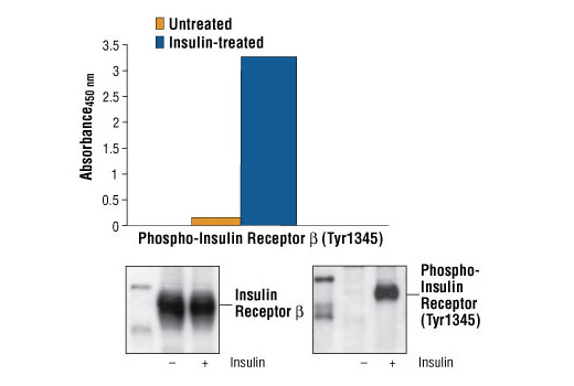
    NOTE: Initial color of positive reaction is blue, which changes to yellow upon addition of STOP solution.

  11. Read results
    1. Visual Determination: Read within 30 min after adding STOP solution.
    2. Spectrophotometric Determination: Wipe underside of wells with a lint-free tissue. Read absorbance at 450 nm within 30 min after adding STOP solution.

posted June 2005

revised November 2013

Protocol Id: 21

Specificity / Sensitivity

CST's PathScan® Phospho-Insulin Receptor β (Tyr1345) Sandwich ELISA Kit #7326 detects phospho-insulin receptor (Tyr1345) protein. As shown in Figure 1, using Phospho-Insulin Receptor β (Tyr1345) Sandwich ELISA Kit #7326, a significant induction of phospho-insulin receptor (Tyr1345) is detected in CHO-IR/IRS-1 cells treated with insulin. The levels of total insulin receptor β (phospho and nonphospho) shown by Western analysis remain unchanged. This kit detects proteins from the indicated species, as determined through in-house testing, but may also detect homologous proteins from other species.

Background

Type I insulin-like growth factor receptor (IGF-IR) is a transmembrane receptor tyrosine kinase that is widely expressed in many cell lines and cell types within fetal and postnatal tissues (1-3). Receptor autophosphorylation follows binding of the IGF-I and IGF-II ligands. Three tyrosine residues within the kinase domain (Tyr1131, Tyr1135, and Tyr1136) are the earliest major autophosphorylation sites (4). Phosphorylation of these three tyrosine residues is necessary for kinase activation (5,6). Insulin receptors (IRs) share significant structural and functional similarity with IGF-I receptors, including the presence of an equivalent tyrosine cluster (Tyr1146/1150/1151) within the kinase domain activation loop. Tyrosine autophosphorylation of IRs is one of the earliest cellular responses to insulin stimulation (7). Autophosphorylation begins with phosphorylation at Tyr1146 and either Tyr1150 or Tyr1151, while full kinase activation requires triple tyrosine phosphorylation (8).

Pathways

Explore pathways related to this product.

Limited Uses

Except as otherwise expressly agreed in a writing signed by a legally authorized representative of CST, the following terms apply to Products provided by CST, its affiliates or its distributors. Any Customer's terms and conditions that are in addition to, or different from, those contained herein, unless separately accepted in writing by a legally authorized representative of CST, are rejected and are of no force or effect.

Products are labeled with For Research Use Only or a similar labeling statement and have not been approved, cleared, or licensed by the FDA or other regulatory foreign or domestic entity, for any purpose. Customer shall not use any Product for any diagnostic or therapeutic purpose, or otherwise in any manner that conflicts with its labeling statement. Products sold or licensed by CST are provided for Customer as the end-user and solely for research and development uses. Any use of Product for diagnostic, prophylactic or therapeutic purposes, or any purchase of Product for resale (alone or as a component) or other commercial purpose, requires a separate license from CST. Customer shall (a) not sell, license, loan, donate or otherwise transfer or make available any Product to any third party, whether alone or in combination with other materials, or use the Products to manufacture any commercial products, (b) not copy, modify, reverse engineer, decompile, disassemble or otherwise attempt to discover the underlying structure or technology of the Products, or use the Products for the purpose of developing any products or services that would compete with CST products or services, (c) not alter or remove from the Products any trademarks, trade names, logos, patent or copyright notices or markings, (d) use the Products solely in accordance with CST Product Terms of Sale and any applicable documentation, and (e) comply with any license, terms of service or similar agreement with respect to any third party products or services used by Customer in connection with the Products.

For Research Use Only. Not for Use in Diagnostic Procedures.
Cell Signaling Technology is a trademark of Cell Signaling Technology, Inc.
PathScan is a trademark of Cell Signaling Technology, Inc.
All other trademarks are the property of their respective owners. Visit our Trademark Information page.