| Cat. # | Size | Qty. | Price |
|---|---|---|---|
| 9038S | 1 Kit (20 assays) |
|
| Product Includes | Quantity | ||||
|---|---|---|---|---|---|
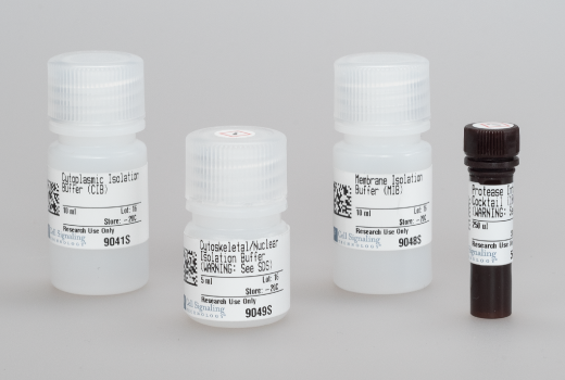
| Cytoplasmic Isolation Buffer (CIB) | 10 ml | ||||
| Membrane Isolation Buffer (MIB) | 10 ml | ||||
| Cytoskeletal/Nuclear Isolation Buffer (CyNIB) | 5 ml | ||||
| Protease Inhibitor Cocktail (100X) 5871 | 250 µl |
Product Information
For western blots, incubate membrane with diluted primary antibody in 5% w/v BSA, 1X TBS, 0.1% Tween® 20 at 4°C with gentle shaking, overnight.
NOTE: Please refer to primary antibody product webpage for recommended antibody dilution.
From sample preparation to detection, the reagents you need for your Western Blot are now in one convenient kit: #12957 Western Blotting Application Solutions Kit
NOTE: Prepare solutions with reverse osmosis deionized (RODI) or equivalent grade water.
Load 20 µl onto SDS-PAGE gel (10 cm x 10 cm).
NOTE: Loading of prestained molecular weight markers (#59329, 10 µl/lane) to verify electrotransfer and biotinylated protein ladder (#7727, 10 µl/lane) to determine molecular weights are recommended.
NOTE: Volumes are for 10 cm x 10 cm (100 cm2) of membrane; for different sized membranes, adjust volumes accordingly.
* Avoid repeated exposure to skin.
posted June 2005
revised June 2020
Protocol Id: 10
For adherent cells
For both adherent and suspension cells
Table 1: Volumes in μl for WCL or buffer at indicated cell numbers.
| Cell Count | ||||
| 2.5 x 10⁶ cells | 5 x 10⁶ cells | 7.5 x 10⁶ cells | 1 x 10⁷ cells | |
| WCL | 50 | 100 | 150 | 200 |
| CIB | 250 | 500 | 750 | 1000 |
| MIB | 250 | 500 | 750 | 1000 |
| CyNIB | 125 | 250 | 375 | 500 |
Protocol Id: 584
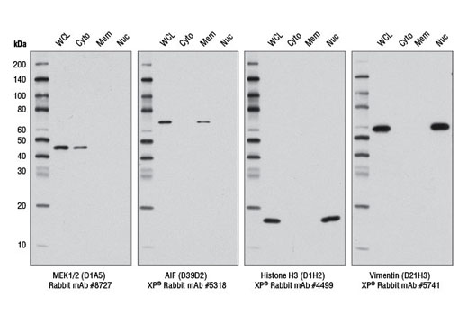
Cellular fractionation allows for the extraction of cellular proteins into distinct compartments. This is achieved by the use of detergents that take advantage of the inherent qualities and composition of different cellular membranes (1). Cellular fractionation has been important for defining the localization of many proteins, observing the translocation of proteins, and determining protein-protein complexes, such as cytoskeletal-associated proteins (2,3). Thus, detergent-based cellular fractionation separates cellular components with greater ease and speed compared to a more laborious density centrifugation method (4).
Except as otherwise expressly agreed in a writing signed by a legally authorized representative of CST, the following terms apply to Products provided by CST, its affiliates or its distributors. Any Customer's terms and conditions that are in addition to, or different from, those contained herein, unless separately accepted in writing by a legally authorized representative of CST, are rejected and are of no force or effect.
Products are labeled with For Research Use Only or a similar labeling statement and have not been approved, cleared, or licensed by the FDA or other regulatory foreign or domestic entity, for any purpose. Customer shall not use any Product for any diagnostic or therapeutic purpose, or otherwise in any manner that conflicts with its labeling statement. Products sold or licensed by CST are provided for Customer as the end-user and solely for research and development uses. Any use of Product for diagnostic, prophylactic or therapeutic purposes, or any purchase of Product for resale (alone or as a component) or other commercial purpose, requires a separate license from CST. Customer shall (a) not sell, license, loan, donate or otherwise transfer or make available any Product to any third party, whether alone or in combination with other materials, or use the Products to manufacture any commercial products, (b) not copy, modify, reverse engineer, decompile, disassemble or otherwise attempt to discover the underlying structure or technology of the Products, or use the Products for the purpose of developing any products or services that would compete with CST products or services, (c) not alter or remove from the Products any trademarks, trade names, logos, patent or copyright notices or markings, (d) use the Products solely in accordance with CST Product Terms of Sale and any applicable documentation, and (e) comply with any license, terms of service or similar agreement with respect to any third party products or services used by Customer in connection with the Products.