| Cat. # | Size | Qty. | Price |
|---|---|---|---|
| 12704T | 1 Kit (4 x 20 microliters) |
|
| Product Includes | Quantity | Applications | Reactivity | MW(kDa) | Isotype |
|---|---|---|---|---|---|
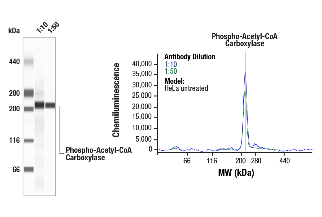
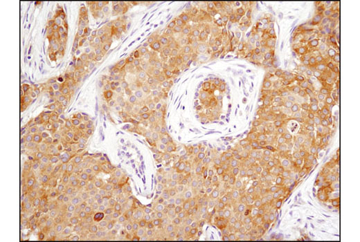
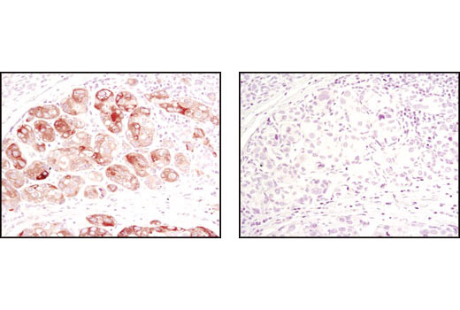
| Phospho-Acetyl-CoA Carboxylase (Ser79) (D7D11) Rabbit mAb 11818 | 20 µl |
|
H M R | 280 | Rabbit IgG |
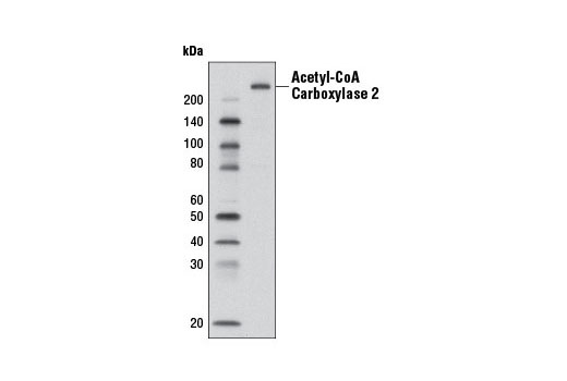
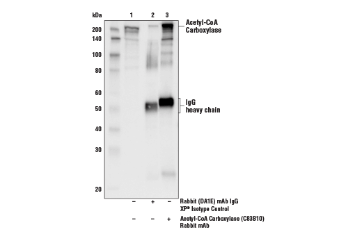
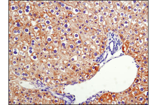
| Acetyl-CoA Carboxylase (C83B10) Rabbit mAb 3676 | 20 µl |
|
H M R Hm | 280 | Rabbit IgG |
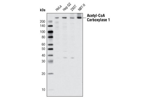
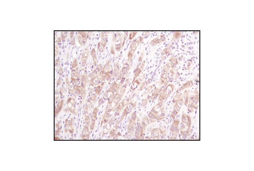
| Acetyl-CoA Carboxylase 1 Antibody 4190 | 20 µl |
|
H M R | 265 | Rabbit |
| Acetyl-CoA Carboxylase 2 (D5B9) Rabbit mAb 8578 | 20 µl |
|
H | 280 | Rabbit IgG |
| Anti-rabbit IgG, HRP-linked Antibody 7074 | 100 µl |
|
Goat |
Product Information
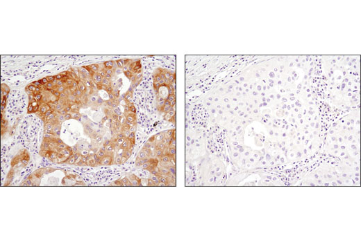
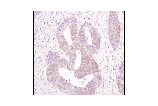
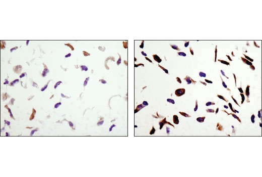
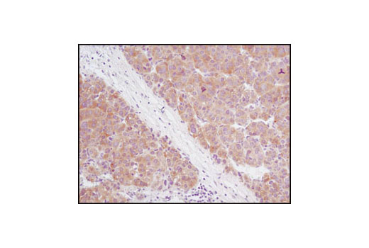
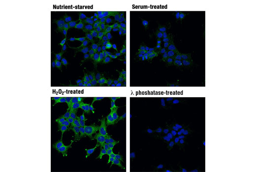
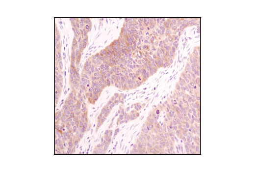
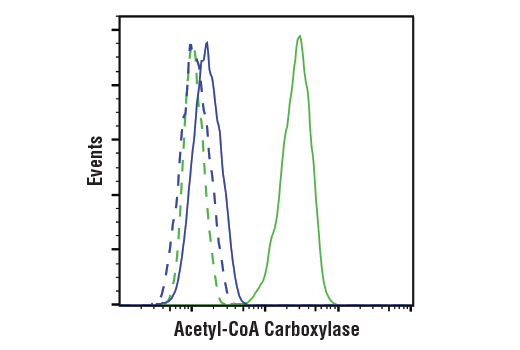
Polyclonal antibodies are produced by immunizing animals with a synthetic peptide corresponding to the sequence of human acetyl-CoA carboxylase 1 protein. Polyclonal antibodies are purified by protein A and peptide affinity chromatography. Monoclonal antibodies are produced by immunizing animals with a synthetic phosphopeptide corresponding to residues surrounding Ser79 of human acetyl-CoA carboxylase protein, or a synthetic peptide corresponding to residues surrounding Ser523 of human acetyl-CoA carboxylase α1, or Val1416 of human acetyl-CoA carboxylase 2 protein.
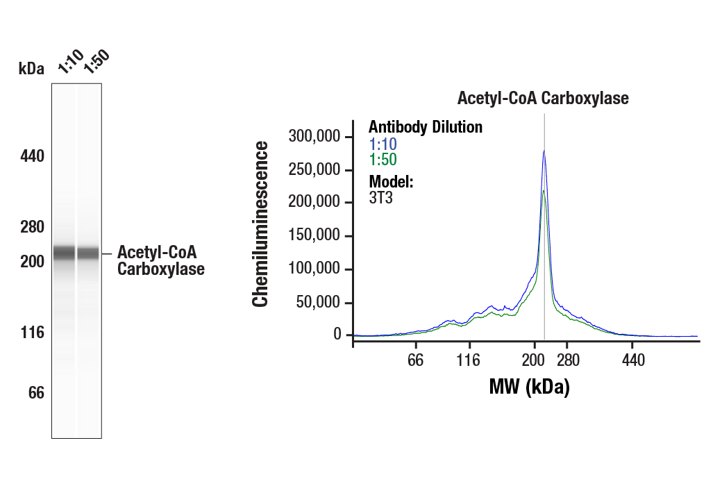
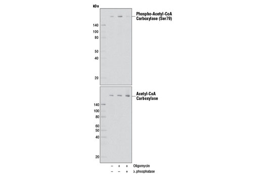
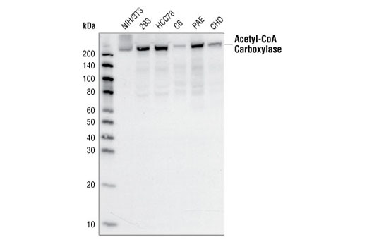
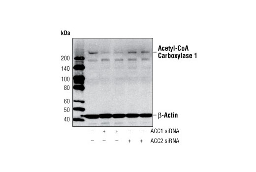
Acetyl-CoA carboxylase (ACC) catalyzes the carboxylation of acetyl-CoA to malonyl-CoA (1). It is the key enzyme in the biosynthesis and oxidation of fatty acids (1). In rodents, the 265 kDa ACC1 (ACCα) form is primarily expressed in lipogenic tissues, while 280 kDa ACC2 (ACCβ) is the main isoform in oxidative tissues (1,2). However, in humans, ACC2 is the predominant isoform in both lipogenic and oxidative tissues (1,2). Phosphorylation by AMPK at Ser79 or by PKA at Ser1200 inhibits the enzymatic activity of ACC (3). ACC is a potential target of anti-obesity drugs (4,5).
Except as otherwise expressly agreed in a writing signed by a legally authorized representative of CST, the following terms apply to Products provided by CST, its affiliates or its distributors. Any Customer's terms and conditions that are in addition to, or different from, those contained herein, unless separately accepted in writing by a legally authorized representative of CST, are rejected and are of no force or effect.
Products are labeled with For Research Use Only or a similar labeling statement and have not been approved, cleared, or licensed by the FDA or other regulatory foreign or domestic entity, for any purpose. Customer shall not use any Product for any diagnostic or therapeutic purpose, or otherwise in any manner that conflicts with its labeling statement. Products sold or licensed by CST are provided for Customer as the end-user and solely for research and development uses. Any use of Product for diagnostic, prophylactic or therapeutic purposes, or any purchase of Product for resale (alone or as a component) or other commercial purpose, requires a separate license from CST. Customer shall (a) not sell, license, loan, donate or otherwise transfer or make available any Product to any third party, whether alone or in combination with other materials, or use the Products to manufacture any commercial products, (b) not copy, modify, reverse engineer, decompile, disassemble or otherwise attempt to discover the underlying structure or technology of the Products, or use the Products for the purpose of developing any products or services that would compete with CST products or services, (c) not alter or remove from the Products any trademarks, trade names, logos, patent or copyright notices or markings, (d) use the Products solely in accordance with CST Product Terms of Sale and any applicable documentation, and (e) comply with any license, terms of service or similar agreement with respect to any third party products or services used by Customer in connection with the Products.