View Featured Offers >>
12784
Nucleus and Nuclear Envelope-Associated Marker Proteins Antibody Sampler Kit
Primary Antibodies
Antibody Sampler Kit

Nucleus and Nuclear Envelope-Associated Marker Proteins Antibody Sampler Kit #12784

Citations (0)
Confocal immunofluorescent analysis of fixed frozen mouse liver using Fibrillarin (C13C3) Rabbit mAb (left, green), DyLight 554 Phalloidin #13054 (right, red), and DAPI #8961 (right, blue).
Confocal immunofluorescent analysis of fixed frozen mouse kidney using Fibrillarin (C13C3) Rabbit mAb (left, green), DyLight 554 Phalloidin #13054 (right, red), and DAPI #8961 (right, blue).
Flow cytometric analysis of Daudi cells (blue) and MJ cells (green) using Lamin A/C (4C11) Mouse mAb (solid lines) or concentration-matched Mouse (G3A1) mAb IgG1 Isotype Control #5415 (dashed lines). Anti-mouse IgG (H+L), F(ab')2 Fragment (Alexa Fluor® 488 Conjugate) #4408 was used as a secondary antibody.
Simple Western™ analysis of lysates (0.1 mg/mL) from HeLa cells using Lamin A/C (4C11) Mouse mAb #4777. The virtual lane view (left) shows the target bands (as indicated) at 1:10 and 1:50 dilutions of primary antibody. The corresponding electropherogram view (right) plots chemiluminescence by molecular weight along the capillary at 1:10 (blue line) and 1:50 (green line) dilutions of primary antibody. This experiment was performed under reducing conditions on the Jess™ Simple Western instrument from ProteinSimple, a BioTechne brand, using the 12-230 kDa separation module.
Western blot analysis of cell lysates from various cell types using LSD1 (C69G12) Rabbit mAb.
Western blot analysis of cell lysates from MCF7 and 293 cells using ESET (C1C12) Rabbit mAb.
Western blot analysis of cell extracts from various cell types using NUP98 (C39A3) Rabbit mAb.
Western blot analysis of extracts from various cell types using Fibrillarin (C13C3) Antibody.
Western blot analysis of HeLa, NIH/3T3, H-4-II-E and COS cell lysates, in addition to 10 ng of recombinant H2A (rH2A) and H2A.Z (rH2A.Z) protein, using Histone H2A.Z Antibody #2718 (upper) and Histone H2A Antibody II #2578 (lower).
Western blot analysis of extracts from various cell lines using Histone H3 (D1H2) XP® Rabbit mAb.
Immunohistochemical analysis of paraffin-embedded human breast carcinoma using Histone H3 (D1H2) XP® Rabbit mAb.
Western blot analysis of extracts from control HeLa cells (lane 1) or HeLa cells with an apparent in-frame truncation mutation in the gene encoding LMNA (lane 2) using Lamin A/C (4C11) Mouse mAb #4777 (upper) or α-actinin (D6F6) XP® Rabbit mAb #6487 (lower). The change in LMNA molecular weight in the mutated HeLa cells is consistent with an in-frame deletion.
After the primary antibody is bound to the target protein, a complex with HRP-linked secondary antibody is formed. The LumiGLO® is added and emits light during enzyme catalyzed decomposition.
After the primary antibody is bound to the target protein, a complex with HRP-linked secondary antibody is formed. The LumiGLO* is added and emits light during enzyme catalyzed decomposition.
Immunohistochemical analysis of paraffin-embedded human breast carcinoma using LSD1 (C69G12) Rabbit mAb.
Confocal immunofluorescent analysis of MCF-7 cells using ESET (C1C12) Rabbit mAb (green). Actin filaments have been labeled with Alexa Fluor® 555 phalloidin (red).
Confocal immunofluorescent analysis of Caki-1 cells using NUP98 (C39A3) Rabbit mAb (green). Actin filaments have been labeled with Alexa Fluor® 555 phalloidin (red).
Confocal immunofluorescent analysis of HeLa cells using Fibrillarin (C13C3) Rabbit mAb (green). Actin filaments have been labeled with Alexa Fluor® 555 phalloidin (red). Blue pseudocolor = DRAQ5® (fluorescent DNA dye).
Confocal immunofluorescent analysis of HeLa cells, using Histone H2A.Z Antibody (green). Actin filaments have been labeled with Alexa Fluor® 555 phalloidin (red).
Immunohistochemical analysis of paraffin-embedded 4T1 syngeneic mammary tumor using Histone H3 (D1H2) XP® Rabbit mAb.
Western blot analysis of extracts from various cell lines using Lamin A/C (4C11) Mouse mAb.
Immunohistochemical analysis of paraffin-embedded human lung carcinoma using LSD1 (C69G12) Rabbit mAb.
Confocal immunofluorescent analysis of HeLa cells using Histone H3 (D1H2) XP® Rabbit mAb (green) and β-Tubulin (9F3) Rabbit mAb (Alexa Fluor® 555 Conjugate) #2116 (red).
Immunohistochemical analysis of paraffin-embedded LL/2 syngeneic tumor using Histone H3 (D1H2) XP® Rabbit mAb.
Western blot analysis of extracts from THP-1 cells, untreated or treated with cycloheximide (CHX, 10 μg/ml, overnight) followed by TNF-α #8902 (20 ng/ml, 4 hr), using Lamin A/C (4C11) Mouse mAb.
Immunohistochemical analysis of paraffin-embedded human stomach carcinoma using LSD1 (C69G12) Rabbit mAb.
Flow cytometric analysis of HeLa cells using Histone H3 (D1H2) XP® Rabbit mAb (solid line) or a concentration-matched Rabbit (DA1E) mAb IgG XP® Isotype Control #3900 (dashed line). Anti-rabbit IgG (H+L), F(ab')2 Fragment (Alexa Fluor® 488 Conjugate) #4412 was used as a secondary antibody.
Immunohistochemical analysis of paraffin-embedded mouse brain using Histone H3 (D1H2) XP® Rabbit mAb.
Immunohistochemical analysis of paraffin-embedded human breast carcinoma using Lamin A/C (4C11) Mouse mAb.
Immunohistochemical analysis of paraffin-embedded rhesus monkey liver using Histone H3 (D1H2) XP® Rabbit mAb.
Immunohistochemical analysis of paraffin-embedded human colon carcinoma using Lamin A/C (4C11) Mouse mAb.
Immunofluorescent analysis of normal rat brain using Lamin A/C (4C11) Mouse mAb (green) and MAP2 Antibody #4542 (red).
Confocal immunofluorescent analysis of HeLa cells using LSD1 (C69G12) Rabbit mAb (green). Actin filaments have been labeled with Alexa Fluor® 555 phalloidin (red).
Confocal immunofluorescent analysis of HeLa cells using Lamin A/C (4C11) Mouse mAb (green). Actin filaments were labeled with DY-554 phalloidin (red).
Flow cytometric analysis of HeLa cells (green) using Lamin A/C (4C11) Mouse mAb (solid lines) or a concentration matched Mouse (G3A1) mAb IgG Isotype Control #5415 (dashed lines). Anti-mouse IgG (H+L), F(ab')2 Fragment (Alexa Fluor® 488 Conjugate) #4408 was used as a secondary antibody.
Chromatin immunoprecipitations were performed with cross-linked chromatin from U-937 cells and either LSD1 (C69G12) Rabbit mAb or Normal Rabbit IgG #2729 using SimpleChIP® Enzymatic Chromatin IP Kit (Magnetic Beads) #9003. The enriched DNA was quantified by real-time PCR SimpleChIP® Human CD11b Promoter Primers #14271, human SPI-1 promoter primers, and SimpleChIP® Human α Satellite Repeat Primers #4486. The amount of immunoprecipitated DNA in each sample is represented as signal relative to the total amount of input chromatin, which is equivalent to one.
Inquiry Info.# 12784

Product Description

The Nucleus and Nuclear Envelope-Associated Marker Proteins Antibody Sampler Kit provides an economical means to evaluate relevant nuclear proteins. This kit contains enough primary antibody to perform at least four western blots per primary antibody.

Specificity / Sensitivity

Each antibody in the Nucleus and Nuclear Envelope-Associated Marker Proteins Antibody Sampler Kit recognizes total endogenous levels of the specific target protein.

Source / Purification

Monoclonal antibody is produced by immunizing animals with a synthetic peptide corresponding to the carboxy terminus of the human ESET protein, residues surrounding Thr298 of human fibrillarin, the carboxy terminus of the human histone H3 protein, a recombinant fragment of human lamin A protein, residues near the amino-terminus of human LSD1 protein, or residues surrounding Pro671 of human NUP98. Polyclonal antibodies are produced by immunizing animals with a synthetic peptide corresponding to the carboxy terminus of human histone H2A.Z. Polyclonal antibodies are purified by protein A and peptide affinity chromatography.

Background

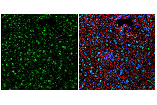
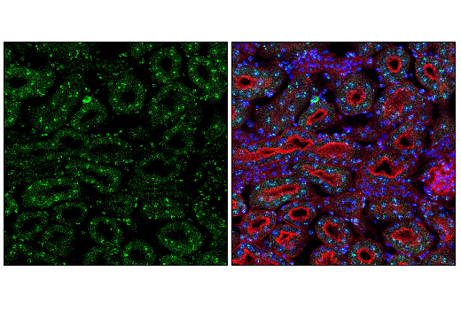
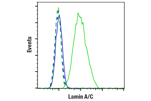
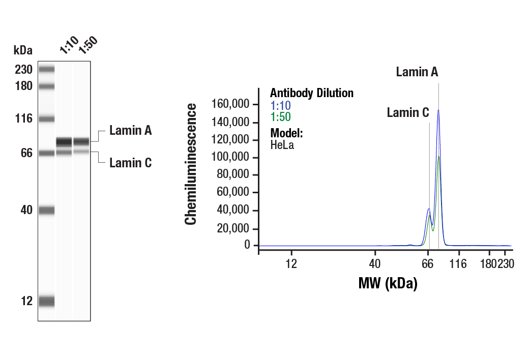
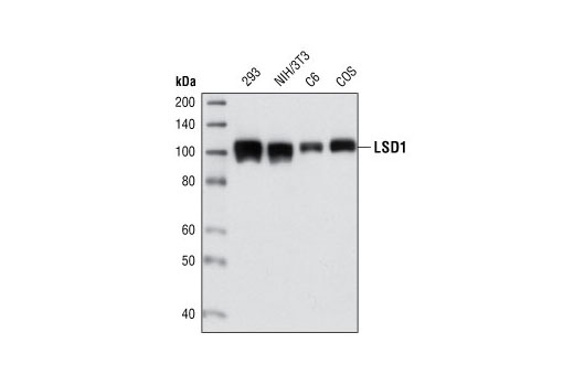
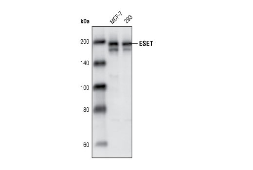
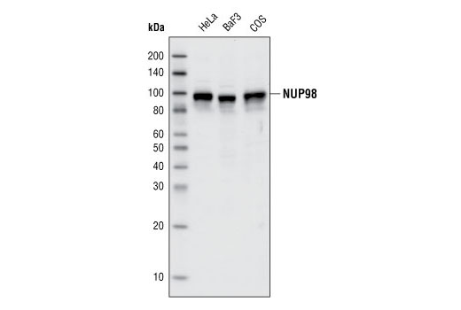
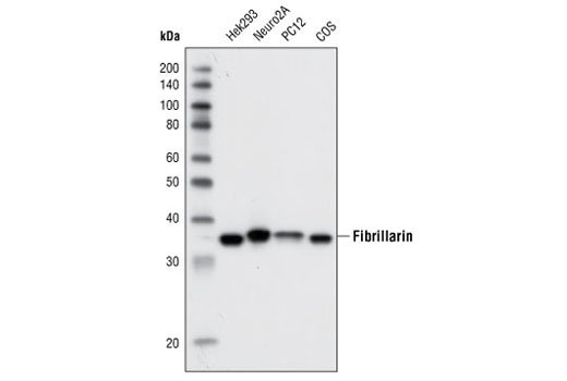
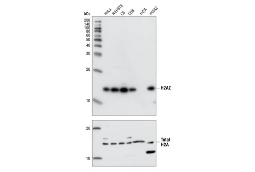
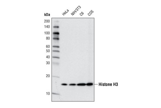
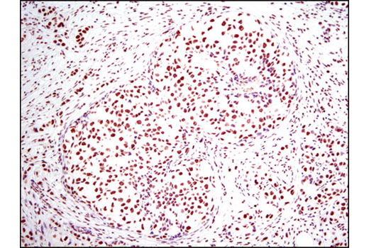
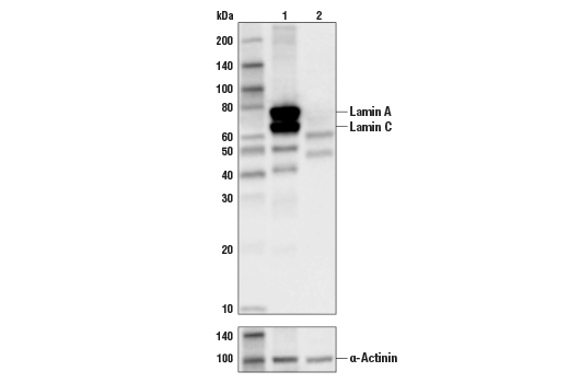
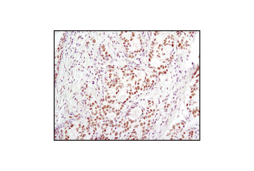
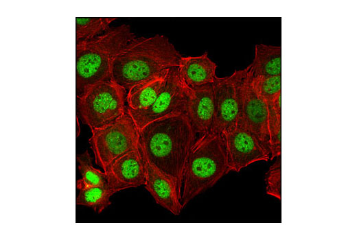
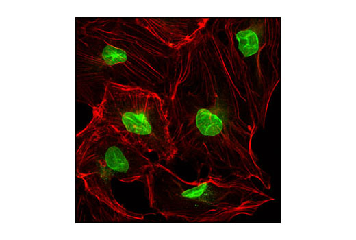
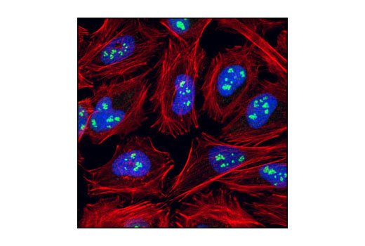
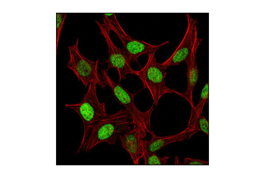
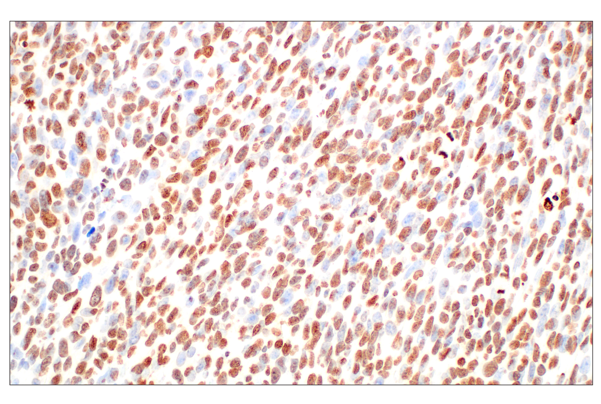
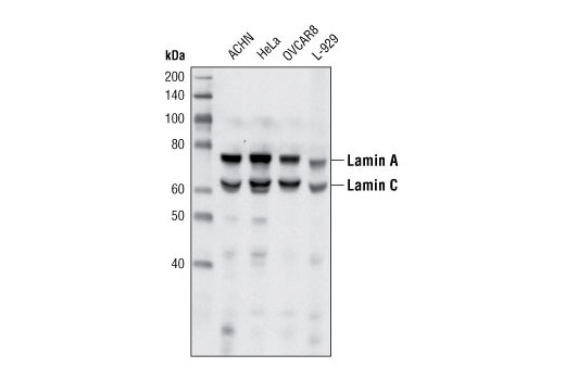
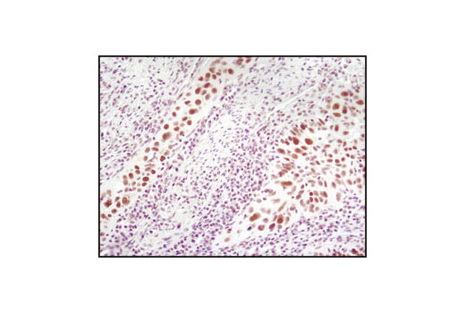
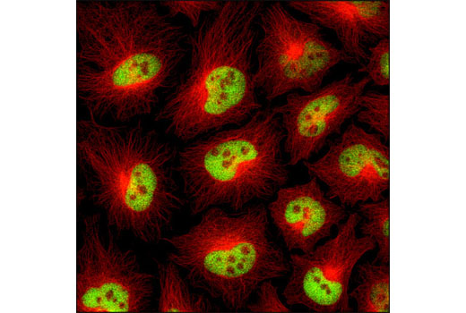
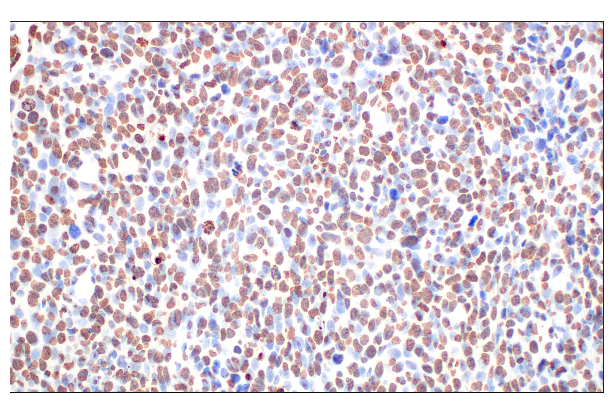
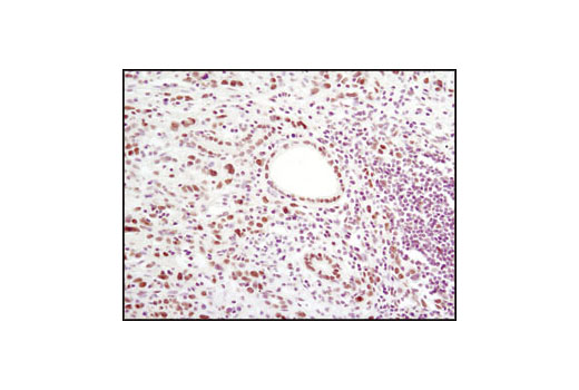
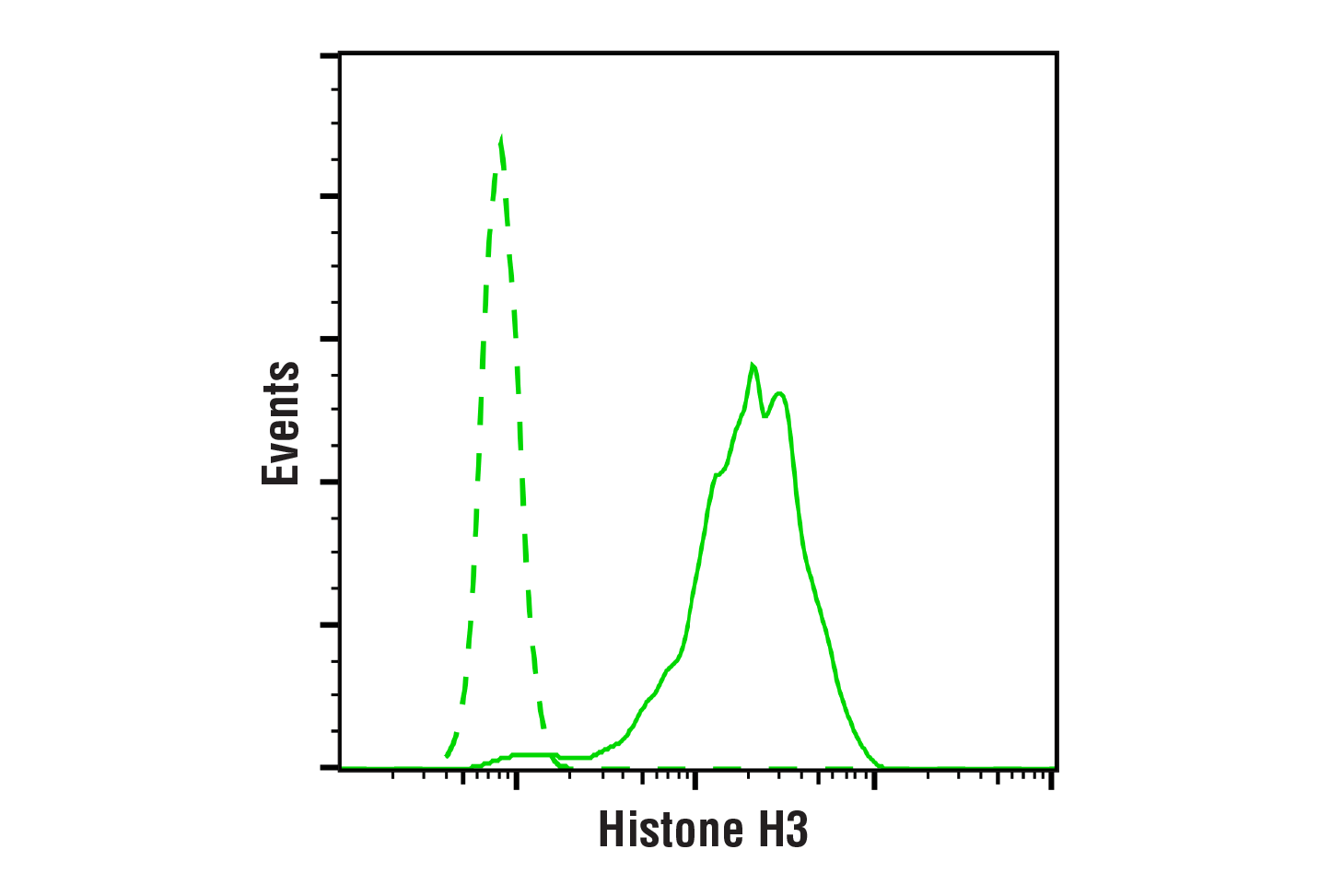
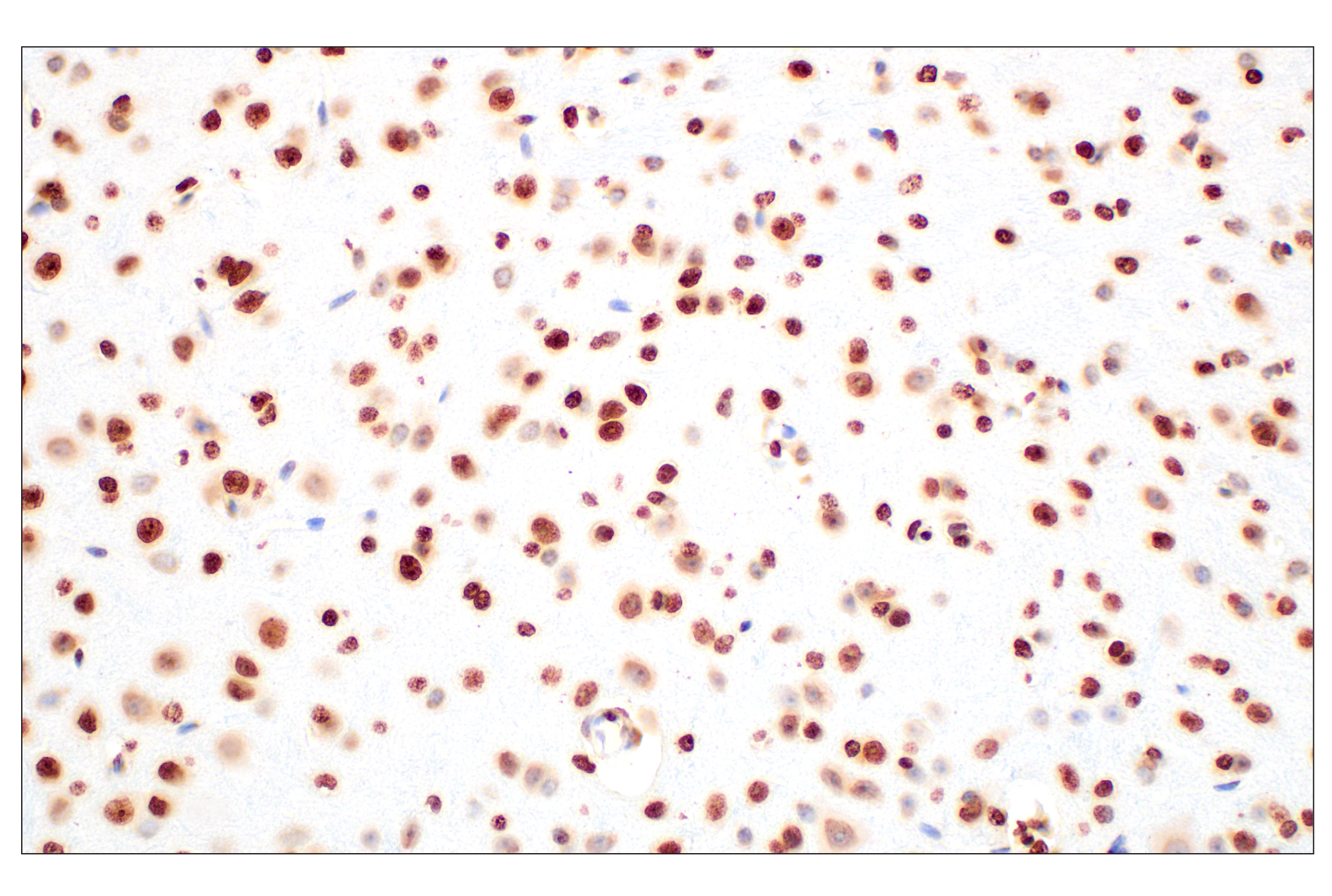
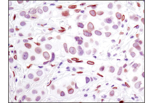
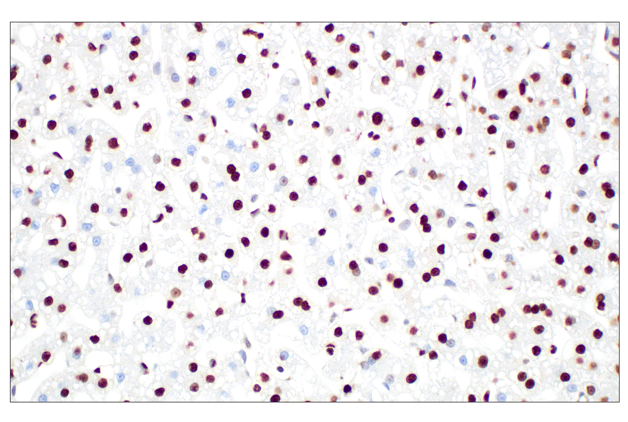
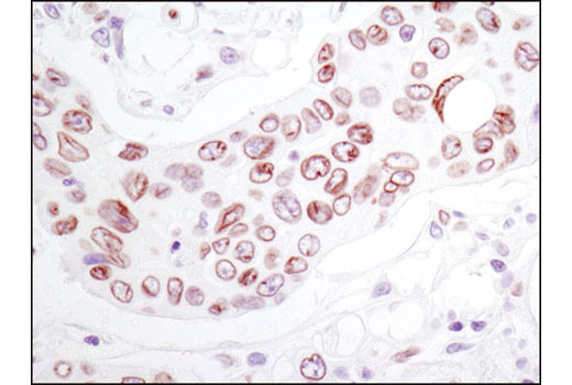
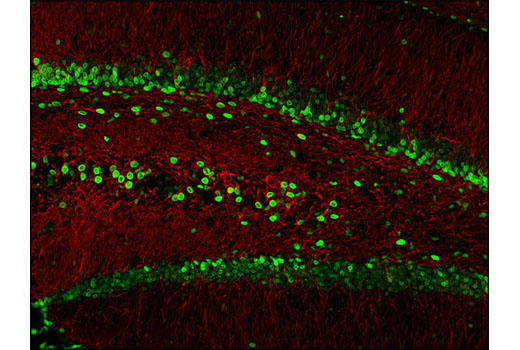
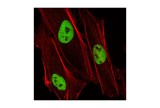
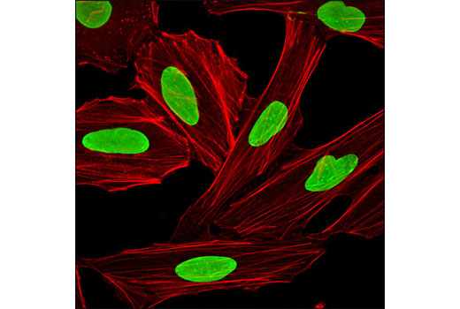
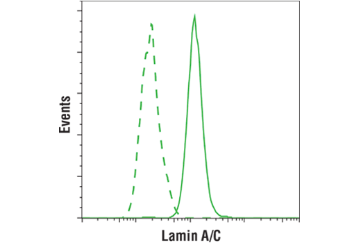
The Nucleus and Nuclear Envelope-Associated Marker Proteins Antibody Sampler Kit contains a variety of antibodies directed against established nuclear proteins (1). Histone H3 and histone H2A.Z are histone family members and components of nucleosomes, the primary building block of chromatin made up of DNA wound around eight core histone proteins. The amino-terminal tails of core histones undergo various post-translational modifications and have a direct effect on the accessibility of chromatin to transcription factors and, therefore, gene expression (2). ESET histone methyltransferase (3) and LSD1 histone demethylase (4) are both regulators of histone methylation and are chromatin-associated. Both NUP98 (5) and lamins (6) are located within the nuclear envelope (also known as the nuclear membrane). NUP98 is a component of the nuclear pore complex. Lamin A and lamin C are fibrous proteins contributing to nuclear structural and transcriptional regulation. Finally, fibrillarin (7) is located in fibrillar regions and Cajal bodies of nucleoli, where it functions to regulate RNA transcription and pre-rRNA processing.

Limited Uses

Except as otherwise expressly agreed in a writing signed by a legally authorized representative of CST, the following terms apply to Products provided by CST, its affiliates or its distributors. Any Customer's terms and conditions that are in addition to, or different from, those contained herein, unless separately accepted in writing by a legally authorized representative of CST, are rejected and are of no force or effect.

Products are labeled with For Research Use Only or a similar labeling statement and have not been approved, cleared, or licensed by the FDA or other regulatory foreign or domestic entity, for any purpose. Customer shall not use any Product for any diagnostic or therapeutic purpose, or otherwise in any manner that conflicts with its labeling statement. Products sold or licensed by CST are provided for Customer as the end-user and solely for research and development uses. Any use of Product for diagnostic, prophylactic or therapeutic purposes, or any purchase of Product for resale (alone or as a component) or other commercial purpose, requires a separate license from CST. Customer shall (a) not sell, license, loan, donate or otherwise transfer or make available any Product to any third party, whether alone or in combination with other materials, or use the Products to manufacture any commercial products, (b) not copy, modify, reverse engineer, decompile, disassemble or otherwise attempt to discover the underlying structure or technology of the Products, or use the Products for the purpose of developing any products or services that would compete with CST products or services, (c) not alter or remove from the Products any trademarks, trade names, logos, patent or copyright notices or markings, (d) use the Products solely in accordance with CST Product Terms of Sale and any applicable documentation, and (e) comply with any license, terms of service or similar agreement with respect to any third party products or services used by Customer in connection with the Products.

For Research Use Only. Not for Use in Diagnostic Procedures.
Cell Signaling Technology is a trademark of Cell Signaling Technology, Inc.
All other trademarks are the property of their respective owners. Visit our Trademark Information page.