| REACTIVITY | H M R Mk |
| SENSITIVITY | Endogenous |
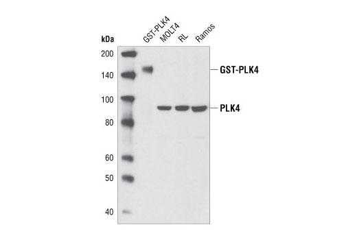
| MW (kDa) | 95 |
| SOURCE | Rabbit |
Product Information
| Application | Dilution |
|---|---|
| Western Blotting | 1:1000 |
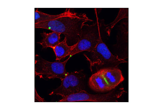
| Immunofluorescence (Immunocytochemistry) | 1:100 |
For western blots, incubate membrane with diluted primary antibody in 5% w/v BSA, 1X TBS, 0.1% Tween® 20 at 4°C with gentle shaking, overnight.
NOTE: Please refer to primary antibody product webpage for recommended antibody dilution.
From sample preparation to detection, the reagents you need for your Western Blot are now in one convenient kit: #12957 Western Blotting Application Solutions Kit
NOTE: Prepare solutions with reverse osmosis deionized (RODI) or equivalent grade water.
Load 20 µl onto SDS-PAGE gel (10 cm x 10 cm).
NOTE: Loading of prestained molecular weight markers (#59329, 10 µl/lane) to verify electrotransfer and biotinylated protein ladder (#7727, 10 µl/lane) to determine molecular weights are recommended.
NOTE: Volumes are for 10 cm x 10 cm (100 cm2) of membrane; for different sized membranes, adjust volumes accordingly.
* Avoid repeated exposure to skin.
posted June 2005
revised June 2020
Protocol Id: 10
Achieve higher quality immunofluorescent images using the efficient and cost-effective, pre-made reagents in our #12727 Immunofluorescence Application Solutions Kit
NOTE: Prepare solutions with reverse osmosis deionized (RODI) or equivalent grade water.
Recommended Fluorochrome-conjugated Anti-Rabbit secondary antibodies:
NOTE: Cells should be grown, treated, fixed and stained directly in multi-well plates, chamber slides or on coverslips.
Aspirate liquid, then cover cells to a depth of 2–3 mm with 4% formaldehyde diluted in 1X PBS.
NOTE: Formaldehyde is toxic, use only in a fume hood.
NOTE: All subsequent incubations should be carried out at room temperature unless otherwise noted in a humid light-tight box or covered dish/plate to prevent drying and fluorochrome fading.
posted November 2006
revised November 2013
Protocol Id: 24
Human, Mouse, Rat, Monkey
Polyclonal antibodies are produced by immunizing animals with a synthetic peptide corresponding to residues surrounding Cys458 of human PLK4. Antibodies were purified by peptide affinity chromatography.
At least four distinct polo-like kinases exist in mammalian cells: PLK1, PLK2, PLK3, and PLK4/SAK (1). PLK1 apparently plays many roles during mitosis, particularly in regulating mitotic entry and exit. The mitosis promoting factor (MPF), cdc2/cyclin B1, is activated by dephosphorylation of cdc2 (Thr14/Tyr15) by cdc25C. PLK1 phosphorylates cdc25C at Ser198 and cyclin B1 at Ser133, causing translocation of these proteins from the cytoplasm to the nucleus (2-5). PLK1 phosphorylation of Myt1 at Ser426 and Thr495 has been proposed to inactivate Myt1, one of the kinases known to phosphorylate cdc2 at Thr14/Tyr15 (6). Polo-like kinases also phosphorylate the cohesin subunit SCC1, causing cohesin displacement from chromosome arms that allow for proper cohesin localization to centromeres (7). Mitotic exit requires activation of the anaphase promoting complex (APC) (8), a ubiquitin ligase responsible for removal of cohesin at centromeres, and degradation of securin, cyclin A, cyclin B1, Aurora A, and cdc20 (9). PLK1 phosphorylation of the APC subunits Apc1, cdc16, and cdc27 has been demonstrated in vitro and has been proposed as a mechanism by which mitotic exit is regulated (10,11).
Substitution of Thr210 with Asp has been reported to elevate PLK1 kinase activity and delay/arrest cells in mitosis, while a Ser137Asp substitution leads to S-phase arrest (12). In addition, while DNA damage has been found to inhibit PLK1 kinase activity, the Thr210Asp mutant is resistant to this inhibition (13). PLK1 has been reported to be phosphorylated in vivo at Ser137 and Thr210 in mitosis; DNA damage prevents phosphorylation at these sites (14).
PLK4/SAK is transcriptionally repressed by p53 and may contribute to p53-mediated apoptosis (12). PLK4 has also been identified as a key regulator of centriole duplication (13-15).
Except as otherwise expressly agreed in a writing signed by a legally authorized representative of CST, the following terms apply to Products provided by CST, its affiliates or its distributors. Any Customer's terms and conditions that are in addition to, or different from, those contained herein, unless separately accepted in writing by a legally authorized representative of CST, are rejected and are of no force or effect.
Products are labeled with For Research Use Only or a similar labeling statement and have not been approved, cleared, or licensed by the FDA or other regulatory foreign or domestic entity, for any purpose. Customer shall not use any Product for any diagnostic or therapeutic purpose, or otherwise in any manner that conflicts with its labeling statement. Products sold or licensed by CST are provided for Customer as the end-user and solely for research and development uses. Any use of Product for diagnostic, prophylactic or therapeutic purposes, or any purchase of Product for resale (alone or as a component) or other commercial purpose, requires a separate license from CST. Customer shall (a) not sell, license, loan, donate or otherwise transfer or make available any Product to any third party, whether alone or in combination with other materials, or use the Products to manufacture any commercial products, (b) not copy, modify, reverse engineer, decompile, disassemble or otherwise attempt to discover the underlying structure or technology of the Products, or use the Products for the purpose of developing any products or services that would compete with CST products or services, (c) not alter or remove from the Products any trademarks, trade names, logos, patent or copyright notices or markings, (d) use the Products solely in accordance with CST Product Terms of Sale and any applicable documentation, and (e) comply with any license, terms of service or similar agreement with respect to any third party products or services used by Customer in connection with the Products.