| Cat. # | Size | Qty. | Price |
|---|---|---|---|
| 4749T | 1 Kit (7 x 20 microliters) |
|
| Product Includes | Quantity | Applications | Reactivity | MW(kDa) | Isotype |
|---|---|---|---|---|---|
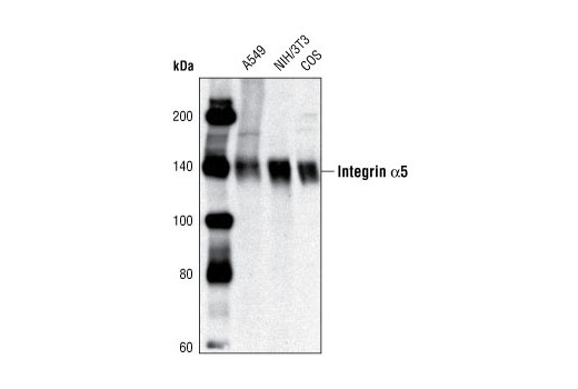
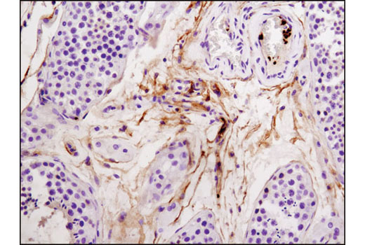
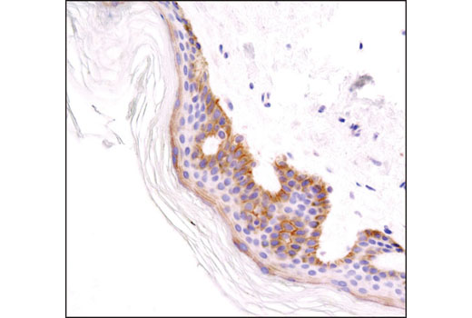
| Integrin α5 Antibody 4705 | 20 µl |
|
H M Mk | 150 | Rabbit |
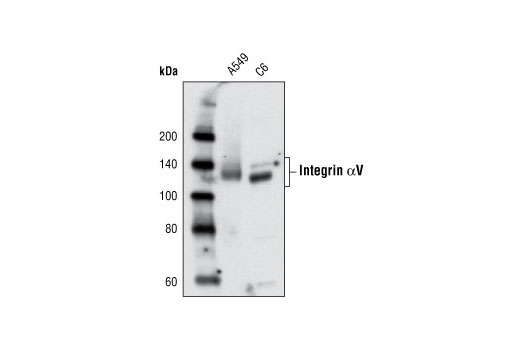
| Integrin αV Antibody 4711 | 20 µl |
|
H R | 135, 140 | Rabbit |
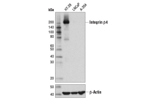
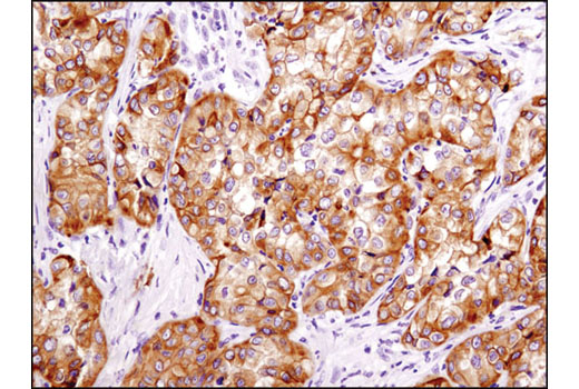
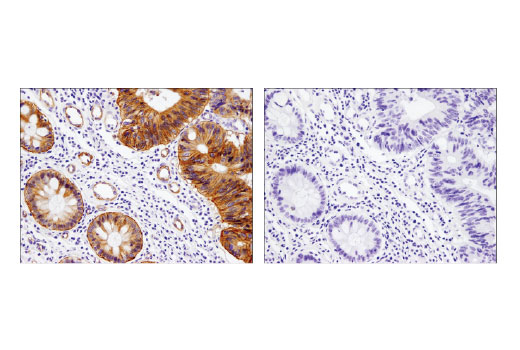
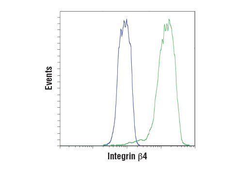
| Integrin β4 (D8P6C) XP® Rabbit mAb 14803 | 20 µl |
|
H | 210 | Rabbit IgG |
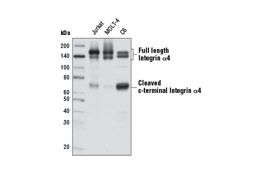
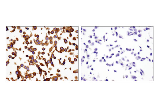
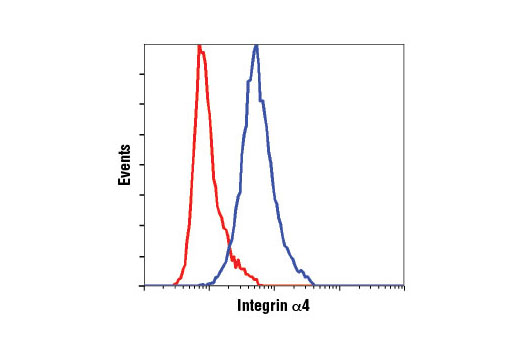
| Integrin α4 (D2E1) XP® Rabbit mAb 8440 | 20 µl |
|
H M R | 70, 140, 150, | Rabbit IgG |
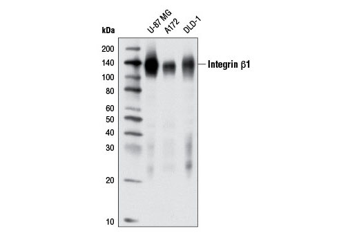
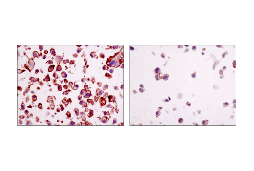
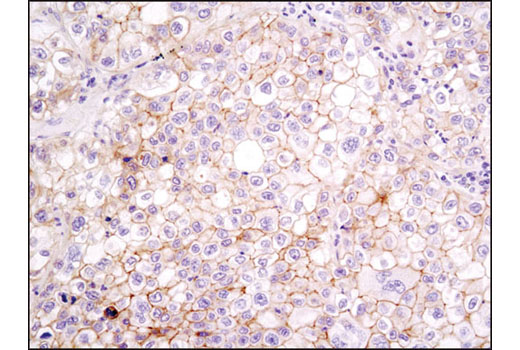
| Integrin β1 (D2E5) Rabbit mAb 9699 | 20 µl |
|
H | 115, 135 | Rabbit IgG |
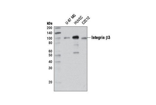
| Integrin β3 (D7X3P) XP® Rabbit mAb 13166 | 20 µl |
|
H M | 100 | Rabbit IgG |
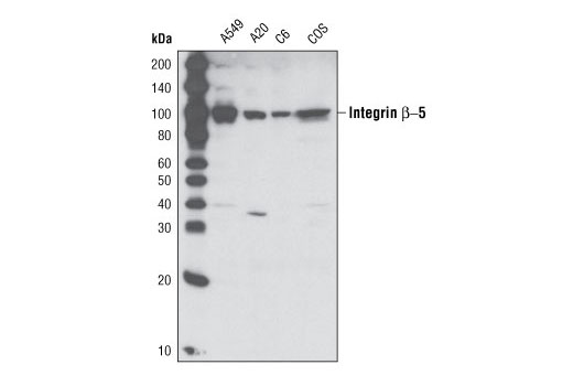
| Integrin β5 (D24A5) Rabbit mAb 3629 | 20 µl |
|
H M R Mk | 90 | Rabbit IgG |
| Anti-rabbit IgG, HRP-linked Antibody 7074 | 100 µl |
|
Rab | Goat |
Product Information
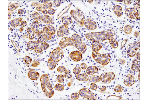
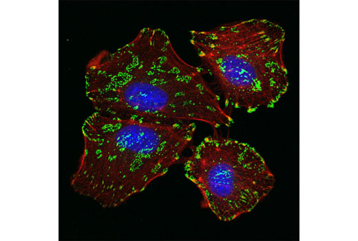
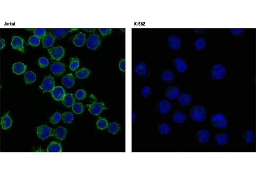
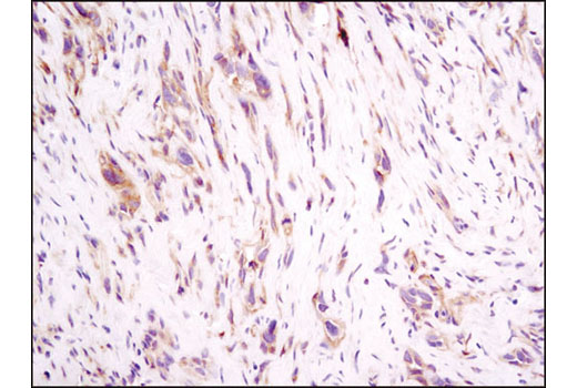
Polyclonal antibodies are produced by immunizing animals with a synthetic peptide corresponding to residues surrounding Ser458 of human integrin α5 protein, Arg108 of human integrin αV. Antibodies are purified by protein A and peptide affinity chromatography. Monoclonal antibodies are produced by immunizing animals with a synthetic peptide sequence corresponding to residues surrounding Ser1027 of human integrin a4 protein, the carboxy terminus of human integrin β4 protein, Pro680 of human integrin b1 protein, Ile114 of human integrin b3 protein, and Lys789 of human β5 protein.
Integrins are α/β heterodimeric cell surface receptors that play a pivotal role in cell adhesion and migration, as well as in growth and survival (1,2). The integrin family contains at least 18 α and 8 β subunits that form 24 known integrins with distinct tissue distribution and overlapping ligand specificities (3). Integrins not only transmit signals to cells in response to the extracellular environment (outside-in signaling), but also sense intracellular cues to alter their interaction with the extracellular environment (inside-out signaling) (1,2).
Explore pathways related to this product.
STRING - Known and Predicted Protein-Protein Interactions.
Except as otherwise expressly agreed in a writing signed by a legally authorized representative of CST, the following terms apply to Products provided by CST, its affiliates or its distributors. Any Customer's terms and conditions that are in addition to, or different from, those contained herein, unless separately accepted in writing by a legally authorized representative of CST, are rejected and are of no force or effect.
Products are labeled with For Research Use Only or a similar labeling statement and have not been approved, cleared, or licensed by the FDA or other regulatory foreign or domestic entity, for any purpose. Customer shall not use any Product for any diagnostic or therapeutic purpose, or otherwise in any manner that conflicts with its labeling statement. Products sold or licensed by CST are provided for Customer as the end-user and solely for research and development uses. Any use of Product for diagnostic, prophylactic or therapeutic purposes, or any purchase of Product for resale (alone or as a component) or other commercial purpose, requires a separate license from CST. Customer shall (a) not sell, license, loan, donate or otherwise transfer or make available any Product to any third party, whether alone or in combination with other materials, or use the Products to manufacture any commercial products, (b) not copy, modify, reverse engineer, decompile, disassemble or otherwise attempt to discover the underlying structure or technology of the Products, or use the Products for the purpose of developing any products or services that would compete with CST products or services, (c) not alter or remove from the Products any trademarks, trade names, logos, patent or copyright notices or markings, (d) use the Products solely in accordance with CST Product Terms of Sale and any applicable documentation, and (e) comply with any license, terms of service or similar agreement with respect to any third party products or services used by Customer in connection with the Products.