| Cat. # | Size | Qty. | Price |
|---|---|---|---|
| 5143T | 1 Kit (5 x 20 microliters) |
|
| Product Includes | Quantity | Applications | Reactivity | MW(kDa) | Isotype |
|---|---|---|---|---|---|
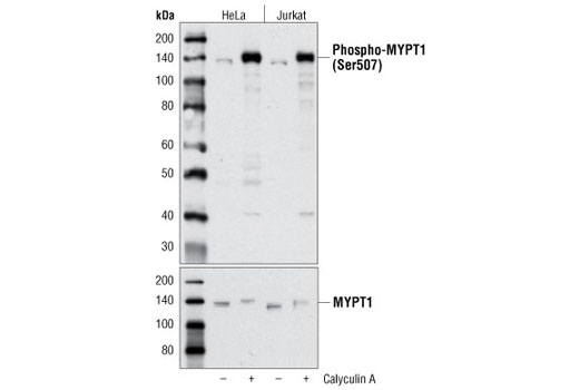
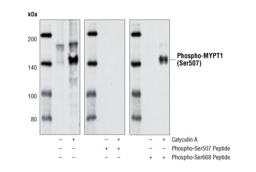
| Phospho-MYPT1 (Ser507) Antibody 3040 | 20 µl |
|
H M R | 140 | Rabbit |
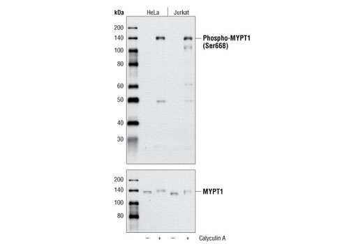
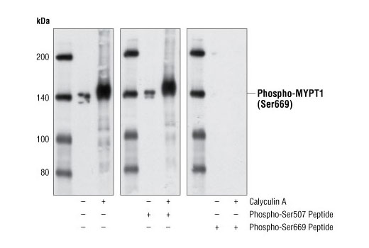
| Phospho-MYPT1 (Ser668) Antibody 3048 | 20 µl |
|
H | 140 | Rabbit |
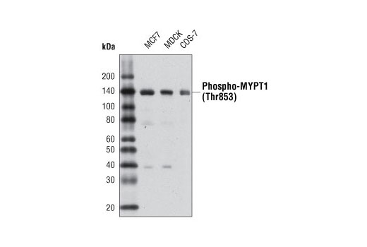
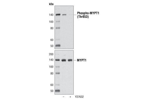
| Phospho-MYPT1 (Thr853) Antibody 4563 | 20 µl |
|
H M R Hm Mk Dg | 140 | Rabbit |
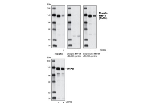
| Phospho-MYPT1 (Thr696) Antibody 5163 | 20 µl |
|
H M R Mk | 140 | Rabbit |
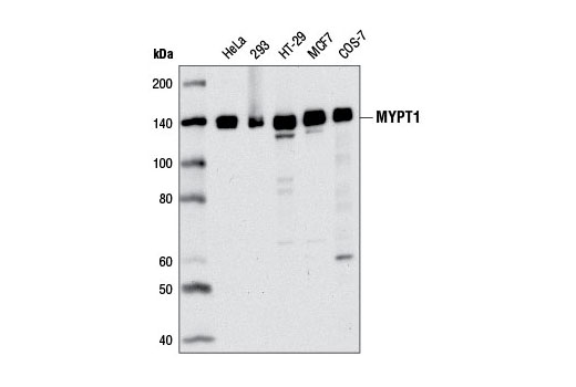
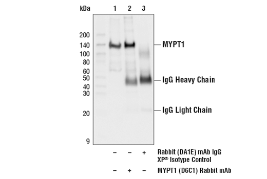
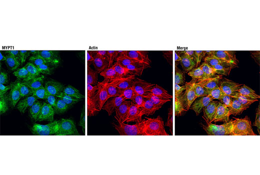
| MYPT1 (D6C1) Rabbit mAb 8574 | 20 µl |
|
H Mk Dg | 140 | Rabbit IgG |
| Anti-rabbit IgG, HRP-linked Antibody 7074 | 100 µl |
|
Rab | Goat |
Product Information
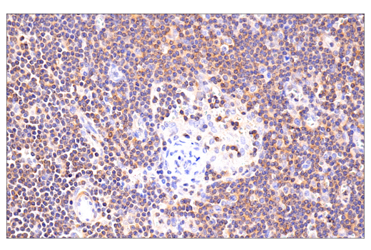
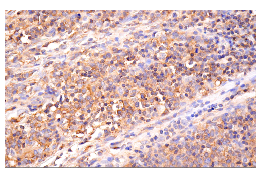
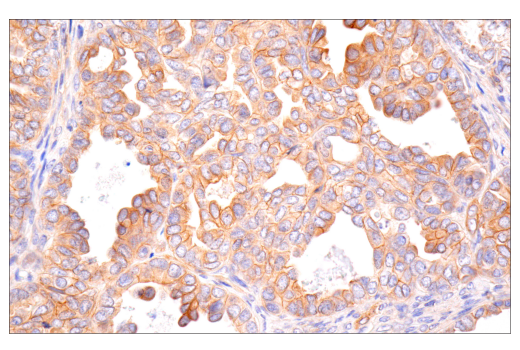
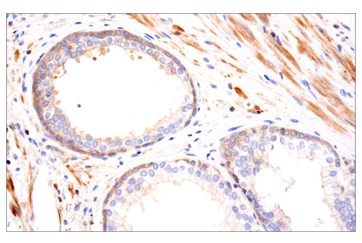
Phospho-specific polyclonal antibodies are produced by immunizing animals with synthetic phosphopeptides corresponding to residues surrounding Ser507, Ser668, Thr853, and Thr696 of human MYPT1. Monoclonal antibody is produced by immunizing animals with a synthetic peptide corresponding to residues near the carboxy terminus of human MYPT1 portein.
Protein phosphatase 1 (PP1) is a ubiquitous eukaryotic protein serine/threonine phosphatase involved in the regulation of various cell functions. Substrate specificity is determined by the binding of a regulatory subunit to the PP1 catalytic subunit (PP1c). It is estimated that over fifty different regulatory subunits exist (1).
The myosin phosphatase holoenzyme is composed of three subunits: PP1c, a targeting/regulatory subunit (MYPT/myosin-binding subunit of myosin phosphatase), and a 20 kDa subunit of unknown function (M20). MYPT binding to PP1cδ alters the conformation of the catalytic cleft and increases enzyme activity and specificity (2). Two MYPT isoforms that are 61% identical have been described. MYPT1 is widely expressed, while MYPT2 expression appears to be exclusive to heart and brain (3). Related family members include MBS85, MYPT3, and TIMAP (4).
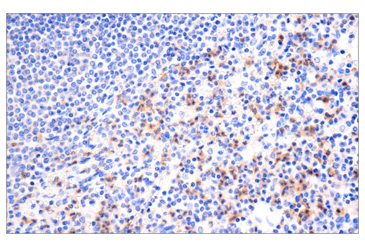
Myosin phosphatase regulates the interaction of actin and myosin in response to signaling through the small GTPase Rho. Rho activity inhibits myosin phosphatase via Rho-associated kinase (ROCK). Phosphorylation of MYPT1 at Thr696 and Thr853 results in phosphatase inhibition and cytoskeletal reorganization (5,6).
Phospho-MYPT1 (Ser507) and phospho-MYPT1 (Ser668) antibodies are directed at sites that were identified at Cell Signaling Technology (CST) using PhosphoScan®, CST's LC-MS/MS platform for modification site discovery. Phosphorylation at these sites was discovered using an Akt substrate antibody. Please visit PhosphoSitePlus®, CST's modification site knowledgebase, at www.phosphosite.org for more information.
Explore pathways related to this product.
STRING - Known and Predicted Protein-Protein Interactions.
Except as otherwise expressly agreed in a writing signed by a legally authorized representative of CST, the following terms apply to Products provided by CST, its affiliates or its distributors. Any Customer's terms and conditions that are in addition to, or different from, those contained herein, unless separately accepted in writing by a legally authorized representative of CST, are rejected and are of no force or effect.
Products are labeled with For Research Use Only or a similar labeling statement and have not been approved, cleared, or licensed by the FDA or other regulatory foreign or domestic entity, for any purpose. Customer shall not use any Product for any diagnostic or therapeutic purpose, or otherwise in any manner that conflicts with its labeling statement. Products sold or licensed by CST are provided for Customer as the end-user and solely for research and development uses. Any use of Product for diagnostic, prophylactic or therapeutic purposes, or any purchase of Product for resale (alone or as a component) or other commercial purpose, requires a separate license from CST. Customer shall (a) not sell, license, loan, donate or otherwise transfer or make available any Product to any third party, whether alone or in combination with other materials, or use the Products to manufacture any commercial products, (b) not copy, modify, reverse engineer, decompile, disassemble or otherwise attempt to discover the underlying structure or technology of the Products, or use the Products for the purpose of developing any products or services that would compete with CST products or services, (c) not alter or remove from the Products any trademarks, trade names, logos, patent or copyright notices or markings, (d) use the Products solely in accordance with CST Product Terms of Sale and any applicable documentation, and (e) comply with any license, terms of service or similar agreement with respect to any third party products or services used by Customer in connection with the Products.