| Cat. # | Size | Qty. | Price |
|---|---|---|---|
| 8340T | 1 Kit (7 x 20 microliters) |
|
| Product Includes | Quantity | Applications | Reactivity | MW(kDa) | Isotype |
|---|---|---|---|---|---|
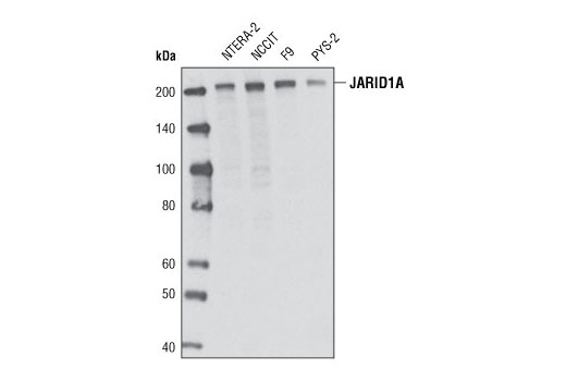
| JARID1A (D28B10) Rabbit mAb 3876 | 20 µl |
|
H M | 200 | Rabbit IgG |
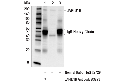
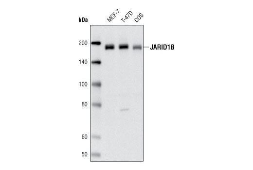
| JARID1B Antibody 3273 | 20 µl |
|
H Mk | 180 | Rabbit |
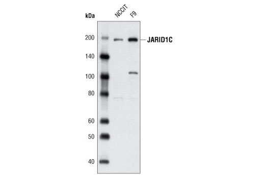
| JARID1C (D29B9) Rabbit mAb 5361 | 20 µl |
|
H M | 180 | Rabbit IgG |
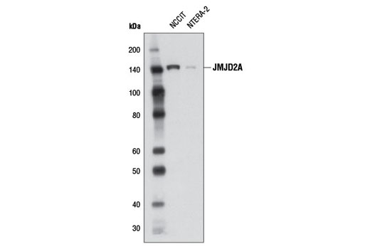
| JMJD2A (C37E5) Rabbit mAb 5328 | 20 µl |
|
H | 150 | Rabbit IgG |
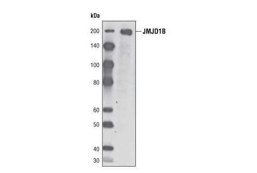
| JMJD1B (6A1-1F5) Mouse mAb 5377 | 20 µl |
|
H M R Mk | 220 | Mouse IgG1 |
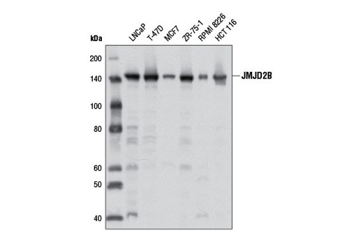
| JMJD2B (D7E6) Rabbit mAb 8639 | 20 µl |
|
H Mk | 150 | Rabbit IgG |
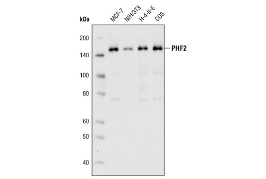
| PHF2 (D45A2) Rabbit mAb 3497 | 20 µl |
|
H M R Mk | 150 | Rabbit IgG |
| Anti-rabbit IgG, HRP-linked Antibody 7074 | 100 µl |
|
Rab | Goat | |
| Anti-mouse IgG, HRP-linked Antibody 7076 | 100 µl |
|
M | Horse |
Product Information
Polyclonal antibodies are produced by immunizing animals with a synthetic peptide and are purified by protein A and peptide affinity chromatography. Monoclonal antibodies are produced by immunizing animals with recombinant human proteins or synthetic peptides.
Jumonji C (JmjC) domain-containing proteins represent the largest class of potential histone demethylase proteins (1). The JmjC domain can catalyze the demethylation of mono-, di-, and tri-methyl lysine residues via an oxidative reaction that requires iron and α-ketoglutarate (1). Based on homology, both humans and mice contain at least 30 such proteins, which can be divided into 7 separate families (1).
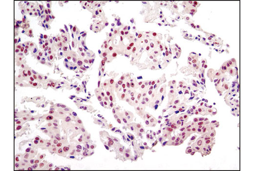
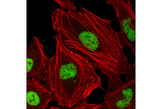
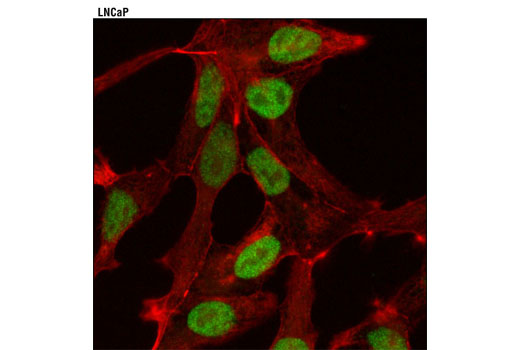
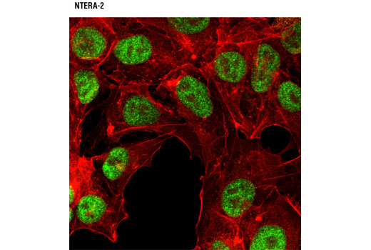
The JMJD2 (Jumonji domain-containing protein 2) family, also known as JHDM3 (JmjC domain-containing histone demethylation protein 3) family, contains four members: JMJD2A/JHDM3A, JMJD2B/JHDM3B, JMJD2C/JHDM3C, and JMJD2D/JHDM3D. In addition to the JmjC domain, these proteins also contain JmjN, PHD and Tudor domains, the latter of which has been shown to bind to methylated histone H3 at Lys4 and Lys9, and methylated histone H4 at Lys20 (2,3). JMJD2 proteins have been shown to demethylate di- and tri-methyl histone H3 at Lys9 and Lys36, and function as both activators and repressors of transcription (4-9). JMJD2A, JMJD2C and JMJD2D function as coactivators of the androgen receptor in prostate tumor cells (5). In contrast, JMJD2A also associates with Rb and N-CoR corepressor complexes and is necessary for transcriptional repression of target genes (6,7). JMJD2B antagonizes histone H3 Lys9 tri-methylation at pericentric heterochromatin (8). JMJD1B is a more widely expressed family member and is frequently deleted in myeloid leukemia (10).
The JARID (Jumonji/AT-rich interactive domain-containing protein) family contains four members: JARID1A (also RBP2 and RBBP2), JARID1B (also PLU-1), JARID1C (also SMCX), and JARID1D (also SMCY) (11). In addition to the JmjC domain, these proteins contain JmjN, BRIGHT, C5HC2 zinc-finger, and PHD domains, the latter of which binds to methylated histone H3 (Lys9) (11). All four JARID proteins demethylate di- and tri-methyl histone H3 Lys4; JARID1B also demethylates mono-methyl histone H3 Lys4 (12-14). JARID1A is a critical RB-interacting protein and is required for Polycomb-Repressive Complex 2 (PRC2)-mediated transcriptional repression during ES cell differentiation (15). A JARID1A-NUP98 gene fusion is associated with myeloid leukemia (16). JARID1B, which interacts with many proteins including c-Myc and HDAC4, may play a role in cell fate decisions by blocking terminal differentiation (17-19). JARID1B is over-expressed in many breast cancers and may act by repressing multiple tumor suppressor genes including BRCA1 and HOXA5 (20,21). JARID1C has been found in a complex with HDAC1, HDAC2, G9a, and REST, which binds to and represses REST target genes in non-neuronal cells (14). JARID1D is largely uncharacterized. PHF2 contains a JmjC domain, which may play a role in histone demethylation (1).
Except as otherwise expressly agreed in a writing signed by a legally authorized representative of CST, the following terms apply to Products provided by CST, its affiliates or its distributors. Any Customer's terms and conditions that are in addition to, or different from, those contained herein, unless separately accepted in writing by a legally authorized representative of CST, are rejected and are of no force or effect.
Products are labeled with For Research Use Only or a similar labeling statement and have not been approved, cleared, or licensed by the FDA or other regulatory foreign or domestic entity, for any purpose. Customer shall not use any Product for any diagnostic or therapeutic purpose, or otherwise in any manner that conflicts with its labeling statement. Products sold or licensed by CST are provided for Customer as the end-user and solely for research and development uses. Any use of Product for diagnostic, prophylactic or therapeutic purposes, or any purchase of Product for resale (alone or as a component) or other commercial purpose, requires a separate license from CST. Customer shall (a) not sell, license, loan, donate or otherwise transfer or make available any Product to any third party, whether alone or in combination with other materials, or use the Products to manufacture any commercial products, (b) not copy, modify, reverse engineer, decompile, disassemble or otherwise attempt to discover the underlying structure or technology of the Products, or use the Products for the purpose of developing any products or services that would compete with CST products or services, (c) not alter or remove from the Products any trademarks, trade names, logos, patent or copyright notices or markings, (d) use the Products solely in accordance with CST Product Terms of Sale and any applicable documentation, and (e) comply with any license, terms of service or similar agreement with respect to any third party products or services used by Customer in connection with the Products.