| Cat. # | Size | Qty. | Price |
|---|---|---|---|
| 9774T | 1 Kit (3 x 20 microliters) |
|
| Product Includes | Quantity | Applications | Reactivity | MW(kDa) | Isotype |
|---|---|---|---|---|---|
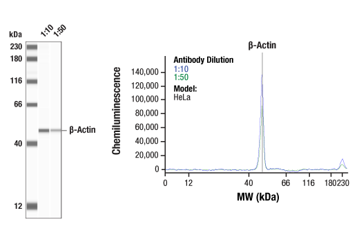
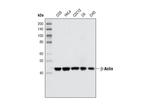
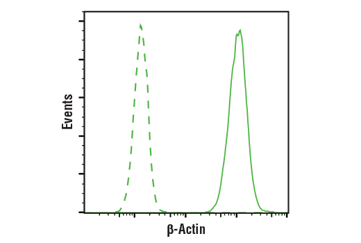
| β-Actin (8H10D10) Mouse mAb 3700 | 20 µl |
|
H M R Hm Mk Dg | 45 | Mouse IgG2b |
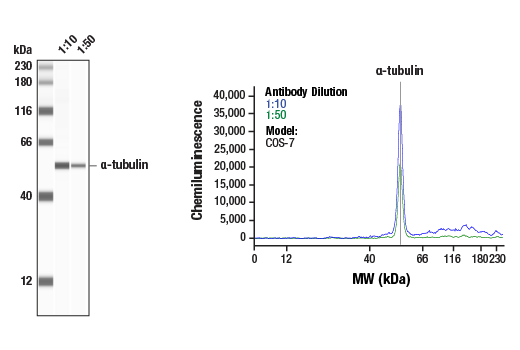
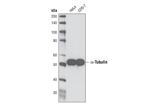
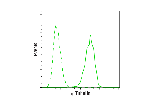
| α-Tubulin (DM1A) Mouse mAb 3873 | 20 µl |
|
H M R Mk | 52 | Mouse IgG1 |
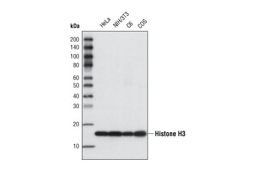
| Histone H3 (96C10) Mouse mAb 3638 | 20 µl |
|
H M R Mk | 17 | Mouse IgG1 |
| Anti-mouse IgG, HRP-linked Antibody 7076 | 100 µl |
|
M | Horse |
Product Information
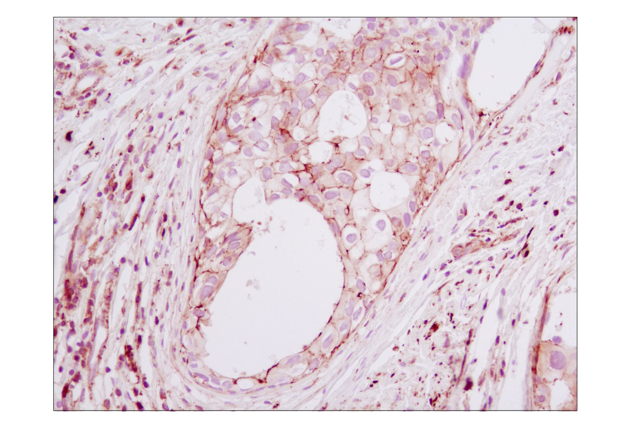
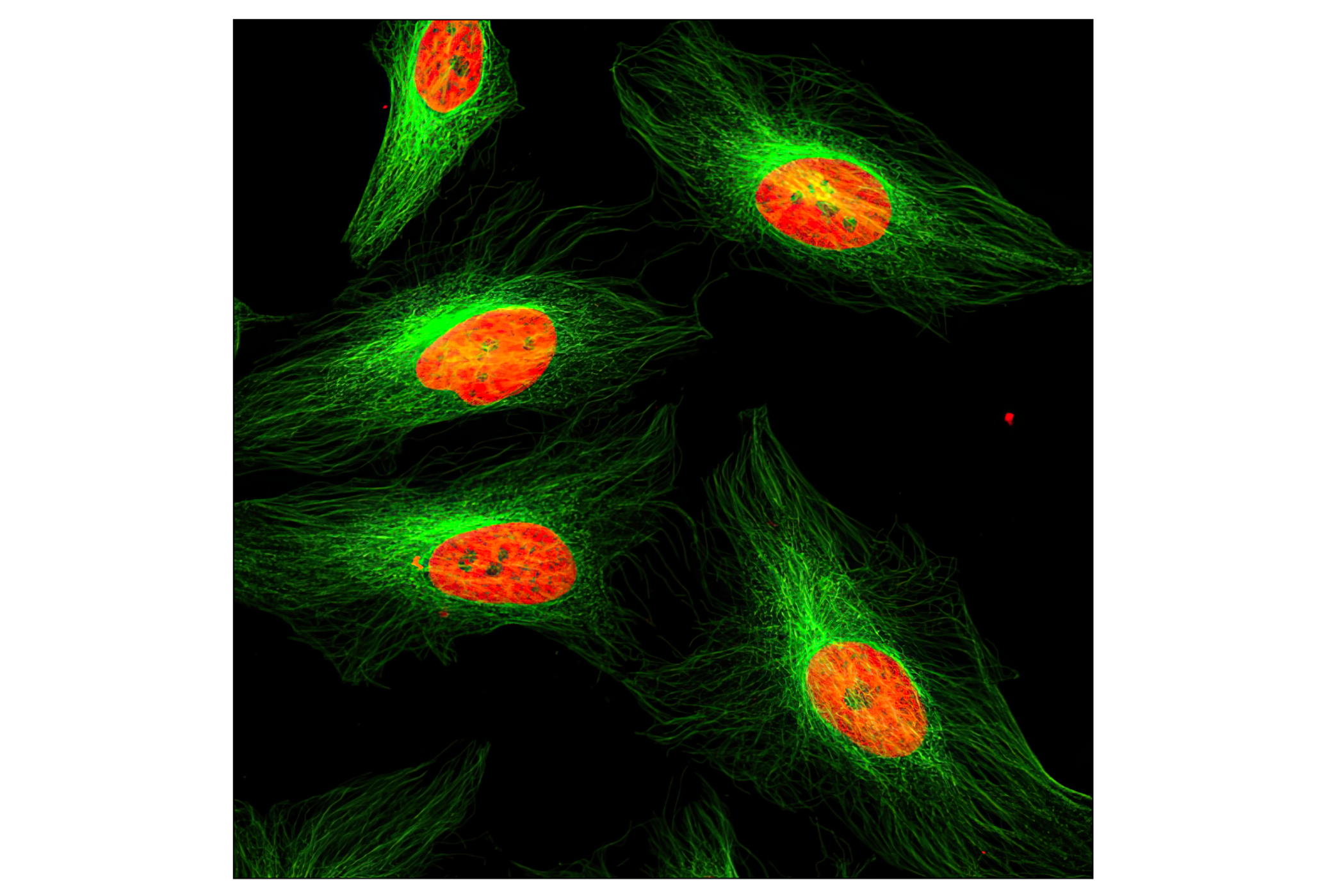
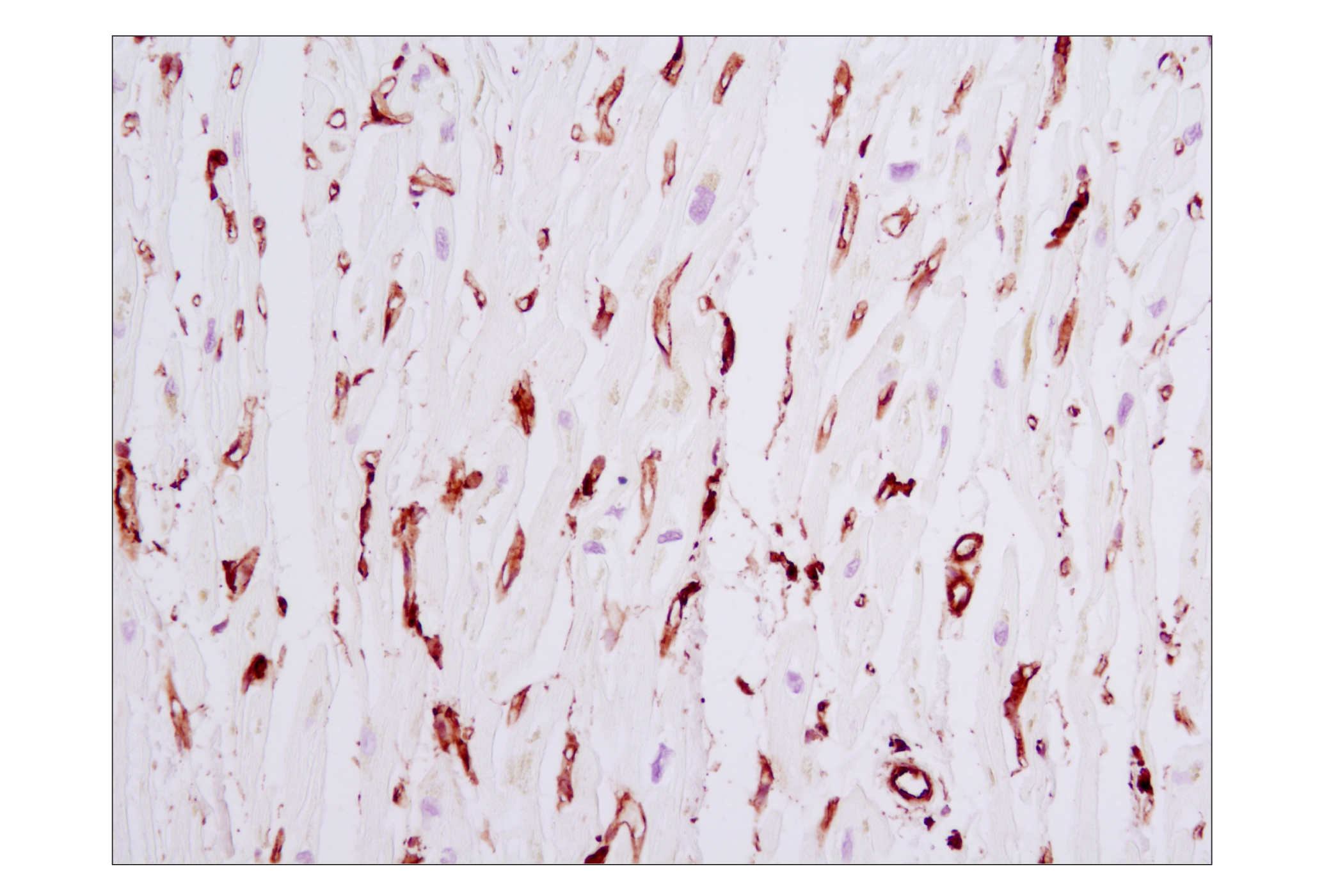
Monoclonal antibodies are produced by immunizing animals with synthetic peptides corresponding to residues near the amino terminus of human β-actin, the carboxy terminus of human histone H3, or with full-length chicken α-tubulin purfied from brain extracts. The epitope of α-Tubulin (DM1A) Mouse mAb recognizes residues surrounding Val440 of human α-tubulin protein.
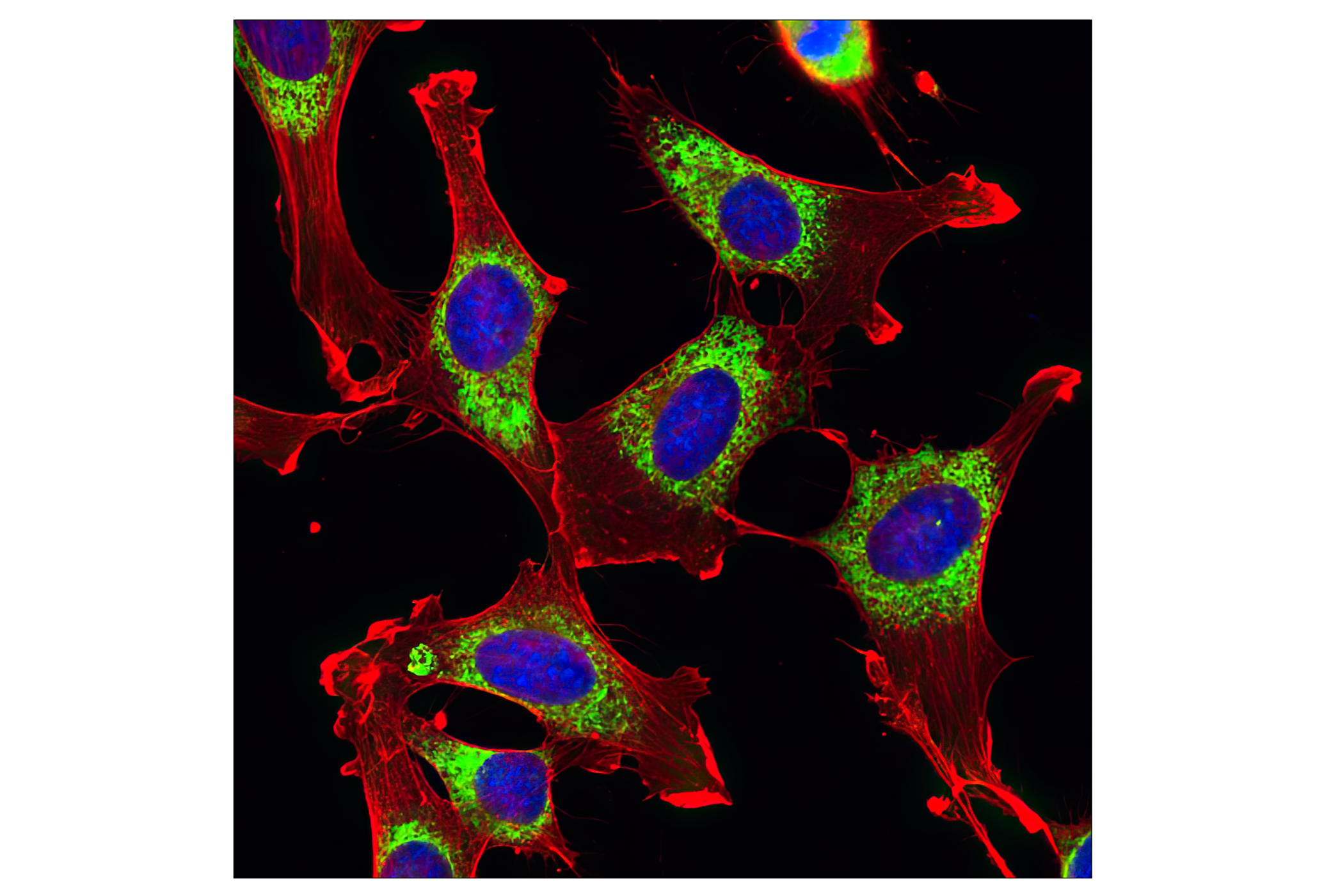
Housekeeping proteins perform numerous basic functions within the cell and are constitutively expressed at high levels in a variety of tissues and cell types. Western blot analysis commonly uses housekeeping proteins such as β-actin, histone H3, and α-tubulin as loading controls. Actin is a ubiquitous protein and a major component of the eukaryotic cytoskeleton. Actin exists mainly as the F-actin fibrous polymer (1). Globular tubulin subunits made up of α- and β-tubulin heterodimers are the building blocks of microtubules, one of three types of cytosolic fibers that comprise the cytoskeleton (2). Histone proteins, including histone H3, make up the primary building block of chromatin known as nucleosomes. Modulation of the chromatin structure plays an important role in the regulation of transcription in eukaryotes (3).
Except as otherwise expressly agreed in a writing signed by a legally authorized representative of CST, the following terms apply to Products provided by CST, its affiliates or its distributors. Any Customer's terms and conditions that are in addition to, or different from, those contained herein, unless separately accepted in writing by a legally authorized representative of CST, are rejected and are of no force or effect.
Products are labeled with For Research Use Only or a similar labeling statement and have not been approved, cleared, or licensed by the FDA or other regulatory foreign or domestic entity, for any purpose. Customer shall not use any Product for any diagnostic or therapeutic purpose, or otherwise in any manner that conflicts with its labeling statement. Products sold or licensed by CST are provided for Customer as the end-user and solely for research and development uses. Any use of Product for diagnostic, prophylactic or therapeutic purposes, or any purchase of Product for resale (alone or as a component) or other commercial purpose, requires a separate license from CST. Customer shall (a) not sell, license, loan, donate or otherwise transfer or make available any Product to any third party, whether alone or in combination with other materials, or use the Products to manufacture any commercial products, (b) not copy, modify, reverse engineer, decompile, disassemble or otherwise attempt to discover the underlying structure or technology of the Products, or use the Products for the purpose of developing any products or services that would compete with CST products or services, (c) not alter or remove from the Products any trademarks, trade names, logos, patent or copyright notices or markings, (d) use the Products solely in accordance with CST Product Terms of Sale and any applicable documentation, and (e) comply with any license, terms of service or similar agreement with respect to any third party products or services used by Customer in connection with the Products.