| Cat. # | Size | Qty. | Price |
|---|---|---|---|
| 9839T | 1 Kit (7 x 20 microliters) |
|
| Product Includes | Quantity | Applications | Reactivity | MW(kDa) | Isotype |
|---|---|---|---|---|---|
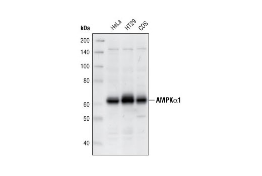
| AMPKα1 Antibody 2795 | 20 µl |
|
H Mk | 62 | Rabbit |
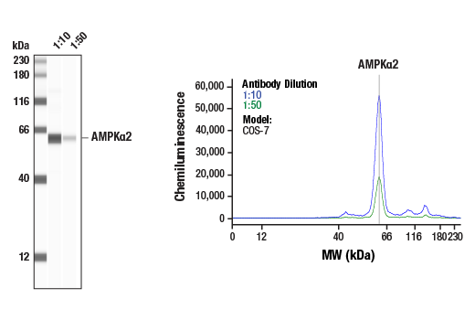
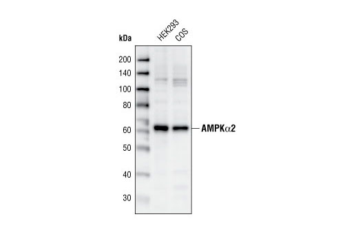
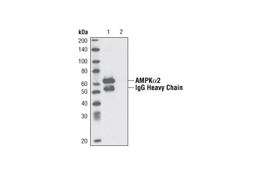
| AMPKα2 Antibody 2757 | 20 µl |
|
H Mk | 62 | Rabbit |
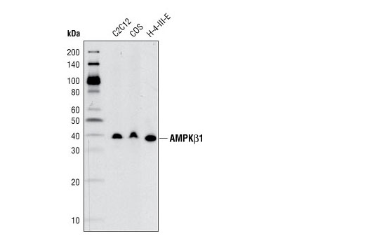
| AMPKβ1 (71C10) Rabbit mAb 4178 | 20 µl |
|
H M R Hm Mk Pg | 38 | Rabbit IgG |
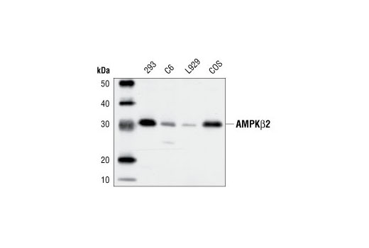
| AMPKβ2 Antibody 4148 | 20 µl |
|
H M R Mk | 30 | Rabbit |
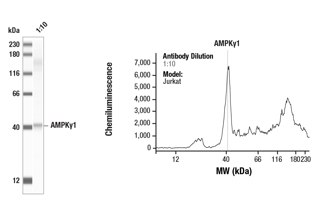
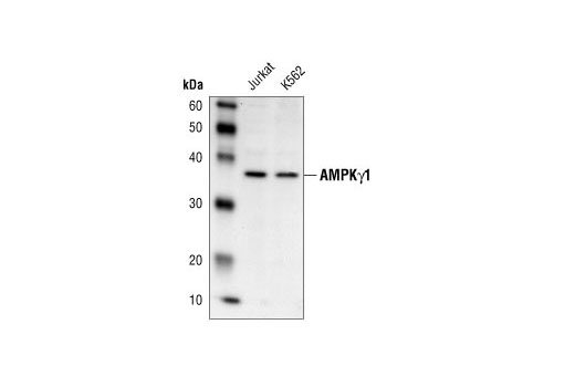
| AMPKγ1 Antibody 4187 | 20 µl |
|
H Mk | 37 | Rabbit |
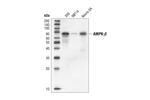
| AMPKγ2 Antibody 2536 | 20 µl |
|
H M R Mk B | 75 | Rabbit |
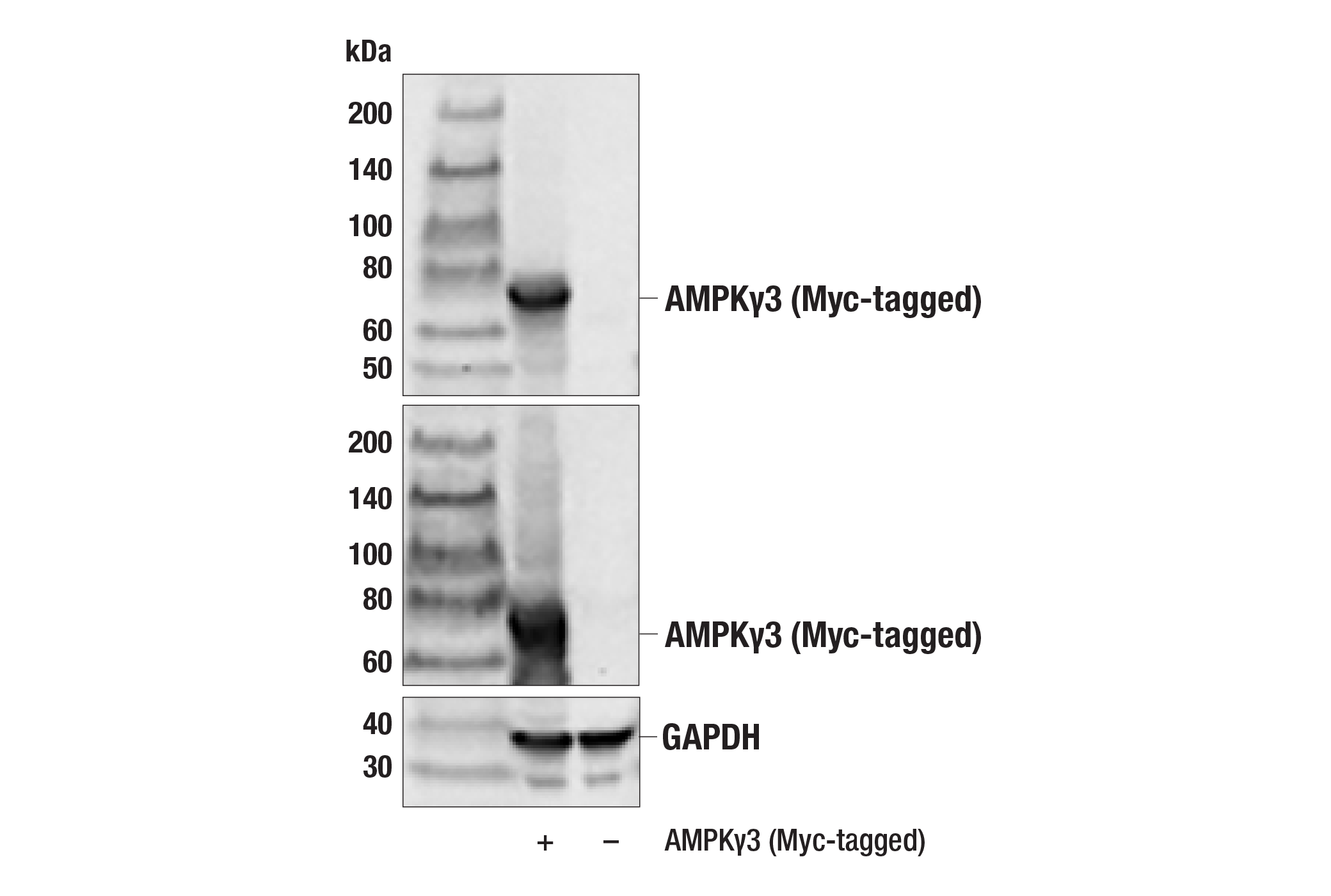
| AMPKγ3 Antibody 2550 | 20 µl |
|
H | 54 | Rabbit |
| Anti-rabbit IgG, HRP-linked Antibody 7074 | 100 µl |
|
Goat |
Product Information
Polyclonal antibodies are produced by immunizing animals with synthetic peptides corresponding to residues surrounding Leu519 near the carboxy terminus of human AMPKα1,
corresponding to residues surrounding Ser500 of human AMPKα2, near the amino terminus of human AMPKγ1, surrounding Ser60 of human AMPKγ2, and corresponding to the sequences of human AMPKβ2 and AMPKγ3. Antibodies are purified by protein A and peptide affinity chromatography. Monoclonal antibody is produced by immunizing animals with synthetic peptides corresponding to residues surrounding Val176 of human AMPKβ1.
AMP-activated protein kinase (AMPK) is highly conserved from yeast to plants and animals and plays a key role in the regulation of energy homeostasis (1). AMPK is a heterotrimeric complex composed of a catalytic α subunit and regulatory β and γ subunits, each of which is encoded by two or three distinct genes (α1, 2; β1, 2; γ1, 2, 3) (2). The kinase is activated by an elevated AMP/ATP ratio due to cellular and environmental stress, such as heat shock, hypoxia, and ischemia (1). The tumor suppressor LKB1, in association with accessory proteins STRAD and MO25, phosphorylates AMPKα at Thr172 in the activation loop, and this phosphorylation is required for AMPK activation (3-5). AMPKα is also phosphorylated at Thr258 and Ser485 (for α1; Ser491 for α2). The upstream kinase and the biological significance of these phosphorylation events have yet to be elucidated (6). The β1 subunit is post-translationally modified by myristoylation and multi-site phosphorylation including Ser24/25, Ser96, Ser101, Ser108, and Ser182 (6,7). Phosphorylation at Ser108 of the β1 subunit seems to be required for AMPK activation, while phosphorylation at Ser24/25 and Ser182 affects AMPK localization (7). Several mutations in AMPKγ subunits have been identified, most of which are located in the putative AMP/ATP binding sites (CBS or Bateman domains). Mutations at these sites lead to reduction of AMPK activity and cause glycogen accumulation in heart or skeletal muscle (1,2). Accumulating evidence indicates that AMPK not only regulates the metabolism of fatty acids and glycogen, but also modulates protein synthesis and cell growth through EF2 and TSC2/mTOR pathways, as well as blood flow via eNOS/nNOS (1).
Explore pathways related to this product.
STRING - Known and Predicted Protein-Protein Interactions.
Except as otherwise expressly agreed in a writing signed by a legally authorized representative of CST, the following terms apply to Products provided by CST, its affiliates or its distributors. Any Customer's terms and conditions that are in addition to, or different from, those contained herein, unless separately accepted in writing by a legally authorized representative of CST, are rejected and are of no force or effect.
Products are labeled with For Research Use Only or a similar labeling statement and have not been approved, cleared, or licensed by the FDA or other regulatory foreign or domestic entity, for any purpose. Customer shall not use any Product for any diagnostic or therapeutic purpose, or otherwise in any manner that conflicts with its labeling statement. Products sold or licensed by CST are provided for Customer as the end-user and solely for research and development uses. Any use of Product for diagnostic, prophylactic or therapeutic purposes, or any purchase of Product for resale (alone or as a component) or other commercial purpose, requires a separate license from CST. Customer shall (a) not sell, license, loan, donate or otherwise transfer or make available any Product to any third party, whether alone or in combination with other materials, or use the Products to manufacture any commercial products, (b) not copy, modify, reverse engineer, decompile, disassemble or otherwise attempt to discover the underlying structure or technology of the Products, or use the Products for the purpose of developing any products or services that would compete with CST products or services, (c) not alter or remove from the Products any trademarks, trade names, logos, patent or copyright notices or markings, (d) use the Products solely in accordance with CST Product Terms of Sale and any applicable documentation, and (e) comply with any license, terms of service or similar agreement with respect to any third party products or services used by Customer in connection with the Products.