| Cat. # | Size | Qty. | Price |
|---|---|---|---|
| 9921T | 1 Kit (9 x 20 microliters) |
|
| Product Includes | Quantity | Applications | Reactivity | MW(kDa) | Isotype |
|---|---|---|---|---|---|
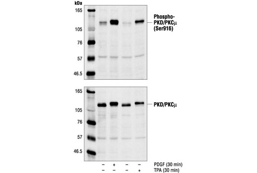
| Phospho-PKD/PKCμ (Ser916) Antibody 2051 | 20 µl |
|
H M R Mk | 115 | Rabbit |
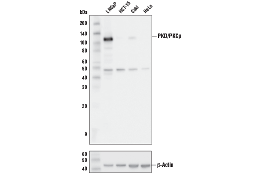
| PKD/PKCμ (D4J1N) Rabbit mAb 90039 | 20 µl |
|
H Mk | 115 | Rabbit IgG |
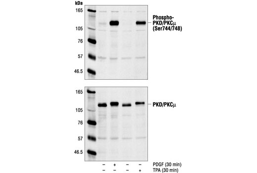
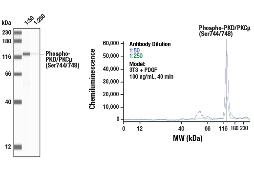
| Phospho-PKD/PKCμ (Ser744/748) Antibody 2054 | 20 µl |
|
H M R Mk | 115 | Rabbit |
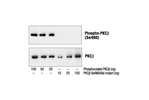
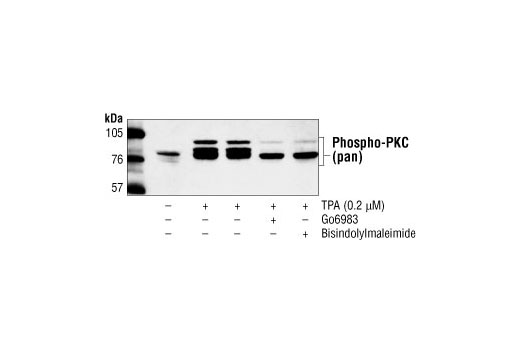
| Phospho-PKC (pan) (βII Ser660) Antibody 9371 | 20 µl |
|
H M R Mk | 78, 80, 82, 85 | Rabbit |
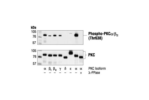
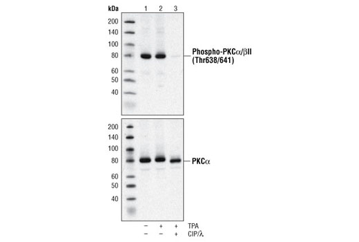
| Phospho-PKCα/β II (Thr638/641) Antibody 9375 | 20 µl |
|
H M Mk | 80, 82 | Rabbit |
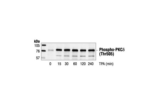
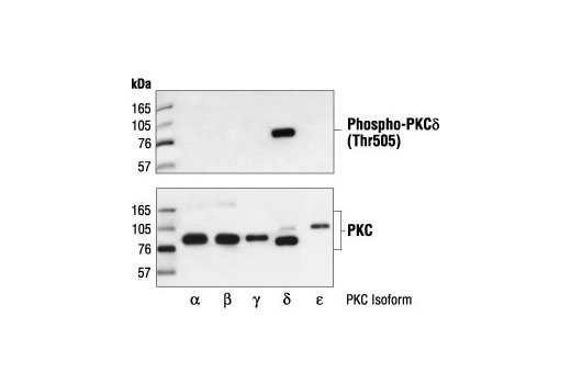
| Phospho-PKCδ (Thr505) Antibody 9374 | 20 µl |
|
H M | 78 | Rabbit |
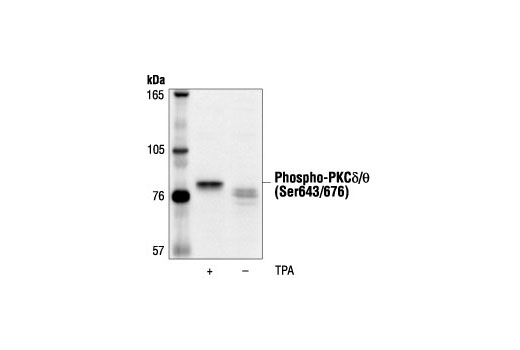
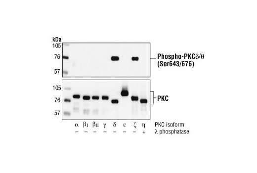
| Phospho-PKCδ/θ (Ser643/676) Antibody 9376 | 20 µl |
|
H M R Mk X | 78 | Rabbit |
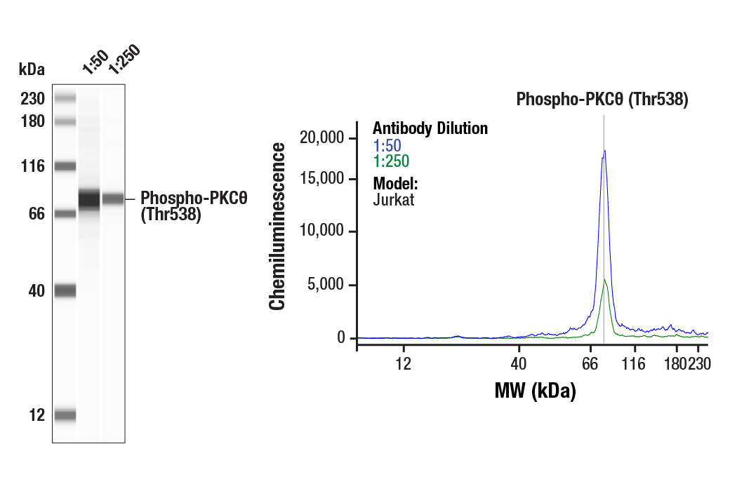
| Phospho-PKCθ (Thr538) Antibody 9377 | 20 µl |
|
H R Mk | 79 | Rabbit |
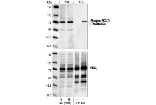
| Phospho-PKCζ/λ (Thr410/403) Antibody 9378 | 20 µl |
|
H M R Mk | 76 | Rabbit |
| Anti-rabbit IgG, HRP-linked Antibody 7074 | 100 µl |
|
Goat |
Product Information
Monoclonal antibody is produced by immunizing animals with a synthetic peptide corresponding to residues near the carboxy terminus of human PKD. Polyclonal antibodies are produced by immunizing animals with synthetic phosphopeptides corresponding to the sequence of the human protein PKCβ II, PKCα, PKCδ, PKCθ, or PKCζ . Antibodies are purified by protein A and peptide affinity chromatography.
Activation of protein kinase C (PKC) is one of the earliest events in a cascade that controls a variety of cellular responses, including secretion, gene expression, proliferation, and muscle contraction (1,2). PKC isoforms belong to three groups based on calcium dependency and activators. Classical PKCs are calcium-dependent via their C2 domains and are activated by phosphatidylserine (PS), diacylglycerol (DAG), and phorbol esters (TPA, PMA) through their cysteine-rich C1 domains. Both novel and atypical PKCs are calcium-independent, but only novel PKCs are activated by PS, DAG, and phorbol esters (3-5). Members of these three PKC groups contain a pseudo-substrate or autoinhibitory domain that binds to substrate-binding sites in the catalytic domain to prevent activation in the absence of cofactors or activators. Control of PKC activity is regulated through three distinct phosphorylation events. Phosphorylation occurs in vivo at Thr500 in the activation loop, at Thr641 through autophosphorylation, and at the carboxy-terminal hydrophobic site Ser660 (2). Atypical PKC isoforms lack hydrophobic region phosphorylation, which correlates with the presence of glutamic acid rather than the serine or threonine residues found in more typical PKC isoforms. The enzyme PDK1 or a close relative is responsible for PKC activation. A recent addition to the PKC superfamily is PKCμ (PKD), which is regulated by DAG and TPA through its C1 domain. PKD is distinguished by the presence of a PH domain and by its unique substrate recognition and Golgi localization (6). PKC-related kinases (PRK) lack the C1 domain and do not respond to DAG or phorbol esters. Phosphatidylinositol lipids activate PRKs, and small Rho-family GTPases bind to the homology region 1 (HR1) to regulate PRK kinase activity (7).
Except as otherwise expressly agreed in a writing signed by a legally authorized representative of CST, the following terms apply to Products provided by CST, its affiliates or its distributors. Any Customer's terms and conditions that are in addition to, or different from, those contained herein, unless separately accepted in writing by a legally authorized representative of CST, are rejected and are of no force or effect.
Products are labeled with For Research Use Only or a similar labeling statement and have not been approved, cleared, or licensed by the FDA or other regulatory foreign or domestic entity, for any purpose. Customer shall not use any Product for any diagnostic or therapeutic purpose, or otherwise in any manner that conflicts with its labeling statement. Products sold or licensed by CST are provided for Customer as the end-user and solely for research and development uses. Any use of Product for diagnostic, prophylactic or therapeutic purposes, or any purchase of Product for resale (alone or as a component) or other commercial purpose, requires a separate license from CST. Customer shall (a) not sell, license, loan, donate or otherwise transfer or make available any Product to any third party, whether alone or in combination with other materials, or use the Products to manufacture any commercial products, (b) not copy, modify, reverse engineer, decompile, disassemble or otherwise attempt to discover the underlying structure or technology of the Products, or use the Products for the purpose of developing any products or services that would compete with CST products or services, (c) not alter or remove from the Products any trademarks, trade names, logos, patent or copyright notices or markings, (d) use the Products solely in accordance with CST Product Terms of Sale and any applicable documentation, and (e) comply with any license, terms of service or similar agreement with respect to any third party products or services used by Customer in connection with the Products.