| Cat. # | Size | Qty. | Price |
|---|---|---|---|
| 9923T | 1 Kit (3 x 20 microliters) |
|
| Product Includes | Quantity | Applications | Reactivity | MW(kDa) | Isotype |
|---|---|---|---|---|---|
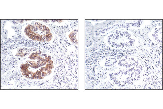
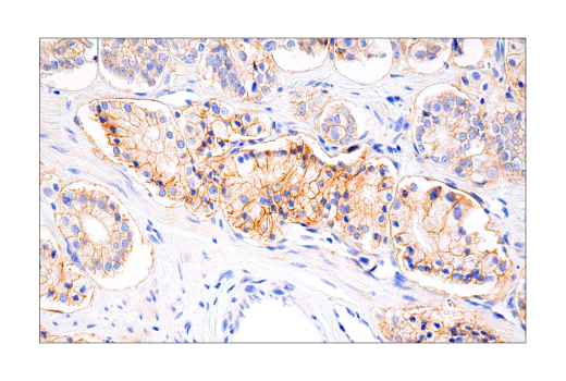
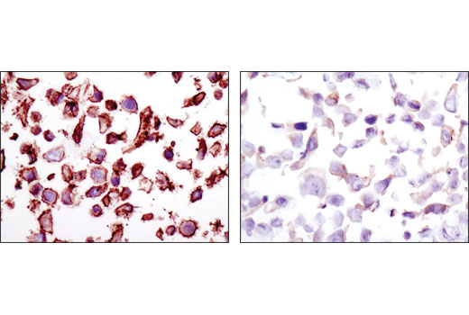
| Phospho-HER2/ErbB2 (Tyr1248) Antibody 2247 | 20 µl |
|
H M | 185 | Rabbit |
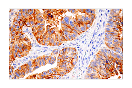
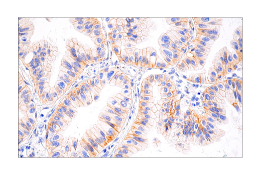
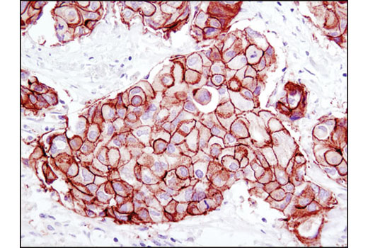
| Phospho-HER2/ErbB2 (Tyr1221/1222) (6B12) Rabbit mAb 2243 | 20 µl |
|
H | 185 | Rabbit IgG |
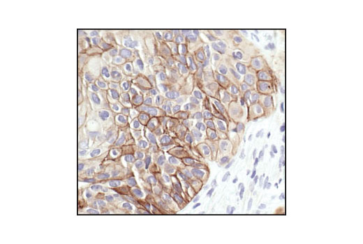
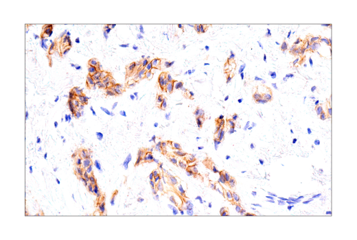
| HER2/ErbB2 (D8F12) XP® Rabbit mAb 4290 | 20 µl |
|
H M | 185 | Rabbit IgG |
| Anti-rabbit IgG, HRP-linked Antibody 7074 | 100 µl |
|
Rab | Goat |
Product Information
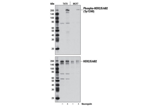
Polyclonal antibodies are produced by immunizing animals with a synthetic phosphopeptide corresponding to residues surrounding Tyr1248. Polyclonal antibodies are purified by protein A and peptide affinity chromatography. Monoclonal antibodies are produced by immunizing animals with synthetic peptides corresponding to residues surrounding tyrosines 1221/1222 and residues near the amino terminus of human ErbB2 protein.
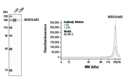
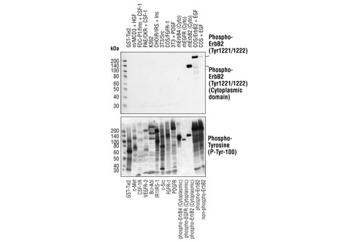
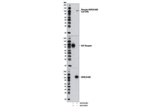
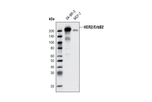
The ErbB2 (HER2) proto-oncogene encodes a 185 kDa transmembrane, receptor-like glycoprotein with intrinsic tyrosine kinase activity (1). While ErbB2 lacks an identified ligand, ErbB2 kinase activity can be activated in the absence of a ligand when overexpressed and through heteromeric associations with other ErbB family members (2). Amplification of the ErbB2 gene and overexpression of its product are detected in almost 40% of human breast cancers (3). Binding of the c-Cbl ubiquitin ligase to ErbB2 at Tyr1112 leads to ErbB2 poly-ubiquitination and enhances degradation of this kinase (4). ErbB2 is a key therapeutic target in the treatment of breast cancer and other carcinomas and targeting the regulation of ErbB2 degradation by the c-Cbl-regulated proteolytic pathway is one potential therapeutic strategy. Phosphorylation of the kinase domain residue Tyr877 of ErbB2 (homologous to Tyr416 of pp60c-Src) may be involved in regulating ErbB2 biological activity. The major autophosphorylation sites in ErbB2 are Tyr1248 and Tyr1221/1222; phosphorylation of these sites couples ErbB2 to the Ras-Raf-MAP kinase signal transduction pathway (1,5).
Explore pathways related to this product.
STRING - Known and Predicted Protein-Protein Interactions.
Except as otherwise expressly agreed in a writing signed by a legally authorized representative of CST, the following terms apply to Products provided by CST, its affiliates or its distributors. Any Customer's terms and conditions that are in addition to, or different from, those contained herein, unless separately accepted in writing by a legally authorized representative of CST, are rejected and are of no force or effect.
Products are labeled with For Research Use Only or a similar labeling statement and have not been approved, cleared, or licensed by the FDA or other regulatory foreign or domestic entity, for any purpose. Customer shall not use any Product for any diagnostic or therapeutic purpose, or otherwise in any manner that conflicts with its labeling statement. Products sold or licensed by CST are provided for Customer as the end-user and solely for research and development uses. Any use of Product for diagnostic, prophylactic or therapeutic purposes, or any purchase of Product for resale (alone or as a component) or other commercial purpose, requires a separate license from CST. Customer shall (a) not sell, license, loan, donate or otherwise transfer or make available any Product to any third party, whether alone or in combination with other materials, or use the Products to manufacture any commercial products, (b) not copy, modify, reverse engineer, decompile, disassemble or otherwise attempt to discover the underlying structure or technology of the Products, or use the Products for the purpose of developing any products or services that would compete with CST products or services, (c) not alter or remove from the Products any trademarks, trade names, logos, patent or copyright notices or markings, (d) use the Products solely in accordance with CST Product Terms of Sale and any applicable documentation, and (e) comply with any license, terms of service or similar agreement with respect to any third party products or services used by Customer in connection with the Products.