| Cat. # | Size | Qty. | Price |
|---|---|---|---|
| 9932T | 1 Kit (8 x 20 microliters) |
|
| Product Includes | Quantity | Applications | Reactivity | MW(kDa) | Isotype |
|---|---|---|---|---|---|
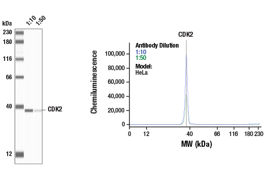
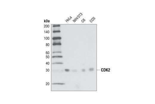
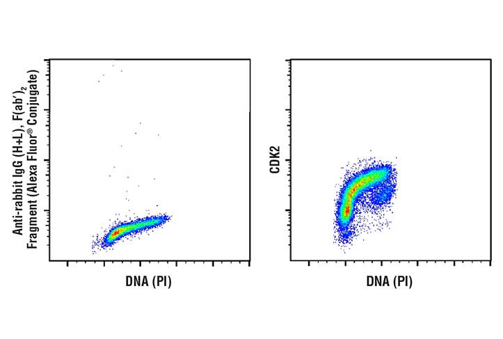
| CDK2 (78B2) Rabbit mAb 2546 | 20 µl |
|
H M R Mk | 33 | Rabbit |
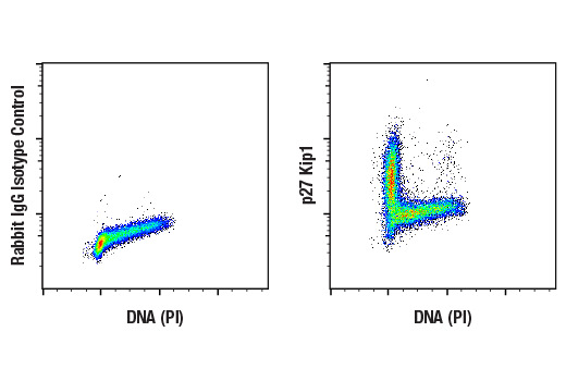
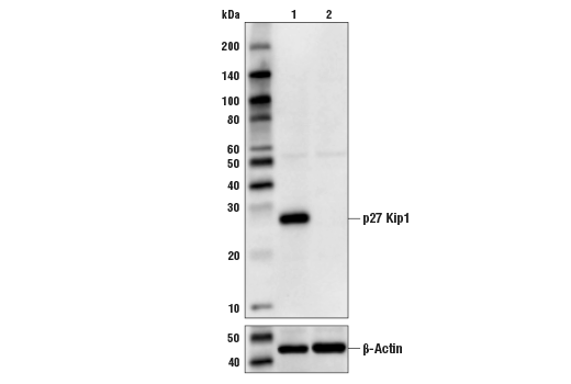
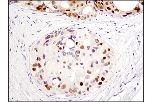
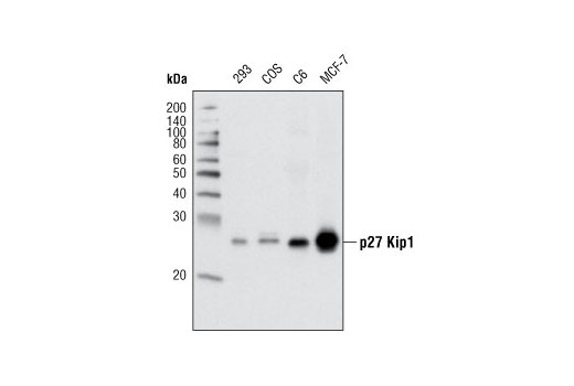
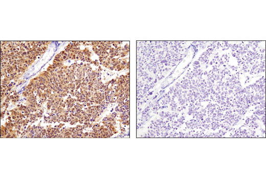
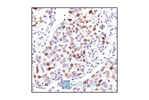
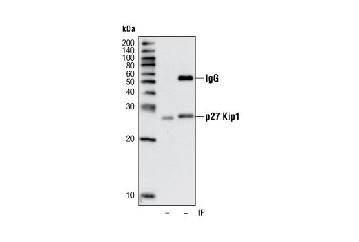
| p27 Kip1 (D69C12) XP® Rabbit mAb 3686 | 20 µl |
|
H R Mk | 27 | Rabbit IgG |
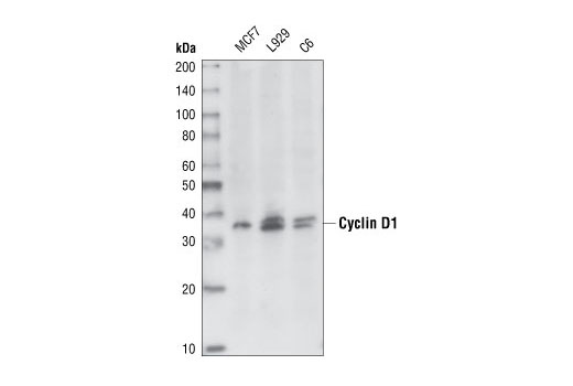
| Cyclin D1 (92G2) Rabbit mAb 2978 | 20 µl |
|
H M R | 36 | Rabbit IgG |
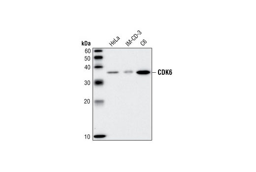
| CDK6 (DCS83) Mouse mAb 3136 | 20 µl |
|
H M R | 36 | Mouse IgG1 |
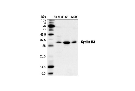
| Cyclin D3 (DCS22) Mouse mAb 2936 | 20 µl |
|
H M R | 31 | Mouse IgG1 |
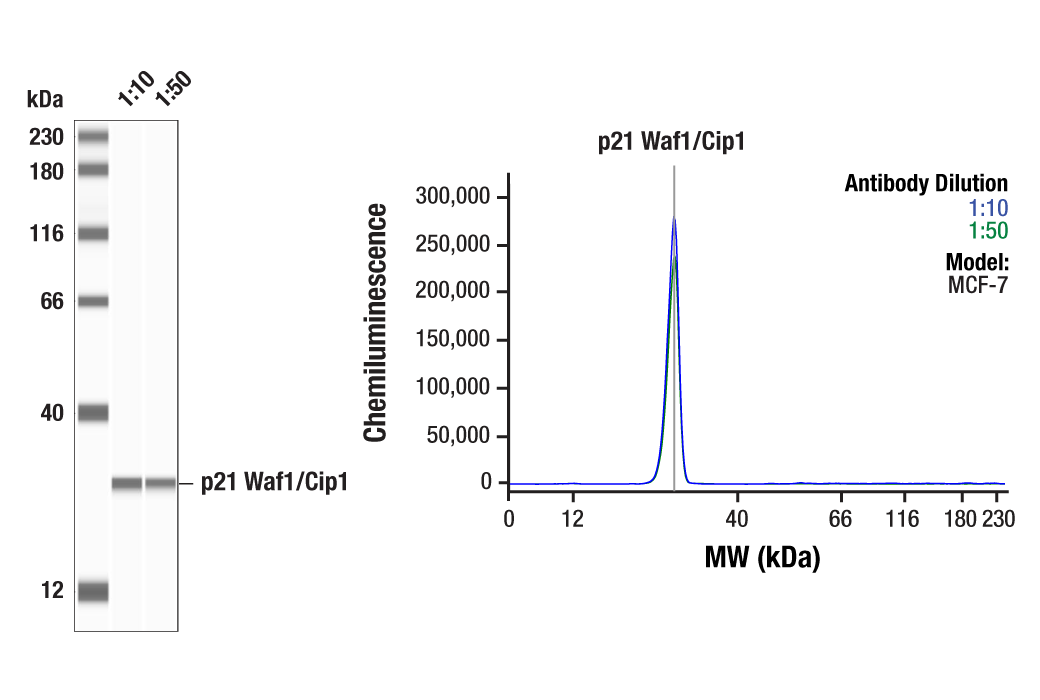
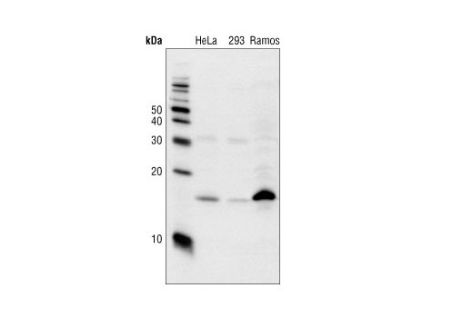
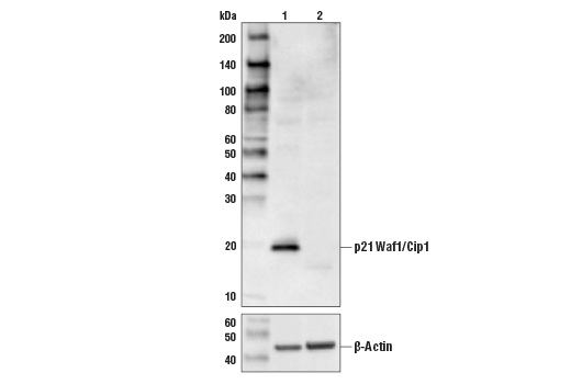
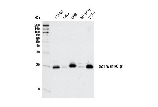
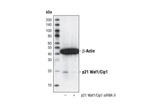
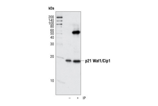
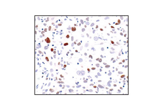
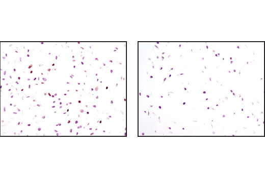
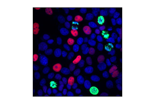
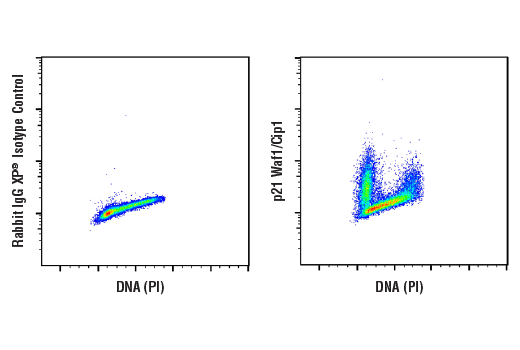
| p21 Waf1/Cip1 (12D1) Rabbit mAb 2947 | 20 µl |
|
H Mk | 21 | Rabbit IgG |
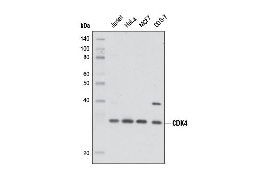
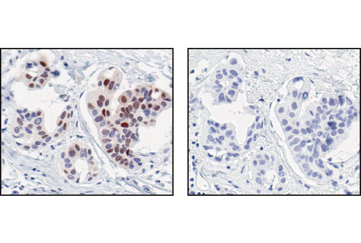
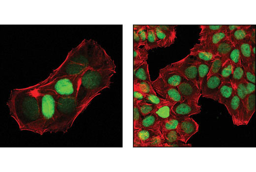
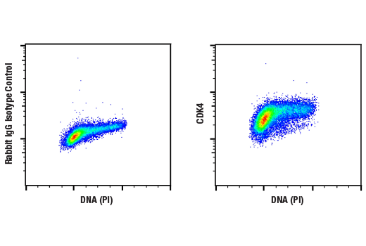
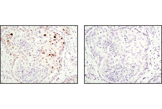
| CDK4 (D9G3E) Rabbit mAb 12790 | 20 µl |
|
H Mk | 30 | Rabbit IgG |
| p18 INK4C (DCS118) Mouse mAb 2896 | 20 µl |
|
H | 18 | Mouse IgG2a |
| Anti-rabbit IgG, HRP-linked Antibody 7074 | 100 µl |
|
Goat | ||
| Anti-mouse IgG, HRP-linked Antibody 7076 | 100 µl |
|
Horse |
Product Information
Polyclonal antibodies are produced by immunizing animals with synthetic peptides and are purified by protein A and peptide affinity chromatography. Monoclonal antibodies are produced by immunizing animals with recombinant human proteins or synthetic peptides.
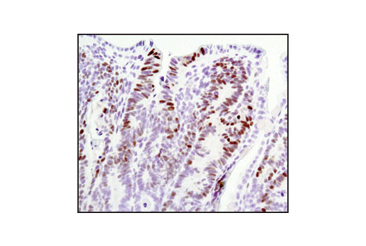
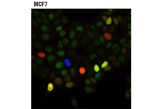
Eukaryotic cell cycle progression is dependent, in part, on the tightly regulated activity of cyclin dependent kinases (CDKs). Cyclin D/CDK4/6 activity occurs in mid-late G1 phase, upstream of CDK2/cyclin E activity. Both of these activities are required for hyperphosphorylation of the retinoblastoma gene product (pRb). pRb phosphorylation allows the release of S phase-promoting transcription factors and is indicative of the cell's commitment to proliferate. This point in the cell cycle is known as the restriction point. Cyclin protein levels oscillate throughout the cell cycle, and their availability is a means of controlling CDK activity and cell proliferation. Cyclin D is degraded through the ubiquitin proteasome pathway in the absence of mitogenic signaling. Ubiquitination of cyclin D1 is enhanced by phosphorylation at Thr286 by glycogen synthase kinase 3b (GSK-3b) (1). p27/Kip1, p57 Kip2 and p21 Waf1/Cip1 are members of the Cip/Kip family of cyclin-dependent kinase inhibitors. They form heterotrimeric complexes with cyclins and CDKs, inhibiting kinase activity and blocking progression through G1/S phase (2). However, p21 may enhance assembly and activity of cyclin D/CDK4/6 complexes (3). Levels of p21 and p27 protein are controlled through ubiquitination and proteasomal degradation (4). Levels of p27 are upregulated in quiescent cells and in cells treated with negative cell cycle regulators. p27 nuclear localization is controlled by Akt-dependent phosphorylation at Thr157 (5). The inhibitors of CDK4 (INK4) family include p15 INK4B, p16 INK4A, p18 INK4C, and p19 INK4D. All INK4 proteins selectively inhibit CDK4/6 activity, either in a binary complex, or in a ternary complex including cyclin D, resulting in inhibition of cell division (6,7).
Except as otherwise expressly agreed in a writing signed by a legally authorized representative of CST, the following terms apply to Products provided by CST, its affiliates or its distributors. Any Customer's terms and conditions that are in addition to, or different from, those contained herein, unless separately accepted in writing by a legally authorized representative of CST, are rejected and are of no force or effect.
Products are labeled with For Research Use Only or a similar labeling statement and have not been approved, cleared, or licensed by the FDA or other regulatory foreign or domestic entity, for any purpose. Customer shall not use any Product for any diagnostic or therapeutic purpose, or otherwise in any manner that conflicts with its labeling statement. Products sold or licensed by CST are provided for Customer as the end-user and solely for research and development uses. Any use of Product for diagnostic, prophylactic or therapeutic purposes, or any purchase of Product for resale (alone or as a component) or other commercial purpose, requires a separate license from CST. Customer shall (a) not sell, license, loan, donate or otherwise transfer or make available any Product to any third party, whether alone or in combination with other materials, or use the Products to manufacture any commercial products, (b) not copy, modify, reverse engineer, decompile, disassemble or otherwise attempt to discover the underlying structure or technology of the Products, or use the Products for the purpose of developing any products or services that would compete with CST products or services, (c) not alter or remove from the Products any trademarks, trade names, logos, patent or copyright notices or markings, (d) use the Products solely in accordance with CST Product Terms of Sale and any applicable documentation, and (e) comply with any license, terms of service or similar agreement with respect to any third party products or services used by Customer in connection with the Products.