View Featured Offers >>
8341
DNA Replication Antibody Sampler Kit
Primary Antibodies
Antibody Sampler Kit

DNA Replication Antibody Sampler Kit #8341

Citations (0)
Western blot analysis of extracts from A431 and K562 cells using RPA70 (C24F2) Antibody.
Western blot analysis of human (HeLa), murine (SV-T2), bovine (BAEC), and porcine (PAE) cell extracts using PCNA (PC10) Mouse mAb.
Western blot analysis of extracts from various cell types using MCM2 (D7G11) XP® Rabbit mAb.
Western blot analysis of extracts from various cell types using MCM7 (D10A11) XP® Rabbit mAb.
Western blot analysis of extracts from various cell types using MCM3 (D47B6) Rabbit mAb.
Western blot analysis of extracts from various cell types using p58 Primase (8D3) Rat mAb.
After the primary antibody is bound to the target protein, a complex with HRP-linked secondary antibody is formed. The LumiGLO® is added and emits light during enzyme catalyzed decomposition.
After the primary antibody is bound to the target protein, a complex with HRP-linked secondary antibody is formed. The LumiGLO* is added and emits light during enzyme catalyzed decomposition.
After the primary antibody is bound to the target protein, a complex with HRP-linked secondary antibody is formed. The LumiGlo® is added and emits light during enzyme catalyzed decomposition.
Western blot analysis of extracts from various cell lines using CDT1 (D10F11) Rabbit mAb.
Western blot analysis, using PCNA (PC10) Mouse mAb (upper) and Beta-Actin Antibody #4967(lower), of HeLa cell extract (lane 1) and PCNA immunoprecipitated from the same extract using PCNA (PC10) Mouse mAb (lane 2).
Immunoprecipitation of MCM2 from HeLa cell lysates using MCM2 (D7G11) XP® Rabbit mAb followed by western blot using the same antibody. Lane 1 is 5% input.
Immunoprecipitation of MCM7 from HeLa cell lysates using MCM7 (D10A11) XP® Rabbit mAb followed by western blot using the same antibody. Lane 1 is 5% input.
Immunoprecipitation of p58 primase from HeLa cell lysates using p58 Primase (8D3) Rat mAb. Western blot analysis was performed using the same antibody.
Immunoprecipitation of CDT1 from HeLa cells using CDT1 (D10F11) Rabbit mAb. Western blot was performed using the same antibody. Lane 1 is 10% input.
Immunohistochemical analysis of paraffin-embedded human breast carcinoma, showing nuclear localization, using PCNA (PC10) Mouse mAb.
Immunohistochemical analysis of paraffin-embedded human colon carcinoma using MCM2 (D7G11) XP® Rabbit mAb.
Immunohistochemical analysis of paraffin-embedded human lung carcinoma using MCM7 (D10A11) XP® Rabbit mAb.
Confocal immunofluorescent analysis of HT-29 and HUVEC cells using CDT1 (D10F11) Rabbit mAb (green). Actin filaments were labeled with DY-554 phalloidin (red). Blue pseudocolor = DRAQ5® #4084 (fluorescent DNA dye).
Immunohistochemical analysis of paraffin-embedded human colon carcinoma, showing nuclear localization, using PCNA (PC10) Mouse mAb.
Immunohistochemical analysis of paraffin-embedded human lung carcinoma using MCM2 (D7G11) XP® Rabbit mAb.
Immunohistochemical analysis of paraffin-embedded human Non-Hodgkin's lymphoma, showing nuclear localization, using PCNA (PC10) Mouse mAb.
Immunohistochemical analysis of paraffin-embedded human small intestine using MCM2 (D7G11) XP® Rabbit mAb.
Confocal immunofluorescent analysis of COS cells using MCM7 (D10A11) XP® Rabbit mAb (green). Actin filaments have been labeled with DY-554 phalloidin (red).
Confocal immunofluorescent analysis of NCI-H460 cells using PCNA (PC10) Mouse mAb (green), Phospho-Histone H3 (Ser10) (D2C8) XP Rabbit mAb (Alexa Fluor 555 Conjugate) (red), and p21 Waf1/Cip1 (12D1) Rabbit mAb (blue).
Confocal immunofluorescent analysis of HeLa cells using MCM2 (D7G11) XP® Rabbit mAb (green). Actin filaments have been labeled with DY-554 phalloidin (red).
Flow cytometric analysis of Jurkat cells using PCNA (PC10) Mouse mAb and Propidium Iodide (PI)/RNase Staining Solution #4087 to measure DNA content. Anti-Mouse IgG (H+L), F(ab’)2 Fragment (Alexa Fluor® 488 Conjugate) #4408 was used as a secondary antibody.
Chromatin immunoprecipitations were performed with cross-linked chromatin from HT-1080 cells treated with IFN-γ (50 ng/ml) for 30 minutes, and either MCM2 (D7G11) XP® Rabbit mAb or Normal Rabbit IgG #2729 using SimpleChIP® Enzymatic Chromatin IP Kit (Magnetic Beads) #9003. The enriched DNA was quantified by real-time PCR using SimpleChIP® Human TAP1 Promoter Primers #5148, human IRF-1 promoter primers, and SimpleChIP® Human α Satellite Repeat Primers #4486. The amount of immunoprecipitated DNA in each sample is represented as signal relative to the total amount of input chromatin, which is equivalent to one.
Inquiry Info.# 8341

Product Description

The DNA Replication Antibody Sampler Kit provides a fast and economical means of evaluating multiple targets regulating DNA replication. The kit contains enough primary antibodies to perform four western blots with each antibody.

Specificity / Sensitivity

All antibodies recognize endogenous levels of the respective target protein.

Source / Purification

Monoclonal antibodies are produced by immunizing animals with recombinant human proteins or synthetic peptides.

Background

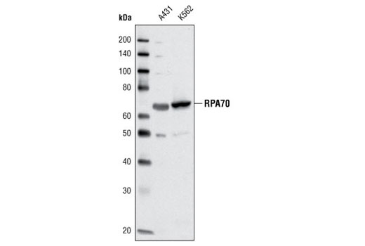
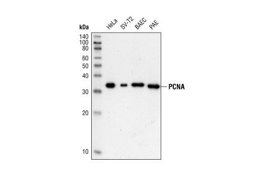
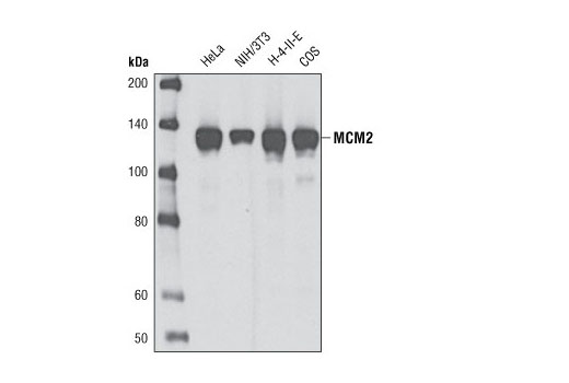
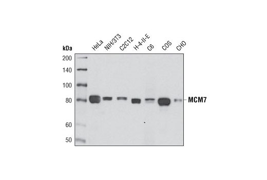
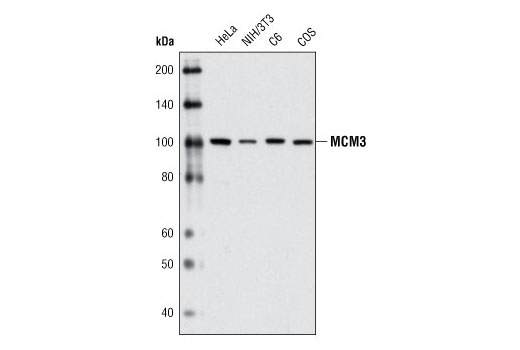
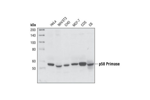
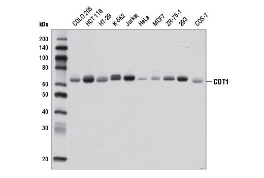
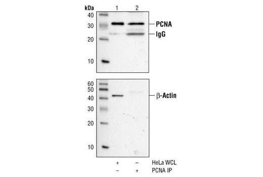
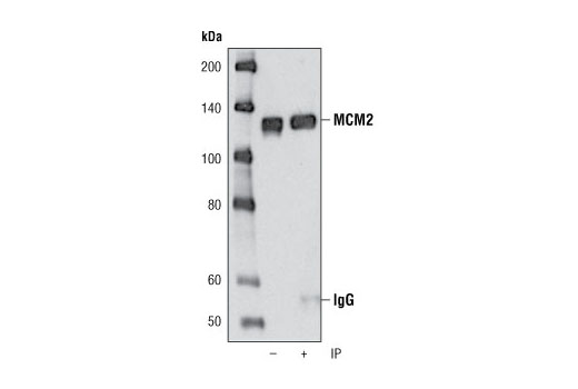
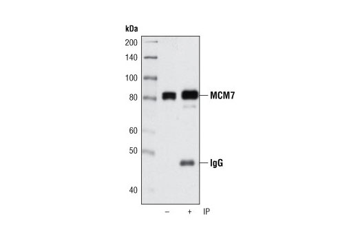
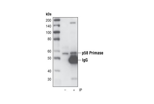
The initiation of DNA replication in mammalian cells is a highly coordinated process that is regulated by several protein complexes. Origins of replication (ORCs), at which replication is initiated, are dispersed throughout the genome. Their activities are regulated via the sequential binding of pre-replication and replication factors that initiate formation of replication forks, the active structures at which DNA is synthesized. The origin recognition complex is thought to be bound to chromatin throughout the cell cycle (1,2). The pre-replication complex (Pre-RC) forms in late mitosis/early G1 phase beginning with the binding of CDT1 and CDC6 to the origin. Together CDT1 and CDC6 promote the loading of the heterohexameric minichromosome maintenance (MCM) complex. This process is referred to as chromatin licensing. Licensing of the chromatin permits the DNA to replicate only once per cell cycle, helping to ensure that genetic alterations and malignant cell growth do not occur (reviewed in 3). The canonical MCM complex proteins (MCM2-7) are a family of six related phospho-proteins that function, in part, as the eukaryotic replicative DNA helicase (3,4). Phosphorylation and ubiquitination of the MCM2, MCM3, MCM4, and MCM6 subunits appears to regulate MCM complex activity and the initiation of DNA synthesis (5-7). MCM proteins are removed during DNA replication, causing chromatin to become unlicensed, inhibiting Pre-RC reformation. In addition to DNA polymerase, initiation of DNA replication requires a pair of primase subunits. DNA Primase activity catalyzes de novo synthesis of an RNA/DNA primer (initiator DNA) on the leading and lagging strands, while polymerase activity extends the initiator DNA (8). The 48 and 58 kDa primase subunits cooperate in the synthesis of small RNA primers. p48 is the catalytically active subunit (9), while p58 couples p48 to the polymerase to allow the transfer of primers to the active site. The p58 subunit may also play a role in regulation of primer length (10,11). Once replication is initiated, Proliferating Cell Nuclear Antigen (PCNA) serves as an accessory factor for DNA polymerases delta and epsilon, acting to tether these polymerases to template DNA during replication. Interactions of PCNA with DNA polymerases increase the processivity of leading strand synthesis. PCNA, a member of DNA sliding clamp family, is a homotrimeric ring complex that encircles and slides along the DNA double helix as the replication fork progresses (12). Multiple proteins involved in DNA replication, DNA repair, and cell cycle control bind to PCNA and regulate DNA synthesis. PCNA is loaded onto the DNA in an ATP-dependent manner by a multiprotein clamp loader, Replication Factor C (RFC) (13). RFC, in turn, associates with DNA via interactions with the single-stranded DNA binding protein complex, Replication Protein A (RPA). The canonical RPA complex is heterotrimeric and composed of RPA1 (RPA70), RPA2 (RPA32), and RPA3 (RPA14) subunits. RPA recognizes and stabilizes single stranded DNA in repair processes and DNA recombination, and plays a role in replication (14-17).

  1. Okuno, Y. et al. (2001) EMBO J 20, 4263-77.
  2. McNairn, A.J. et al. (2005) Exp Cell Res 308, 345-56.
  3. Forsburg, S.L. (2004) Microbiol Mol Biol Rev 68, 109-31.
  4. Johnson, A. and O'Donnell, M. (2005) Annu Rev Biochem 74, 283-315.
  5. Charych, D.H. et al. (2008) J Cell Biochem 104, 1075-86.
  6. Masai, H. et al. (2006) J Biol Chem 281, 39249-61.
  7. Lin, D.I. et al. (2008) Proc Natl Acad Sci U S A 105, 8079-84.
  8. Shiratori, A. et al. (1995) Genomics 28, 350-3.
  9. Copeland, W.C. (1997) Protein Expr Purif 9, 1-9.
  10. Copeland, W.C. and Wang, T.S. (1993) J Biol Chem 268, 26179-89.
  11. Arezi, B. and Kuchta, R.D. (2000) Trends Biochem Sci 25, 572-6.
  12. Bowman, G.D. et al. (2004) Nature 429, 724-30.
  13. Zhang, G. et al. (1999) Proc Natl Acad Sci U S A 96, 1869-74.
  14. Sakaguchi, K. et al. (2009) FEBS J 276, 943-63.
  15. Zou, Y. et al. (2006) J Cell Physiol 208, 267-73.
  16. Wold, M.S. (1997) Annu Rev Biochem 66, 61-92.
  17. Binz, S.K. et al. DNA Repair (Amst) 3, 1015-24.

Limited Uses

Except as otherwise expressly agreed in a writing signed by a legally authorized representative of CST, the following terms apply to Products provided by CST, its affiliates or its distributors. Any Customer's terms and conditions that are in addition to, or different from, those contained herein, unless separately accepted in writing by a legally authorized representative of CST, are rejected and are of no force or effect.

Products are labeled with For Research Use Only or a similar labeling statement and have not been approved, cleared, or licensed by the FDA or other regulatory foreign or domestic entity, for any purpose. Customer shall not use any Product for any diagnostic or therapeutic purpose, or otherwise in any manner that conflicts with its labeling statement. Products sold or licensed by CST are provided for Customer as the end-user and solely for research and development uses. Any use of Product for diagnostic, prophylactic or therapeutic purposes, or any purchase of Product for resale (alone or as a component) or other commercial purpose, requires a separate license from CST. Customer shall (a) not sell, license, loan, donate or otherwise transfer or make available any Product to any third party, whether alone or in combination with other materials, or use the Products to manufacture any commercial products, (b) not copy, modify, reverse engineer, decompile, disassemble or otherwise attempt to discover the underlying structure or technology of the Products, or use the Products for the purpose of developing any products or services that would compete with CST products or services, (c) not alter or remove from the Products any trademarks, trade names, logos, patent or copyright notices or markings, (d) use the Products solely in accordance with CST Product Terms of Sale and any applicable documentation, and (e) comply with any license, terms of service or similar agreement with respect to any third party products or services used by Customer in connection with the Products.

For Research Use Only. Not for Use in Diagnostic Procedures.
Cell Signaling Technology is a trademark of Cell Signaling Technology, Inc.
All other trademarks are the property of their respective owners. Visit our Trademark Information page.