| REACTIVITY | H |
| SENSITIVITY | Endogenous |
| MW (kDa) | 142 |
| Source/Isotype | Mouse IgG1 |
Product Information
| Application | Dilution |
|---|---|
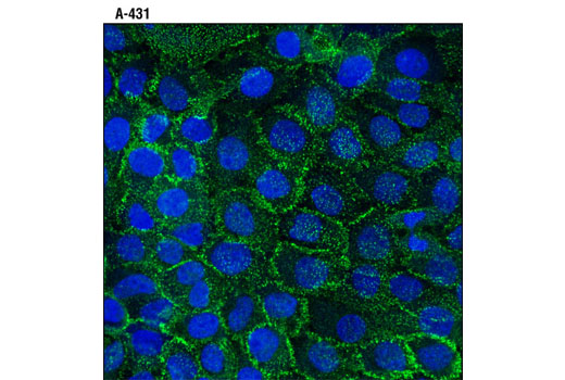
| Immunofluorescence (Immunocytochemistry) | 1:100 |
Achieve higher quality immunofluorescent images using the efficient and cost-effective, pre-made reagents in our #12727 Immunofluorescence Application Solutions Kit
NOTE: Prepare solutions with reverse osmosis deionized (RODI) or equivalent grade water.
Recommended Fluorochrome-conjugated Anti-Mouse secondary antibodies:
NOTE: Cells should be grown, treated, fixed and stained directly in multi-well plates, chamber slides or on coverslips.
Aspirate liquid, then cover cells to a depth of 2–3 mm with 4% formaldehyde diluted in 1X PBS.
NOTE: Formaldehyde is toxic, use only in a fume hood.
NOTE: All subsequent incubations should be carried out at room temperature unless otherwise noted in a humid light-tight box or covered dish/plate to prevent drying and fluorochrome fading.
posted November 2006
revised November 2013
Protocol Id: 148
Human
Monoclonal antibody is produced by immunizing animals with a recombinant protein specific to the amino terminus of human DSG2 protein.
Desmosomes are a class of intracellular junction that tightly link adjacent cells in mechanically stressed tissues such as the epithelium and myocardium (1). They derive their characteristic strength from the protein desmoplakin, which acts as a tether by binding the cytoplasmic component of the desmosome at its N terminus (2), while its C terminus is anchored to the intermediate-filament cytoskeleton (3). Desmogleins and desmocollins belong to the superfamily of cadherin proteins, the "glue" of the desmosome, as they are essential for strong cell-cell contacts (4). There are 4 types of desmogleins in humans, DSG1-4, and 3 types of desmocollins, DSC1-3. DSG2 is expressed in all desmosome bearing tissues, while other desmosome cadherin proteins have more specialized tissue expression. Research studies have shown that aberrant expression due to mutation of DSG2 is associated with arrhythmogenic right ventricular cardiomyopathy (5,6). Research has also shown that mutation and altered expression of DSG1 and 3 have been associated with autoimmune disorders such as Pemphigus, inherited disorders such as defective hair-follicle differentiation, and striate palmoplantar keratoderma, an epidermal-thickening disease (reviewed in 7).
Except as otherwise expressly agreed in a writing signed by a legally authorized representative of CST, the following terms apply to Products provided by CST, its affiliates or its distributors. Any Customer's terms and conditions that are in addition to, or different from, those contained herein, unless separately accepted in writing by a legally authorized representative of CST, are rejected and are of no force or effect.
Products are labeled with For Research Use Only or a similar labeling statement and have not been approved, cleared, or licensed by the FDA or other regulatory foreign or domestic entity, for any purpose. Customer shall not use any Product for any diagnostic or therapeutic purpose, or otherwise in any manner that conflicts with its labeling statement. Products sold or licensed by CST are provided for Customer as the end-user and solely for research and development uses. Any use of Product for diagnostic, prophylactic or therapeutic purposes, or any purchase of Product for resale (alone or as a component) or other commercial purpose, requires a separate license from CST. Customer shall (a) not sell, license, loan, donate or otherwise transfer or make available any Product to any third party, whether alone or in combination with other materials, or use the Products to manufacture any commercial products, (b) not copy, modify, reverse engineer, decompile, disassemble or otherwise attempt to discover the underlying structure or technology of the Products, or use the Products for the purpose of developing any products or services that would compete with CST products or services, (c) not alter or remove from the Products any trademarks, trade names, logos, patent or copyright notices or markings, (d) use the Products solely in accordance with CST Product Terms of Sale and any applicable documentation, and (e) comply with any license, terms of service or similar agreement with respect to any third party products or services used by Customer in connection with the Products.