Product Information
Monoclonal antibody is produced by immunizing animals with synthetic phosphopeptides corresponding to residues surrounding Ser15 of human p53, Thr180/Tyr182 of human p38 MAPK, Ser345 of human Chk1, Thr183/Tyr185 of human SAPK/JNK, Ser139 of human histone H2A.X, Tyr15 of human cdc2, or the carboxy terminus of human p21. Polyclonal antibodies are produced by immunizing animals with a synthetic phosphopeptide corresponding to residues surrounding Thr68 of human Chk2. Polyclonal antibodies are purified by protein A and peptide affinity chromatography.
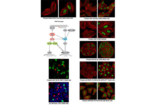
Cell cycle control involves an ordered series of cellular signaling events designed to maintain the integrity and proper function of cells and biological pathways. Cells have evolved complex mechanisms, collectively termed the DNA damage checkpoint, to modulate cell cycle progression in response to genomic insult. Damage to DNA caused by either internal or external sources such as UV light, ionizing radiation, genotoxic agents, etc. initiates a cascade of events that blocks cell cycle progression to either allow time to repair damaged DNA, or activate cell death pathways if too much damage has been incurred. Monitoring the signaling components of the DNA damage pathway is important to aid in understanding the aberrant cellular signaling associated with unchecked cell cycle control in disease states such as cancer. Cell cycle progression can be affected by the DNA damage checkpoint at the G1/S transition, during S phase, or at the G2/M phase transition. At G2/M, cdc2/cyclin B activity acts as a master regulator of entry into mitosis (1). The cdc25C phosphatase removes inhibitory phosphorylation on Thr14 and Tyr15 of cdc2, allowing for maximal activation of cdc2/cyclin B (1,2). When DNA damage occurs, a signaling cascade is activated that inhibits the ability of cdc25 to activate cdc2/cyclin B. A proximal event in the signaling pathway is localization of Ser139-phosphorylated histone H2A.X to sites of DNA damage at subnuclear foci (3). Histone H2A.X and other mediators recruit additional signaling molecules to the site of the damage, activating the ATM/ATR kinases, the central mediators of the DNA damage response. ATM is primarily activated by double strand breaks of DNA (4,5), while ATR is activated by a variety of DNA lesions and replication stresses (6). ATM/ATR in turn initiate two parallel cascades that inactivate the cdc2/cyclin B complex (7). The first cascade rapidly inhibits progression into mitosis through the activation of the Chk kinases (Chk1 for ATR and Chk2 for ATM), which phosphorylate and inactivate cdc25, preventing activation of cdc2/cyclin B (8-10). The more long-term second cascade involves phosphorylation of the tumor suppressor protein p53, leading to either cell cycle arrest and DNA repair or apoptosis through regulation of p53 downstream effectors, including the tumor suppressor protein p21 Waf1/Cip1 (11). Upon DNA damage, p53 is phosphorylated at a number of sites and up-regulates p21 transcription via a p53 responsive element. p21 expression can block cell cycle progression by inhibiting a subset of the cyclin-dependent kinases including cdc2 (12,13). In addition to canonical ATM/ATR checkpoint signaling, the SAPK/JNK and p38 MAP kinase pathways are activated by a variety of cellular stresses including inflammatory cytokines, UV light, and growth factors (14,15). These stress-activated pathways contribute to G2/M checkpoint control through activation of p53 and other MAPK substrates, such as MAPKAPK-2, which can directly affect components of the checkpoint cascade, such as cdc25 (8).
Except as otherwise expressly agreed in a writing signed by a legally authorized representative of CST, the following terms apply to Products provided by CST, its affiliates or its distributors. Any Customer's terms and conditions that are in addition to, or different from, those contained herein, unless separately accepted in writing by a legally authorized representative of CST, are rejected and are of no force or effect.
Products are labeled with For Research Use Only or a similar labeling statement and have not been approved, cleared, or licensed by the FDA or other regulatory foreign or domestic entity, for any purpose. Customer shall not use any Product for any diagnostic or therapeutic purpose, or otherwise in any manner that conflicts with its labeling statement. Products sold or licensed by CST are provided for Customer as the end-user and solely for research and development uses. Any use of Product for diagnostic, prophylactic or therapeutic purposes, or any purchase of Product for resale (alone or as a component) or other commercial purpose, requires a separate license from CST. Customer shall (a) not sell, license, loan, donate or otherwise transfer or make available any Product to any third party, whether alone or in combination with other materials, or use the Products to manufacture any commercial products, (b) not copy, modify, reverse engineer, decompile, disassemble or otherwise attempt to discover the underlying structure or technology of the Products, or use the Products for the purpose of developing any products or services that would compete with CST products or services, (c) not alter or remove from the Products any trademarks, trade names, logos, patent or copyright notices or markings, (d) use the Products solely in accordance with CST Product Terms of Sale and any applicable documentation, and (e) comply with any license, terms of service or similar agreement with respect to any third party products or services used by Customer in connection with the Products.