| Cat. # | Size | Qty. | Price |
|---|---|---|---|
| 8648T | 1 Kit (5 x 20 microliters) |
|
| Product Includes | Quantity | Applications | Reactivity | MW(kDa) | Isotype |
|---|---|---|---|---|---|
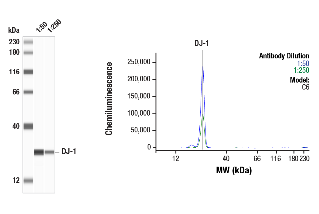
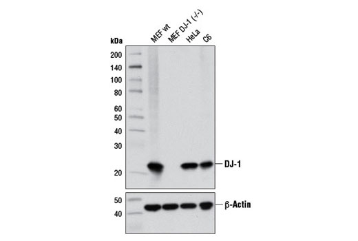
| DJ-1 (D29E5) XP® Rabbit mAb 5933 | 20 µl |
|
H M R Hm Mk | 22 | Rabbit IgG |
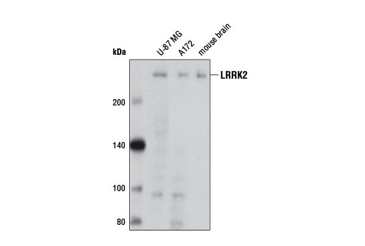
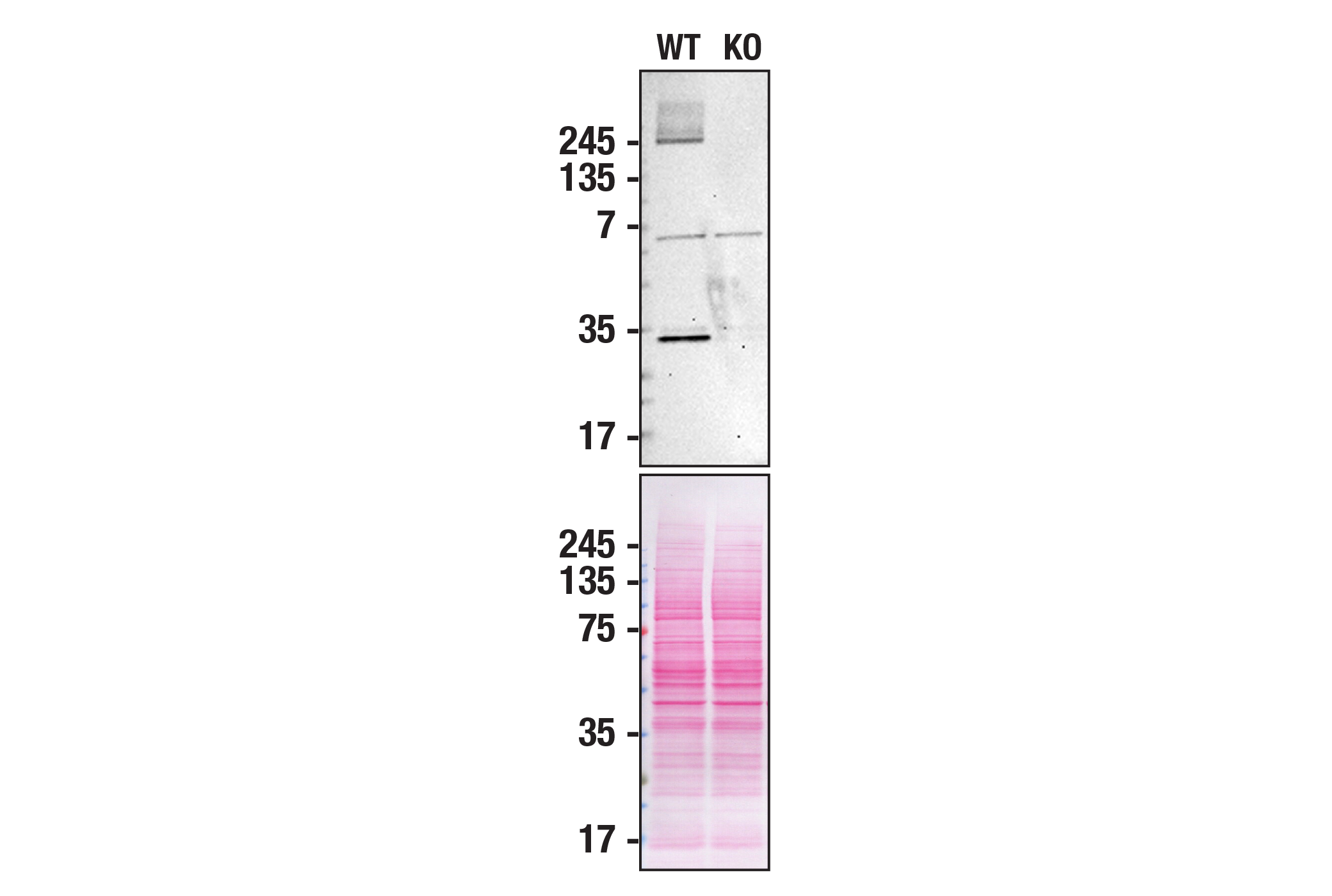
| LRRK2 (D18E12) Rabbit mAb 13046 | 20 µl |
|
H M R | 290 | Rabbit IgG |
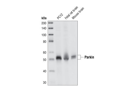
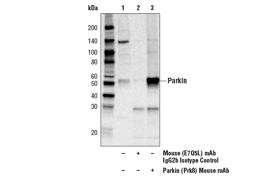
| Parkin (Prk8) Mouse mAb 4211 | 20 µl |
|
H M R | 50 | Mouse IgG2b |
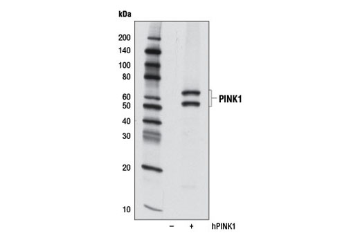
| PINK1 (D8G3) Rabbit mAb 6946 | 20 µl |
|
H | 60, 50 | Rabbit IgG |
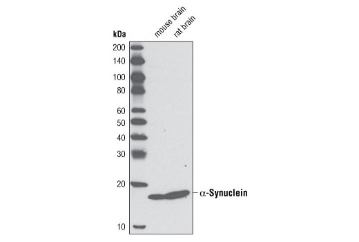
| α-Synuclein (D37A6) Rabbit mAb 4179 | 20 µl |
|
M R | 18 | Rabbit IgG |
| Anti-rabbit IgG, HRP-linked Antibody 7074 | 100 µl |
|
Goat | ||
| Anti-mouse IgG, HRP-linked Antibody 7076 | 100 µl |
|
Horse |
Product Information
Monoclonal antibodies are produced by immuninzing animals with a recombinant protein specific to the carboxy terminus of Parkin protein, a synthetic peptide corresponding to residues surrounding Lys148 of human DJ-1 protein, a synthetic peptide corresponding to residues surrounding Pro2080 of human LRRK2 protein, a synthetic peptide corresponding to residues surrounding Pro140 of human PINK1 protein, or a synthetic peptide corresponding to residues surrounding Glu105 of mouse α-synuclein protein.
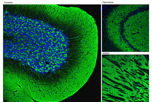
Parkinson’s disease (PD), the second most common neurodegenerative disease after Alzheimer’s, is a progressive movement disorder characterized by rigidity, tremors, and postural instability. The pathological hallmark of PD is progressive loss of dopaminergic neurons in the substantia nigra of the ventral midbrain and the presence of intracellular Lewy bodies in surviving neurons of the brain stem (1). Research studies have shown that various genes and loci (α-synuclein/PARK1 and 4, parkin/PARK2, UCH-L1/PARK5, PINK1/PARK6, DJ-1/PARK7, LRRK2/PARK8, synphilin-1, and NR4A2) are genetically linked to PD (2).
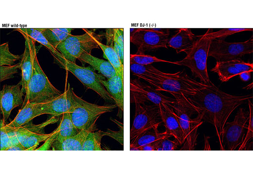
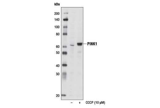
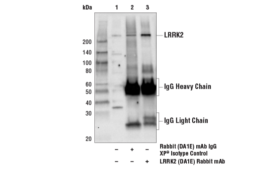
α-Synuclein, a 140 amino acid protein expressed abundantly in the brain, is a major component of aggregates found in Lewy bodies (3). Parkin is involved in protein degradation through the ubiquitin-proteasome pathway, and investigators have shown that mutations in Parkin cause early onset of PD (4). In the case of autosomal recessive juvenile Parkinsonism (AR-JP), deletions have been found on chromosome 6 in the Parkin gene (5). PTEN induced putative kinase 1 (PINK1) is a mitochondrial serine/threonine kinase involved in the normal function and integrity of mitochondria, as well as a reduction of cytochrome c release from mitochondria (6-8). PINK1 phosphorylates Parkin and promotes its translocation to mitochondria (7). Mutations of PINK1 are associated with loss of protective function, mitrochondrial dysfunction, aggregation of α-synuclein, and proteasome dysfunction (6,8). DJ-1 is involved in multiple cellular functions; it has been shown to cooperate with Ras to increase cell transformation, to regulate transcription of the androgen receptor, and may function as an indicator of oxidative stress, while loss-of-function mutations in DJ-1 cause early onset of PD (9-12). Dopamine D2 receptor-mediated functions are greatly impaired in DJ-1 (-/-) mice, resulting in reduced long-term depression (13). Leucine-rich repeat kinase 2 (LRRK2) contains amino-terminal leucine-rich repeats (LRR), a Ras-like small GTP binding protein-like (ROC) domain, an MLK protein kinase domain, and a carboxy-terminal WD40-repeat. At least 20 LRRK2 mutations have been linked to PD (14). The most prevalent mutation, G2019S, causes increased LRRK2 kinase activity, leading to progressive neurite loss and decreased neuronal survival (15).
Except as otherwise expressly agreed in a writing signed by a legally authorized representative of CST, the following terms apply to Products provided by CST, its affiliates or its distributors. Any Customer's terms and conditions that are in addition to, or different from, those contained herein, unless separately accepted in writing by a legally authorized representative of CST, are rejected and are of no force or effect.
Products are labeled with For Research Use Only or a similar labeling statement and have not been approved, cleared, or licensed by the FDA or other regulatory foreign or domestic entity, for any purpose. Customer shall not use any Product for any diagnostic or therapeutic purpose, or otherwise in any manner that conflicts with its labeling statement. Products sold or licensed by CST are provided for Customer as the end-user and solely for research and development uses. Any use of Product for diagnostic, prophylactic or therapeutic purposes, or any purchase of Product for resale (alone or as a component) or other commercial purpose, requires a separate license from CST. Customer shall (a) not sell, license, loan, donate or otherwise transfer or make available any Product to any third party, whether alone or in combination with other materials, or use the Products to manufacture any commercial products, (b) not copy, modify, reverse engineer, decompile, disassemble or otherwise attempt to discover the underlying structure or technology of the Products, or use the Products for the purpose of developing any products or services that would compete with CST products or services, (c) not alter or remove from the Products any trademarks, trade names, logos, patent or copyright notices or markings, (d) use the Products solely in accordance with CST Product Terms of Sale and any applicable documentation, and (e) comply with any license, terms of service or similar agreement with respect to any third party products or services used by Customer in connection with the Products.