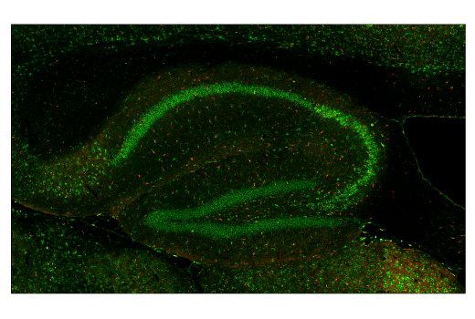
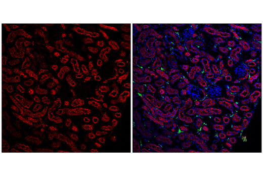
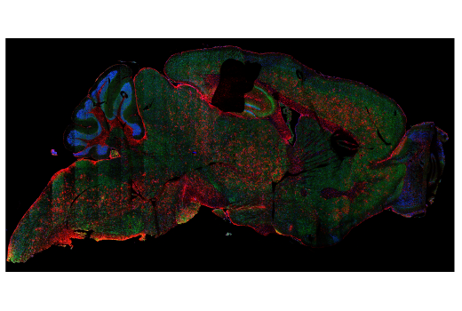
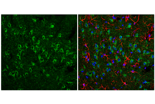
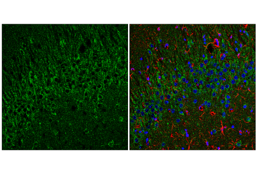
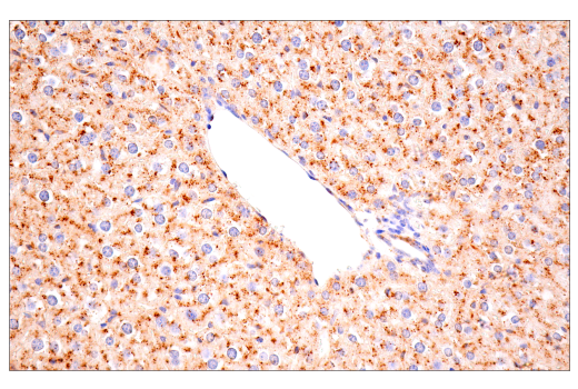
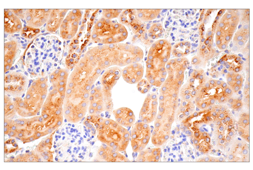
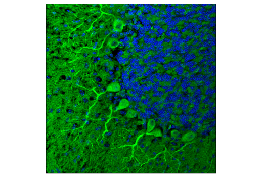
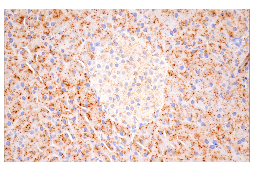
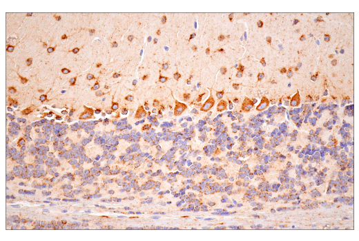
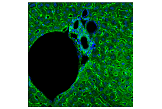
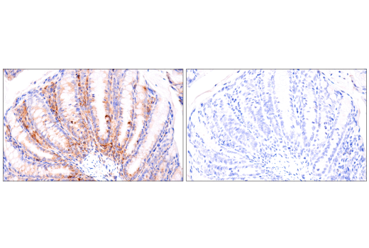
Immunohistochemical analysis of paraffin-embedded mouse cerebellum using Cathepsin D (E7Z4L) XP® Rabbit mAb.
| Cat. # | Size | Qty. | Price |
|---|---|---|---|
| 61381T | 1 Kit (5 x 20 microliters) |
|
| Product Includes | Quantity | Applications | Reactivity | MW(kDa) | Isotype |
|---|---|---|---|---|---|
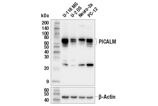
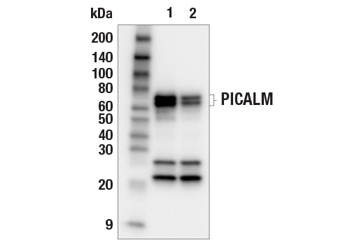
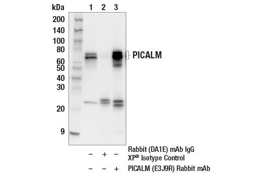
| PICALM (E3J9R) Rabbit mAb 26765 | 20 µl |
|
H M R | 68, 70 | Rabbit IgG |
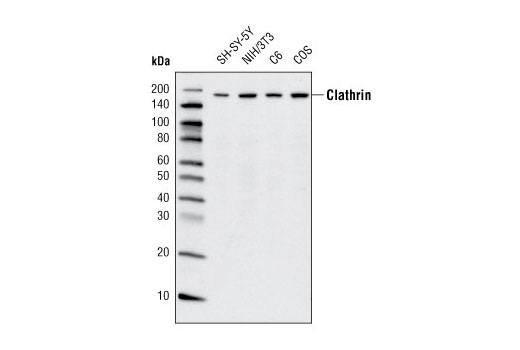
| Clathrin Heavy Chain (D3C6) XP® Rabbit mAb 4796 | 20 µl |
|
H M R Mk | 190 | Rabbit IgG |
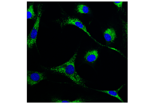
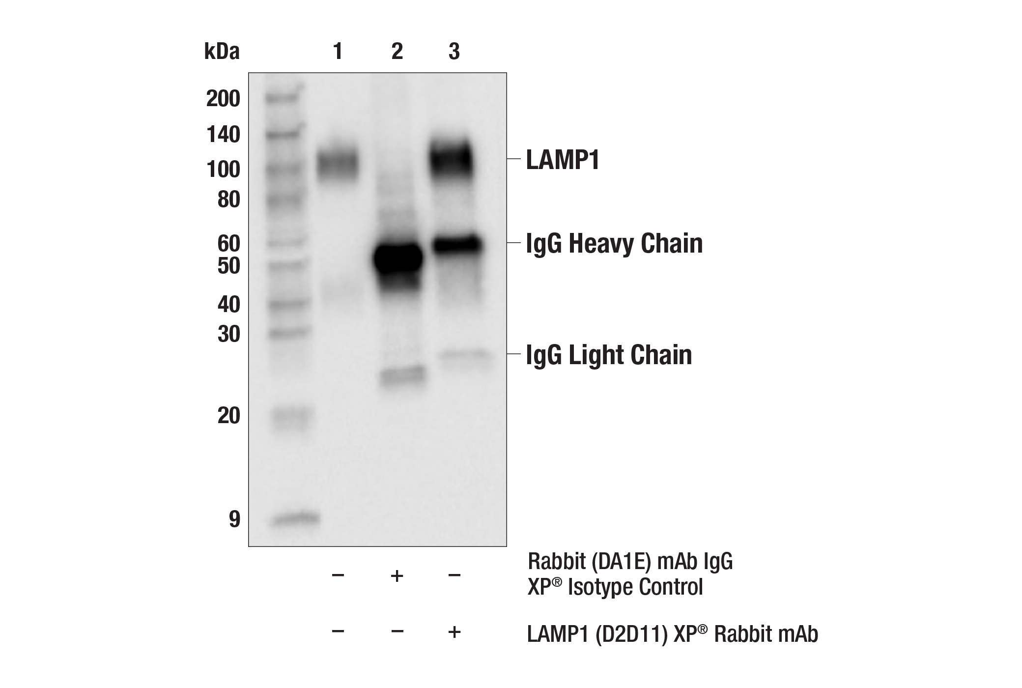
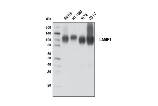
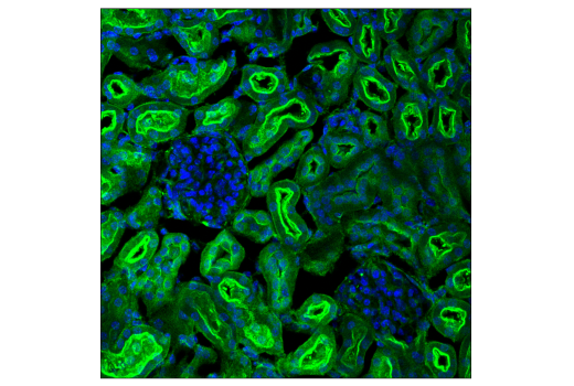
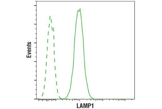
| LAMP1 (D2D11) XP® Rabbit mAb 9091 | 20 µl |
|
H Mk | 42 (non-glycosylated), 90-120 (glycosylated) | Rabbit IgG |
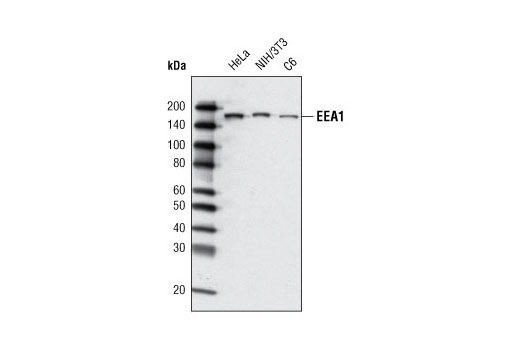
| EEA1 (C45B10) Rabbit mAb 3288 | 20 µl |
|
H M R | 170 | Rabbit IgG |
| Cathepsin D (E7Z4L) XP® Rabbit mAb 88239 | 20 µl |
|
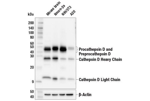
M | 46, 43, 28 | Rabbit IgG |
| Anti-rabbit IgG, HRP-linked Antibody 7074 | 100 µl |
|
Goat |
Product Information
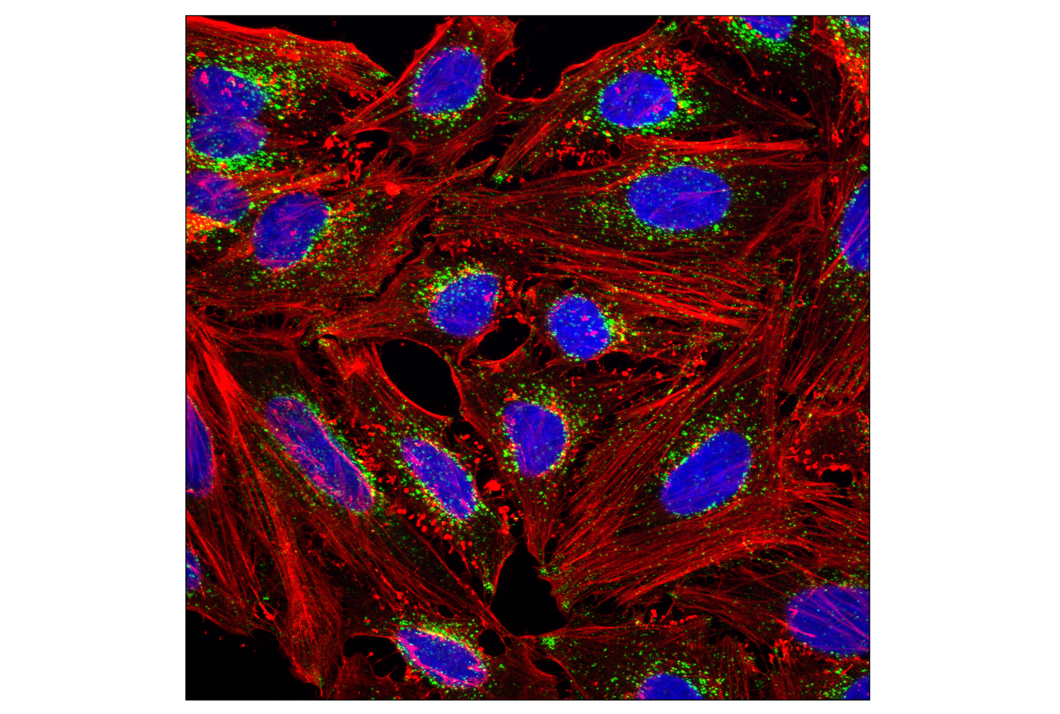
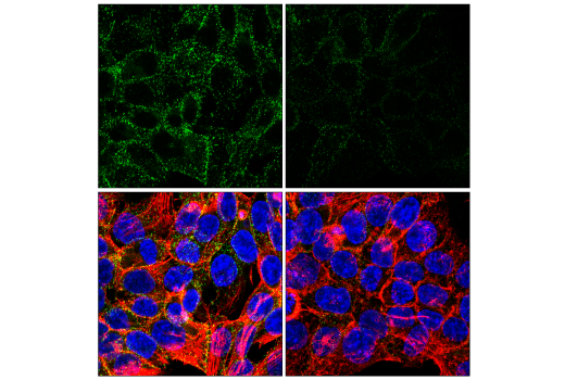
The antibodies in this kit serve to characterize phosphatidylinositol-binding clathrin assembly protein (PICALM)-mediated lysosomal maturation, as endo-lysosomal systems are important for normal physiology and prevention of common late-onset neurodegenerative diseases such as Alzheimer's disease (AD).
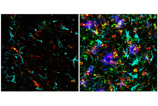
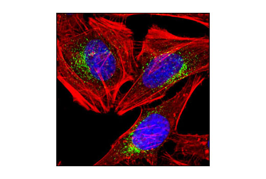
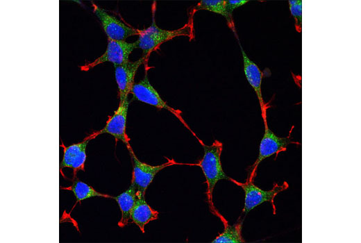
PICALM is a clathrin-binding protein involved in the endo-lysosomal pathway, where it has been genetically associated with AD (1,2). Clathrin is a triskelion-shaped protein that plays a major role in the formation of coated vesicles (1). Formation of these vesicles is critical for shaping the cell membrane to promote intracellular trafficking for multiple membrane trafficking pathways in the cell, including the trans-Golgi network as well as transport to and from the cell membrane and endosomal compartments. PICALM disruption increases the number of early endosomes, which is linked to exacerbated tau aggregation (2). Early endosome antigen 1 (EEA1) is an early endosomal marker and a Rab5 effector protein essential for early endosomal membrane fusion and trafficking (3,4). Lysosome-associated membrane protein 1 (LAMP1) is an abundant lysosomal membrane protein involved in regulating lysosomal motility during lysosome-phagosome fusion (5,6). Cathepsin D (CSTD) is a ubiquitously expressed lysosomal aspartyl protease involved in the normal degradation of proteins (7). Mutations in PICALM were shown to cause lysosomal enzymes and membrane proteins to be mis-trafficked and accumulated; for example, immature forms of CTSD accumulate abnormally within endosomes. These changes correlate with reduced turnover of lysosomal cargoes generated by autophagy and endocytosis (2).
Explore pathways related to this product.
STRING - Known and Predicted Protein-Protein Interactions.
Except as otherwise expressly agreed in a writing signed by a legally authorized representative of CST, the following terms apply to Products provided by CST, its affiliates or its distributors. Any Customer's terms and conditions that are in addition to, or different from, those contained herein, unless separately accepted in writing by a legally authorized representative of CST, are rejected and are of no force or effect.
Products are labeled with For Research Use Only or a similar labeling statement and have not been approved, cleared, or licensed by the FDA or other regulatory foreign or domestic entity, for any purpose. Customer shall not use any Product for any diagnostic or therapeutic purpose, or otherwise in any manner that conflicts with its labeling statement. Products sold or licensed by CST are provided for Customer as the end-user and solely for research and development uses. Any use of Product for diagnostic, prophylactic or therapeutic purposes, or any purchase of Product for resale (alone or as a component) or other commercial purpose, requires a separate license from CST. Customer shall (a) not sell, license, loan, donate or otherwise transfer or make available any Product to any third party, whether alone or in combination with other materials, or use the Products to manufacture any commercial products, (b) not copy, modify, reverse engineer, decompile, disassemble or otherwise attempt to discover the underlying structure or technology of the Products, or use the Products for the purpose of developing any products or services that would compete with CST products or services, (c) not alter or remove from the Products any trademarks, trade names, logos, patent or copyright notices or markings, (d) use the Products solely in accordance with CST Product Terms of Sale and any applicable documentation, and (e) comply with any license, terms of service or similar agreement with respect to any third party products or services used by Customer in connection with the Products.