View Featured Offers >>
8345
Tumor Suppressor Inactivation Antibody Sampler Kit
Primary Antibodies
Antibody Sampler Kit

Tumor Suppressor Inactivation Antibody Sampler Kit #8345

Citations (0)
Simple Western™ analysis of lysates (1 mg/mL) from COS-7 cells using p53 (7F5) Rabbit mAb #2527. The virtual lane view (left) shows the target band (as indicated) at 1:10 and 1:50 dilutions of primary antibody. The corresponding electropherogram view (right) plots chemiluminescence by molecular weight along the capillary at 1:10 (blue line) and 1:50 (green line) dilutions of primary antibody. This experiment was performed under reducing conditions on the Jess™ ​​​​​​​ Simple Western instrument from ProteinSimple, a BioTechne brand, using the 12-230 kDa separation module.
Flow cytometric analysis of Jurkat cells using p27 Kip1 (D69C12) XP® Rabbit mAb (right) or concentration-matched Rabbit (DA1E) mAb IgG XP® Isotype Control #3900 (left) and Propidium Iodide (PI)/RNase Staining Solution #4087 to measure DNA content. Anti-rabbit IgG (H+L), F(ab')2 Fragment (Alexa Fluor® 488 Conjugate) #4412 was used as a secondary antibody.
Western blot analysis of extracts from 293 and COS cells, using p53 (7F5) Rabbit mAb.
Western blot analysis from control HeLa cells (lane 1) or p27 Kip1 knockout HeLa cells (lane 2) using p27 Kip1 (D69C12) XP Rabbit mAb #3686 (upper) or B-Actin (D6A8) Rabbit mAb #8457 (lower). The absence of signal in the p27 Kip1 knockout HeLa cells confirms specificity of the antibody for p27 Kip1.
Western blot analysis of extracts from various cell lines using COPS5 Antibody.
After the primary antibody is bound to the target protein, a complex with HRP-linked secondary antibody is formed. The LumiGLO® is added and emits light during enzyme catalyzed decomposition.
Western blot analysis of extracts from COS, NIH3T3, PC12, and SK-N-MC cells, using Smad4 Antibody.
Immunohistochemical analysis of paraffin-embedded human breast carcinoma, using p53 (7F5) Rabbit mAb.
Western blot analysis of extracts from various cell types using p27 Kip1 (D69C12) XP® Rabbit mAb.
Western blot analysis of extracts from 293T cells, either mock-transfected (-) or transfected with a Myc/DDK-tagged human COPS5 cDNA expression construct (+), using COPS5 Antibody.
Western blot analysis of extracts from NIH/3T3 cells, transfected with 100 nM SignalSilence® Control siRNA (Unconjugated) #6568 (-) or SignalSilence® Smad4 siRNA I (Mouse Specific) (+), using Smad4 Antibody #9515 (upper) or β-Actin (D6A8) Rabbit mAb #8457 (lower). The Smad4 Antibody confirms silencing of Smad4 expression, while the β-Actin (D6A8) Rabbit mAb is used as a loading control.
Immunohistochemical analysis of paraffin-embedded human colon carcinoma, using p53 (7F5) Rabbit mAb.
Immunoprecipitation of p27 Kip1 from 293 cells using p27 Kip1 (D69C12) XP® Rabbit mAb. Western analysis was performed using the same antibody. Lane 1 is 5% input.
Confocal immunofluorescent analysis of U-2 OS cells using COPS5 Antibody (green). Blue pseudocolor = DRAQ5® #4084 (fluorescent DNA dye).
Chromatin immunoprecipitations were performed with cross-linked chromatin from 4 x 10⁶ HaCaT cells treated with Human TGF-β3 #3706 (7ng/ml) for 1 h and either 20 μl of Smad4 Antibody or 2 μl of Normal Rabbit IgG #2729 using SimpleChIP® Enzymatic Chromatin IP Kit (Magnetic Beads) #9003. The enriched DNA was quantified by real-time PCR using SimpleChIP® Human CDKN1A Intron 1 Primers #4669, SimpleChIP® Human ID1 Promoter Primers #5139, and SimpleChIP® Human α Satellite Repeat Primers #4486. The amount of immunoprecipitated DNA in each sample is represented as signal relative to the total amount of input chromatin, which is equivalent to one.
Immunohistochemical analysis of paraffin-embedded HT-29 (left) and SaOs-2 (right) cells, using p53 (7F5) Rabbit mAb. Note the lack of staining in p53-negative SaOs-2 cells.
Confocal immunofluorescent analysis of MCF-7 cells using p27 Kip1 (D69C12) XP® Rabbit mAb (green). Actin filaments have been labeled with DY-554 phalloidin (red).
Confocal Immunofluorescent analysis of HT-29 cells using p53 (7F5) Rabbit mAb (green). Actin filaments have been labeled with DY-554 phalloidin (red).
Flow cytometric analysis of HT-29 cells using p53 (7F5) Rabbit mAb (solid line) compared to concentration-matched Rabbit (DA1E) mAb IgG XP® Isotype Control #3900 (dashed line). Anti-rabbit IgG (H+L), F(ab')2 Fragment (Alexa Fluor® 488 Conjugate) #4412 was used as a secondary antibody.
Chromatin immunoprecipitations were performed with cross-linked chromatin from HCT116 cells treated with UV (100 J/m2 followed by a 3 hour recovery) and either p53 (7F5) Rabbit mAb or Normal Rabbit IgG #2729 using SimpleChIP® Enzymatic Chromatin IP Kit (Magnetic Beads) #9003. The enriched DNA was quantified by real-time PCR using SimpleChIP® Human CDKN1A Promoter Primers #6449, human MDM2 intron 2 primers, and SimpleChIP® Human α Satellite Repeat Primers #4486. The amount of immunoprecipitated DNA in each sample is represented as signal relative to the total amount of input chromatin, which is equivalent to one.
Inquiry Info.# 8345

Product Description

The Tumor Suppressor Inactivation Antibody Sampler Kit provides a fast and economical means of evaluating the role of COPS5 in the inhibition of the tumor suppressors p27 Kip1, p53, and Smad4. The kit contains enough primary antibody to perform four western blot experiments with each primary antibody.

Specificity / Sensitivity

COPS5 Antibody recognizes endogenous levels of total COPS5 protein. This antibody does not cross-react with PSMD14/POH1. p27 Kip1 (D69C12) XP® Rabbit mAb detects endogenous levels of total p27 Kip1 protein. p53 (7F5) Rabbit mAb detects endogenous levels of total p53 protein. This antibody binding has been mapped to the amino terminal region of human p53 protein. Smad4 Antibody detects endogenous levels of total Smad4 protein.

Source / Purification

Monoclonal antibodies are produced by immunizing animals with a synthetic peptide corresponding to residues near the amino terminus of human p27 Kip1 protein or with a full-length human p53 fusion protein. Polyclonal antibodies are produced by immunizing animals with a synthetic peptide corresponding to residues near the amino terminus of human COPS5 protein or to the residues surrounding Pro278 of human Smad4 protein. Polyclonal antibodies are purified by protein A and peptide affinity chromatography.

Background

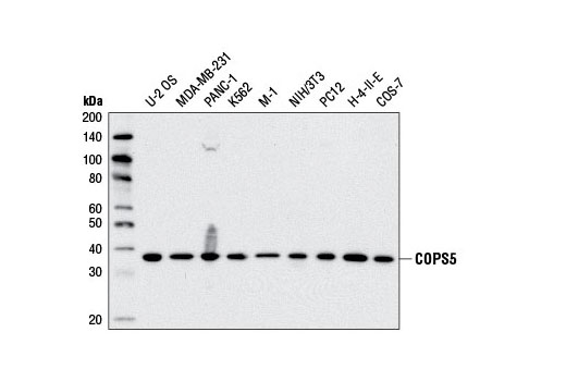
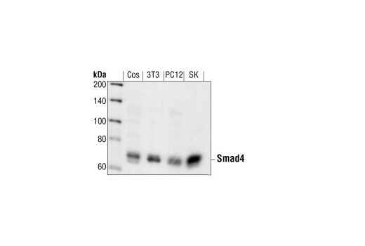
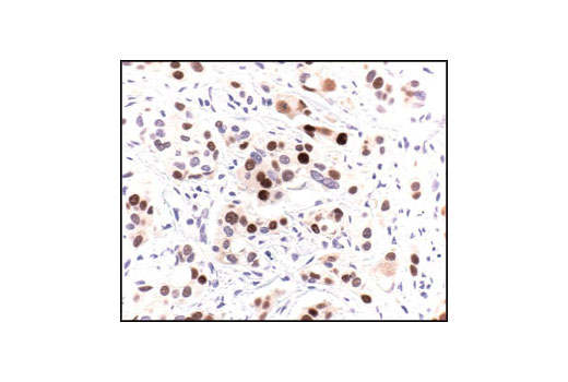
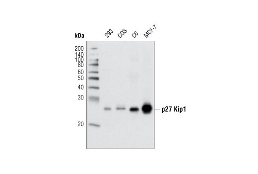
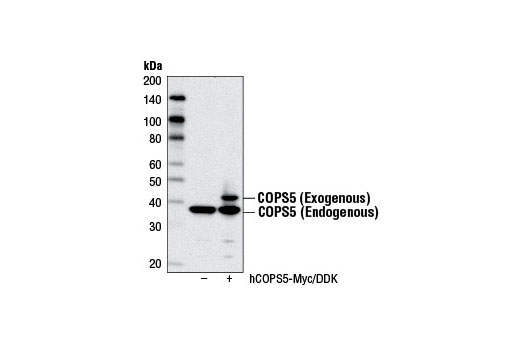
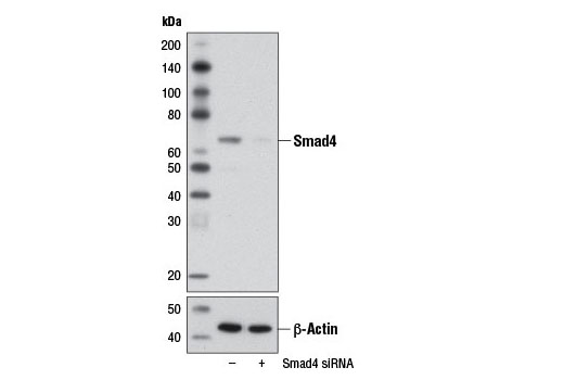
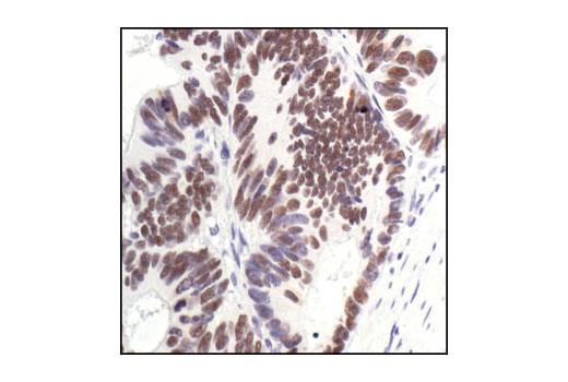
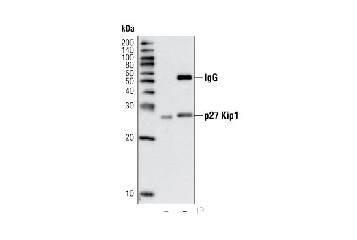
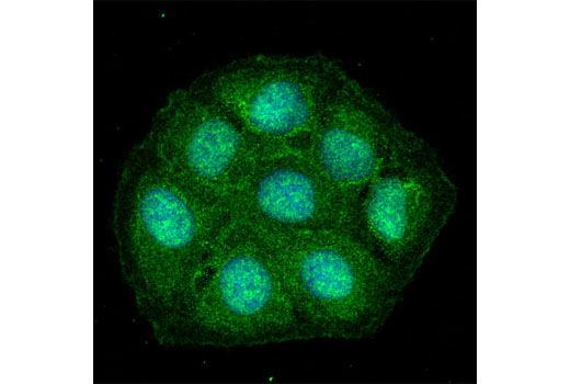
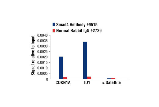
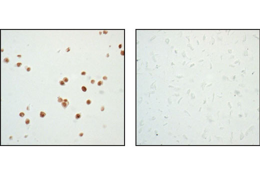
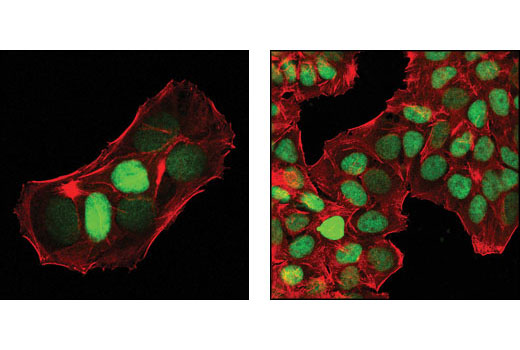
COPS5/CSN5/Jab1 (c-Jun activation domain-binding protein-1) was originally identified as a transcriptional coactivator of c-Jun and subsequently discovered to be a fifth component and integral part of the CSN (1). As the catalytic center of the CSN, COPS5 is able to integrate multiple functions of the CSN complex such as cell cycle control, transcription, and DNA damage response by regulating the activity of CRLs through deneddylation of cullins (2). Indeed, COPS5 harbors an Mpr1-Pad1-N-terminal (MPN) domain with an embedded Jab1/CSN5 MPN domain metalloenzyme (JAMM) motif that is essential for the CSN isopeptidase activity responsible for deneddylation of CRLs. COPS5 is an evolutionarily conserved 38 kDa protein in humans, mice, fission yeast, and plants, which suggests that it is critical to cell survival and proliferation. A role for COPS5 as a positive regulator of cellular proliferation is supported by evidence that it functionally inactivates several key tumor suppressors such as p53, RUNX3, Smad4, and p27 Kip1 through altered subcellular localization, degradation, and deneddylation (3-7). These findings are underscored by the observation that COPS5 overexpression has been identified in a number of different tumor types and has been implicated in the initiation and progression of several types of cancer (8). Moreover, COPS5 deficient mice display an embryonically lethal phenotype highlighted by elevated expression of COPS5 targets such as p53 and p27 (9,10).

Limited Uses

Except as otherwise expressly agreed in a writing signed by a legally authorized representative of CST, the following terms apply to Products provided by CST, its affiliates or its distributors. Any Customer's terms and conditions that are in addition to, or different from, those contained herein, unless separately accepted in writing by a legally authorized representative of CST, are rejected and are of no force or effect.

Products are labeled with For Research Use Only or a similar labeling statement and have not been approved, cleared, or licensed by the FDA or other regulatory foreign or domestic entity, for any purpose. Customer shall not use any Product for any diagnostic or therapeutic purpose, or otherwise in any manner that conflicts with its labeling statement. Products sold or licensed by CST are provided for Customer as the end-user and solely for research and development uses. Any use of Product for diagnostic, prophylactic or therapeutic purposes, or any purchase of Product for resale (alone or as a component) or other commercial purpose, requires a separate license from CST. Customer shall (a) not sell, license, loan, donate or otherwise transfer or make available any Product to any third party, whether alone or in combination with other materials, or use the Products to manufacture any commercial products, (b) not copy, modify, reverse engineer, decompile, disassemble or otherwise attempt to discover the underlying structure or technology of the Products, or use the Products for the purpose of developing any products or services that would compete with CST products or services, (c) not alter or remove from the Products any trademarks, trade names, logos, patent or copyright notices or markings, (d) use the Products solely in accordance with CST Product Terms of Sale and any applicable documentation, and (e) comply with any license, terms of service or similar agreement with respect to any third party products or services used by Customer in connection with the Products.

For Research Use Only. Not for Use in Diagnostic Procedures.
Cell Signaling Technology is a trademark of Cell Signaling Technology, Inc.
All other trademarks are the property of their respective owners. Visit our Trademark Information page.