View Featured Offers >>

Altered Cell Signaling

Abnormal cell-cell communication, for example disrupted presynaptic input, as well as disrupted intracellular signaling contribute to the pathogenesis of neurodegenerative disease. Understanding the signal transduction pathways that regulate gene expression will help efforts to develop therapeutic interventions.

Start with these targets

CREB

CREB signaling is a cellular transcription factor that plays an important role in the formation of memories. Perturbed signaling has been observed in the brains of Alzheimer’s disease mouse models, suggesting CREB signaling may be disrupted in human Alzheimer’s disease brains as well. Disturbances in CREB function may also contribute to the development and progression of Huntington’s disease.
Products
CREB (48H2) Rabbit mAb #9197 – W, IP, IHC-P, IHC-F, IF-F, IF-IC, ChIP, ChIP-seq, F
CREB (48H2) Rabbit mAb #9197

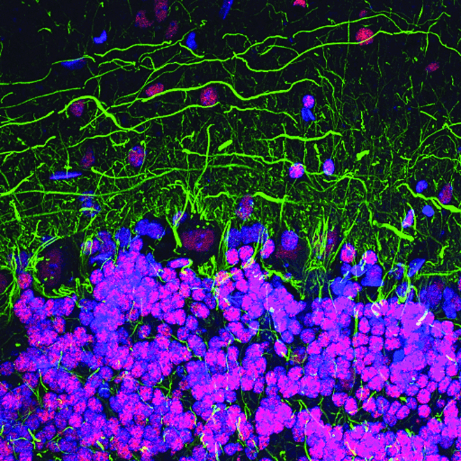
CREB (48H2) Rabbit mAb #9197: Confocal immunofluorescent analysis of mouse cerebellum labeled with CREB (48H2) Rabbit mAb (red) and Neurofilament-L (DA2) Mouse mAb #2835 (green). Blue pseudocolor =DRAQ5 #4084 (fluorescent DNA dye).

Phospho-CREB (Ser133)

CREB is a cellular transcription factor activated when phosphorylated on Ser133. pCREB (Ser133) levels are reduced in the prefrontal cortex of patients with Alzheimer’s disease, indicating a dysfunction in CREB signaling. Reduced pCREB levels in the peripheral blood mononuclear cells (PBMCs) of patients with Alzheimer’s disease correlate with pCREB levels observed in postmortem Alzheimer’s disease brains, suggesting pCREB expression in PBMCs may be a potential biomarker for disease progression.
Products
Phospho-CREB (Ser133) (87G3) Rabbit mAb #9198 – W, IHC-P, IF-IC, IF-F, ChIP, ChIP-seq, F
Phospho-CREB (Ser133) (87G3) Rabbit mAb #9198

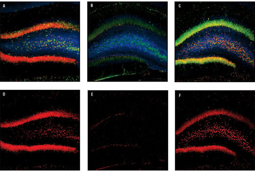
Phospho-CREB (Ser133) (87G3) Rabbit mAb #9198: Conofocal immunofluorescent images of rat dentate gyrus, either sham-operated (left) or 15 min ischemia followed by 30 min (center) and 4 h (right) reperfusion, labeled with Phospho-CREB (Ser133) (87G3) Rabbit mAb (red), Neurofilament-L (DA2) Mouse mAb #2835 (blue) and Phospho-S6 Ribosomal Protein (Ser235/236) (2F9) Rabbit mAb (Alexa Fluor® 488 Conjugate) #4854.

GSK-3β

GSK-3β is known to interact with Tau, beta-amyloid (Aβ), and α-synuclein and is implicated in the pathogenesis of Alzheimer’s disease and Parkinson’s disease. It is one of the kinases responsible for Tau hyperphosphorylation, resulting in neurofibrillary tangles. GSK-3β regulates several critical cellular events, such as axonal transport, microtubule dynamics, apoptosis, and inflammation, making GSK-3β a potential therapeutic target.

Products
GSK-3β (D5C5Z) XP® Rabbit mAb #12456 – W, IP, IHC-P, IF-IC, F
GSK-3β (D5C5Z) XP<sup>®</sup> Rabbit mAb #12456

GSK-3β (D5C5Z) XP® Rabbit mAb #12456: Immunohistochemical analysis of paraffin-embedded MEF cell pellets, wild type (left), GSK-3α (-/-) (middle) and GSK-3β (-/-) (right) using GSK-3β (D5C5Z) XP® Rabbit mAb. (MEF wild type, GSK-3β (-/-), and GSK-3α (-/-) cells were kindly provided by Dr. Jim Woodgett, University of Toronto, Canada).

Phospho-GSK-3β (Ser9)

Phosphorylation of GSK-3β on Ser9 inactivates the protein, influencing its ability to regulate glycogen synthesis in response to insulin. In Alzheimer’s disease mouse models, GSK-3β Ser9 phosphorylation may also reduce APP processing by β-secretase, decreasing Aβ production.
Phospho-GSK-3β (Ser9) (D85E12) XP<sup>®</sup> Rabbit mAb #5558

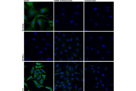
Phospho-GSK-3β (Ser9) (D85E12) XP® Rabbit mAb #5558: Confocal immunofluorescent analysis of wild type mouse embryonic fibroblasts (MEFs) (top row), GSK-3β (-/-) MEFs (middle row) , or PC-3 cells (bottom row), untreated (left), LY294002- and Wortmannin-treated (#9901 and #9951 respectively; center) or lambda phosphatase-treated (right), using Phospho-GSK-3β (Ser9) (D85E12) XP® Rabbit mAb (green). Actin filaments were labeled with DY-554 phalloidin (red). Blue pseudocolor = DRAQ5 #4084 (fluorescent DNA dye). (MEF wild type and GSK-3β (-/-) cells were kindly provided by Dr. Jim Woodgett, University of Toronto, Canada).