View Featured Offers >>

Altered Epigenetics

Epigenetic regulation, including aberrant DNA methylation and histone modifications, have been linked to Alzheimer’s disease, Huntington’s disease, Parkinson’s disease, multiple sclerosis, and amyotrophic lateral sclerosis. However, the exact effects on disease progression are unclear. Brain health is heavily reliant on epigenetic mechanisms, and loss of chromatin dynamics is observed in neurodegenerative diseases. Modifying the environment and targeting sites of potential risk for epigenetic changes are growing areas in the development of therapies against neurodegeneration.

Start with these targets

5-hmC

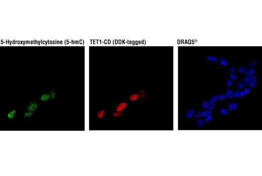
5-hydroxymethylcytosine (5-hmC) is a novel modified cytosine oxidized from 5-mC (5-methylcytosine) by the Tet protein family. The pattern of 5-hmC throughout development is essential for proper neurodevelopment and neurological function. Dysregulation leads to neurodegenerative diseases like Alzheimer’s disease and Huntington’s disease; however, the exact mechanism of action remains to be determined.

Products
5-Hydroxymethylcytosin (5-hmC) (HMC31) Mouse mAb #51660 – Dot blot, MeDIP, IF-IC
5-Hydroxymethylcytosin (5-hmC) (HMC31) Mouse mAb #51660

5-Hydroxymethylcytosin (5-hmC) (HMC31) Mouse mAb #51660: Confocal immunofluorescent analysis of 293T cells transfected with a construct expressing DDK-tagged TET1 catalytic domain (TET1-CD) using 5-Hydroxymethylcytosine (5-hmC) (HMC31) Mouse mAb (green) and DYKDDDDK Tag Antibody #2368 (red). Blue pseudocolor = DRAQ5® #4084 (fluorescent DNA dye). As expected, 293T cells expressing TET1-CD (red) exhibit inccreased levels of 5-hydroxymethylcytosine (green).

5-mC

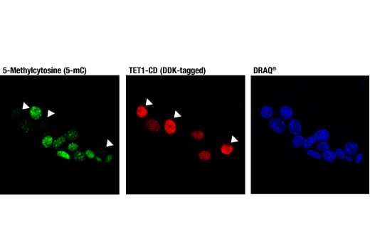
5-methylcytosine (5-mC) is the most common state of cytosine in the brain after the unmodified state. As cells age, total genomic 5-mC content decreases in the brain. Decreased global 5-mC has been observed in Alzheimer’s disease neurons and dysregulation of 5-mC may contribute to progression of amyotrophic lateral sclerosis and Parkinson’s disease.

Products
5-Methylcytosine (5-mC) (D3S2Z) Rabbit mAb #28692 – Dot blot, MeDIP, IF-IC
5-Methylcytosine (5-mC) (D3S2Z) Rabbit mAb #28692

5-Methylcytosine (5-mC) (D3S2Z) Rabbit mAb #28692: Confocal immunofluorescent analysis of 293T cells transfected with a construct expressing DYKDDDDK-tagged TET1 catalytic domain (TET1-CD) using 5-Methylcytosine (5-mC) (D3S2Z) Rabbit mAb (green) and DYKDDDDK Tag (9A3) Mouse mAb #8146 (red). Blue pseudocolor = DRAQ5® #4084 (fluorescent DNA dye). As expected, 293T cells expressing TET1-CD (red) exhibit decreased levels of 5-methylcytosine (green).

HDAC2

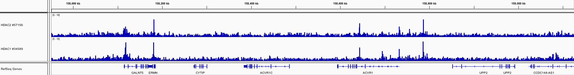
HDAC2 is a class I histone deacetylase that typically leads to gene repression. Deletion of HDAC2 in mouse Alzheimer’s disease models results in improved cognition and decreased amyloid load. Increased HDAC2 expression has also been observed in Alzheimer’s disease patients. HDAC2 is implicated in Huntington’s disease and multiple sclerosis as well.

Products
HDAC2 (D6S5P) Rabbit mAb #57156 – W, IP, ChIP, ChIP-seq, IF-IC
HDAC2 (D6S5P) Rabbit mAb #57156

HDAC2 (D6S5P) Rabbit mAb #57156: Chromatin immunoprecipitations were performed with cross-linked chromatin from K-562 cells and either HDAC2 (D6S5P) Rabbit mAb or HDAC1 (D5C6U) XP® Rabbit mAb #34589, using SimpleChIP® Plus Enzymatic Chromatin IP Kit (Magnetic Beads) #9005. DNA Libraries were prepared using DNA Library Prep Kit for Illumina (ChIP-seq, CUT&RUN) #56795. HDAC2 and HDAC1 are known to have similar binding pattern on chromatin. The figure shows binding of both HDAC2 and HDAC1 across ACVR1 gene. For additional ChIP-seq tracks, please download the product data sheet.

HDAC6

HDAC6 is a class II histone deacetylase with increased expression in the cortex and hippocampus of patients with Alzheimer’s disease. HDAC6 co-localizes with Tau proteins and correlates with Tau phosphorylation. Decreasing HDAC6 levels may result in improved cognition.

Products
HDAC6 (D2E5) Rabbit mAb #7558 – W, IP, IHC-P, IF-IC, F
HDAC6 (D2E5) Rabbit mAb #7558

HDAC6 (D2E5) Rabbit mAb #7558: Confocal immunofluorescent analysis of A549 cells, untreated (left) or treated with MG132 (5 μM, 24 hr; right), using HDAC6 (D2E5) Rabbit mAb (green). Actin filaments were labeled with DY-554 phalloidin (red). Blue pseudocolor = DRAQ5® #4084 (fluorescent DNA dye).

p300

p300 is a histone acetyltransferase that plays a role in the chromatin acetylation that is modulated in response to neuronal activity. Neuronal histone acetylation levels are lower in Alzheimer’s disease mouse models, and activation of amyloid precursor protein-dependent signaling results in reduced histone acetyltransferase levels in primary neuronal cultures. p300 also plays a role in Huntington’s disease and Parkinson’s disease.

Products
p300 (D8Z4E) Rabbit mAb #86377 – W, IP, IHC-P, IF-IC
p300 (D8Z4E) Rabbit mAb #86377

p300 (D8Z4E) Rabbit mAb #86377: Confocal immunofluorescent analysis of 293T cells (left, positive) and HCT-15 cells (right, negative) using p300 (D8Z4E) Rabbit mAb (green). Actin filaments were labeled with DyLight™ 554 Phalloidin #13054 (red).