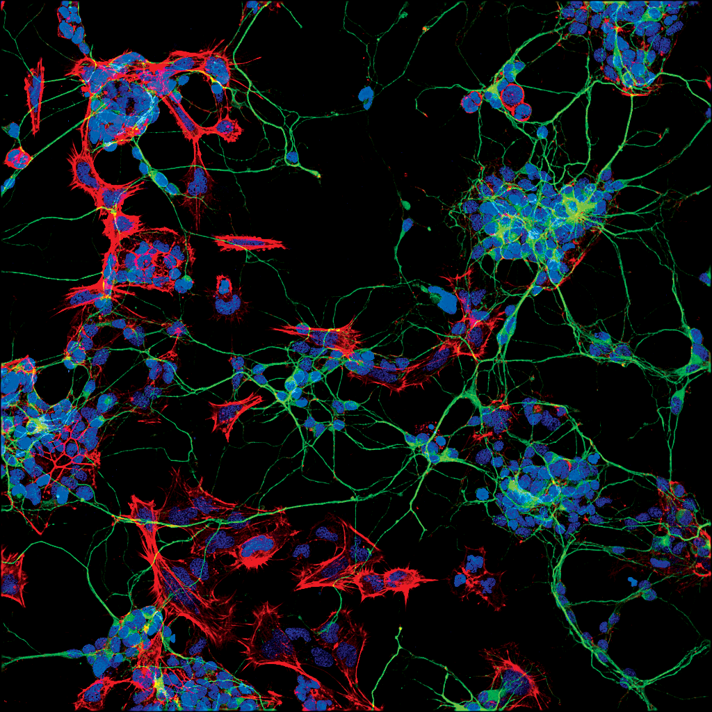
Studying the brain and nervous system requires examination not only of neurons, but also of microglia, oligodendrocytes, and astrocytes. The key to visualizing and identifying each of these cell types lies in using antibodies that target protein biomarkers specifically expressed and localized within these cells.
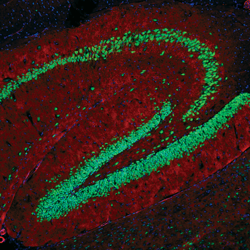
Neurons are electrically excitable cells in the Central Nervous System (CNS) and Peripheral Nervous System (PNS) that send and receive electrical impulses and neurochemical signals to process information. Neurons drive activity in the brain, spinal cord, and peripheral sensory and motor systems. They are highly specialized to quickly transmit electrical signals and neurotransmitters across synapses. Although there is a wide variety of neuronal types and morphologies in the nervous system, signals are typically received on the cell body or dendrites and sent out along the axon. Neurons are often identified by observing the expression of cell-specific intracellular proteins, such as NeuN, β3-Tubulin, and UCHL1.
View a full list of neuronal cell markers. Many of these products are also available conjugated to fluorophores across the spectrum.
Neuronal Nuclei (NeuN) is a pre-mRNA alternative splicing regulator that was first identified in mammals by generating a monoclonal antibody that successfully targeted an antigen, which turned out to be Fox-3, in the nuclei of neurons. Antibodies to this protein are commonly used to label nuclei in the majority of neurons in vertebrates.
β3-tubulin (TUBB3) is one of six β-tubulin isoforms that comprise the major components of microtubules in neurons. It is highly expressed during fetal and postnatal development and plays a critical role in proper axon guidance, maturation, and maintenance. Antibodies against this cell marker stain the cytoskeleton of all neurons.
Microtubule-associated protein 2 (MAP2) is a neuronal phosphoprotein that regulates the structure and stability of microtubules, neuronal morphogenesis, cytoskeleton dynamics, and organelle trafficking in axons and dendrites. Isoforms of MAP2 are expressed in the perikarya and dendrites of neurons, making antibodies that target MAP2 useful tools to highlight the dendritic projections of neurons.
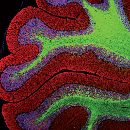
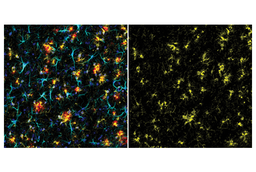
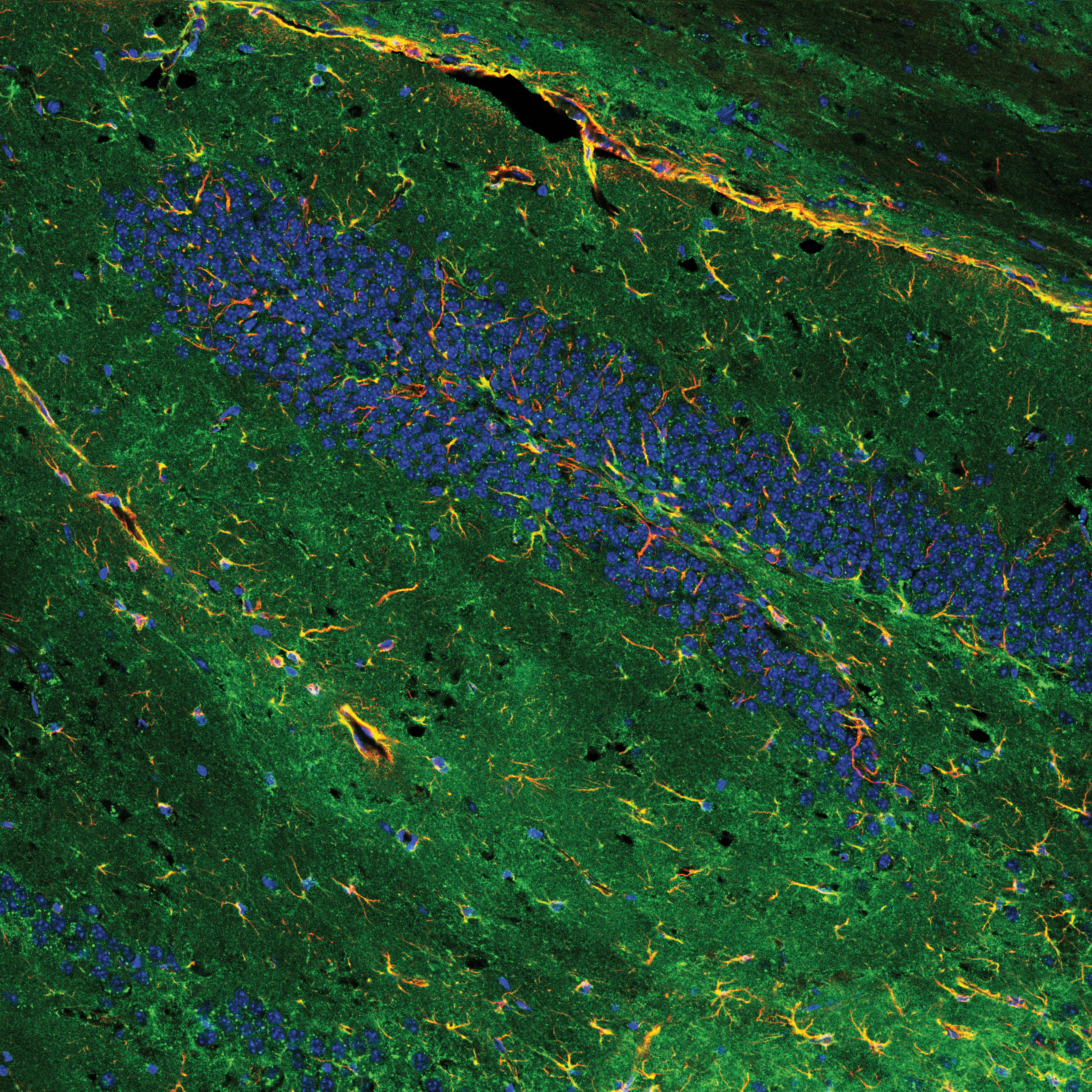
Astrocytes are the most prevalent type of glial cell in the CNS and are found within the brain and spinal cord. In a healthy nervous system, astrocytes play essential roles in development, regulation of blood flow (by supporting endothelial cells in the blood brain barrier), synaptic transmission and function, and energy and metabolism (by providing nutrients to neurons and synthesizing certain neurotransmitters). The loss or abnormal function of astrocytes is implicated in a wide variety of neurodegenerative disease processes. Chronic activation of astrocytes results in the formation of lesions similar to those observed in Alzheimer’s Disease and Huntington’s Disease. Some useful astrocyte markers include GFAP and ALDH1L1.
Glial fibrillary acidic protein (GFAP) is a class-III intermediate filament that makes up the cytoskeleton of astrocytes in the CNS. GFAP establishes and maintains astrocyte morphology and is important for mitosis as well as neuron-astrocyte communication. Antibodies that detect GFAP are often used to label astrocytes and reveal their distinctive morphologies in the brain.
Aldehyde dehydrogenase 1 family member L1 (ALDH1L1) is a key enzyme in folate metabolism and, as such, plays an important role in the regulation of cellular metabolism and proliferation. Downregulation of ALDH1L1 has been observed in tumors, leading to decreased suppression of cancer cell proliferation. Antibodies that target ALDH1L1 label the cytoplasm of astrocytes in the brain, effectively staining the cell body and processes of these cells.
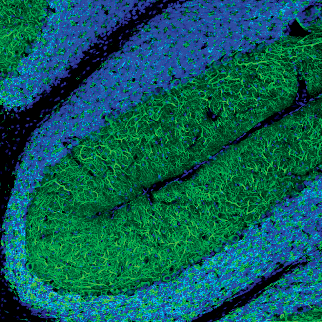
Oligodendrocytes are highly specialized glial cells which produce myelin, a lipid-rich substance that provides a protective sheath around axons and improves the conduction velocity of signals between neurons. They are characterized by the expression of the myelin family proteins: Myelin Oligodendrocyte Glycoprotein (MOG), Myelin-Associated Glycoprotein (MAG), and Myelin Basic Protein (MBP). Oligodendrocyte progenitor cells exist in the brain to facilitate regeneration of cells due to injury. However, the breakdown of myelin and inability to regenerate fully myelinated oligodendrocytes is correlated with several neurodegenerative diseases, including Alzheimer’s Disease (AD), Parkinson’s Disease (PD), Amyotrophic Lateral Sclerosis (ALS), and Multiple Sclerosis (MS).
Myelin basic protein (MBP) is one of the most abundant proteins in the CNS and plays an important role in the myelination of nerve cells. As a key component of myelin sheaths that surround axons, MBP contributes to the adhesion of the cytosolic membranes of compacted myelin. This is crucial to facilitate the conduction of neuronal impulses. MBP antibodies are often used to stain myelin sheaths of oligodendrocytes and Schwann cells in both the CNS and PNS.
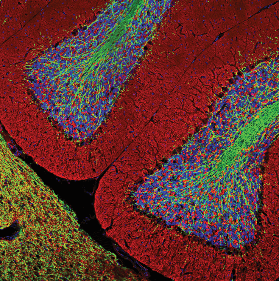
Myelin proteolipid protein (PLP1) is the major membrane bound phospholipid protein that is enriched in oligodendrocytes of the CNS. It plays a crucial role in the formation and maintenance of the multilamellar structure of myelin. Antibodies against this cell marker stain the myelin of oligodendrocytes in the CNS.
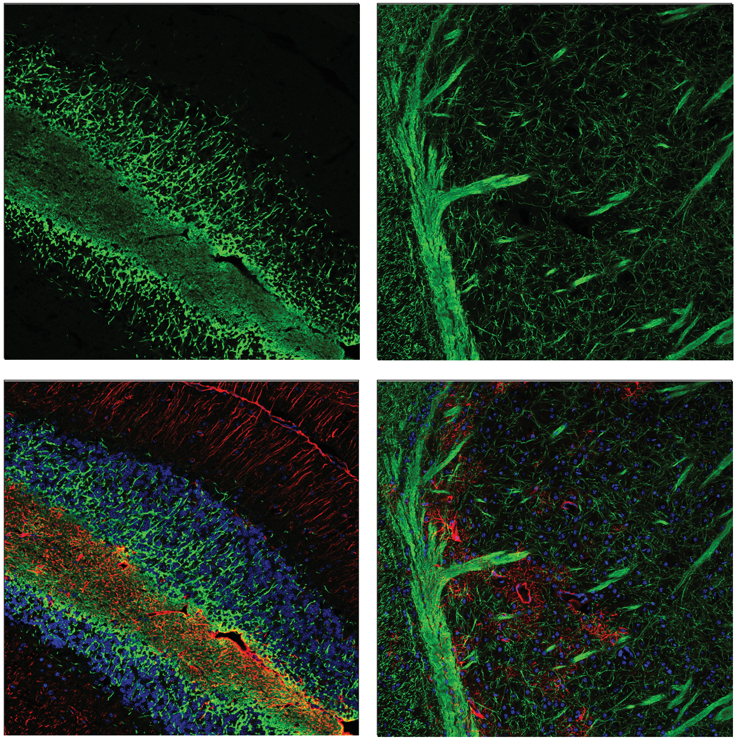
CNPase (2', 3’-cyclic nucleotide 3'-phosphodiesterase) is an enzyme that catalyzes the hydrolysis of 2’, 3’-cyclic nucleotides. It plays an early role in oligodendrocyte differentiation and may help generate the compacted myelin that surrounds axons. CNPase is enriched in oligodendrocytes and Schwann cells and antibodies that target this enzyme are commonly used to label myelin in the CNS and PNS.