View Featured Offers >>

Western Blotting Protocol (Fluorescent)

For fluorescent western blotting, we highly recommend using the TrueBlack® Fluorescent Western Blot Blocking Buffer Kit #40683. This kit is specifically formulated to increase the specificity and sensitivity of the fluorescent western blot assay by blocking non-specific interaction between fluorophore-labeled antibodies and the western blotting membrane. This results in less background and brighter target signal when using the TrueBlack® Fluorescent Western Blot Blocking Buffer Kit compared to the standard antibody diluent and blocking buffers. If using this kit, please use the kit-specific protocol on the product webpage or the datasheet.

FIGURE 1

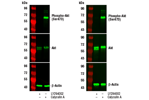
Western blot analysis of extracts from Jurkat cells, untreated (-) or treated with LY294002 #9901 or Calyculin A #9902 (+), using Phospho-Akt (Ser473) (D9E) XP® Rabbit mAb #4060 (upper), Akt (pan) (C67E7) Rabbit mAb #4691 (middle), or β-Actin (D6A8) Rabbit mAb #8457 (lower). The western blot membranes were treated with our standard antibody diluent and blocking buffers (left) or with the TrueBlack® Fluorescent Western Blot Blocking Buffer Kit (right). Increased antibody sensitivity and decreased background fluorescence can be seen when using the TrueBlack® Fluorescent Western Blot Blocking Buffer Kit.

NOTE: Two-color western blots require primary antibodies from different species and appropriate secondary antibodies labeled with different dyes. Overlap of epitopes may cause interference and should be considered in two color western blots. If the primary antibodies require different primary antibody incubation buffers, test each primary individually in both buffers to determine the optimal one for the dual-labeling experiment.

For western blots, incubate membrane with diluted primary antibody in either 5% w/v BSA or nonfat dry milk, 1X TBS, 0.1% Tween® 20 at 4°C with gentle shaking, overnight. Please refer to primary antibody product webpage for recommended primary antibody dilution buffer and recommended antibody dilution.

A. Solutions and Reagents

NOTE: Prepare solutions with reverse osmosis deionized (RODI) or equivalent grade water.

  1. 20X Phosphate Buffered Saline (PBS): (#9808) To prepare 1 L 1X PBS: add 50 ml 20X PBS to 950 ml dH2O, mix.
  2. 10X Tris Buffered Saline (TBS): (#12498) To prepare 1 L 1X TBS: add 100 ml 10X TBS to 900 ml dH2O, mix.
  3. 1X SDS Sample Buffer: Blue Loading Pack (#7722) or Red Loading Pack (#7723) Prepare fresh 3X reducing loading buffer by adding 1/10 volume 30X DTT to 1 volume of 3X SDS loading buffer. Dilute to 1X with dH2O.
  4. 10X Tris-Glycine SDS Running Buffer: (#4050) To prepare 1 L 1X running buffer: add 100 ml 10X running buffer 900 ml dH2O, mix.
  5. 10X Tris-Glycine Transfer Buffer: (#12539) To prepare 1 L 1X Transfer Buffer: add 100 ml 10X Transfer Buffer 200 ml methanol + 700 ml dH2O, mix.
  6. 10X Tris Buffered Saline with Tween® 20 (TBST-10X): (#9997) To prepare 1 L 1X TBST: add 100 ml 10X TBST to 900 ml dH2O, mix.
  7. Nonfat Dry Milk: (#9999).
  8. Blocking Buffer: 1X TBS with 5% w/v nonfat dry milk; for 150 ml, add 7.5 g nonfat dry milk to 150 ml 1X TBS and mix well. Tween® 20 should not be present in the Blocking Buffer because it is auto-fluorescent and increases non-specific background. After the blocking step, Tween® 20 can be reintroduced to subsequent diluent buffers.
  9. Wash Buffer: 1X TBST.
  10. Bovine Serum Albumin (BSA): (#9998).
  11. Primary Antibody Dilution Buffer: 1X TBST with 5% BSA or 5% nonfat dry milk as indicated on primary antibody datasheet; for 20 ml, add 1.0 g BSA or nonfat dry milk to 20 ml 1X TBST and mix well.
  12. Secondary Antibody Dilution Buffer: 1X TBST with 5% nonfat dry milk; for 20 ml, add 1.0 g nonfat dry milk to 20 ml 1X TBST and mix well. (Secondary antibodies; anti-rabbit #5151 and #5366; anti-mouse #5257 and #5470).
  13. Prestained Protein Marker, Broad Range (11-190 kDa): (#13953).
  14. Blotting Membrane and Paper: (#12369) This protocol has been optimized for nitrocellulose membranes (recommended). Pore size 0.2 µm is generally recommended.

B. Protein Blotting

A general protocol for sample preparation.

  1. Treat cells by adding fresh media containing regulator for desired time.
  2. Aspirate media from cultures; wash cells with cold 1X PBS; aspirate.
  3. Lyse cells by adding 1X SDS sample buffer (100 µl per well of 6-well plate or 500 µl per plate of 10 cm diameter plate). Immediately scrape the cells off the plate and transfer the extract to a microcentrifuge tube. Keep on ice.
  4. Sonicate for 10–15 sec to complete cell lysis and shear DNA (to reduce sample viscosity).
  5. Heat a 20 µl sample to 95–100°C for 5 min; cool on ice.
  6. Microcentrifuge for 5 min.
  7. Load 20 µl onto SDS-PAGE gel (10 cm x 10 cm).

    NOTE: Loading of prestained molecular weight markers (#59329, 10 µl/lane) is recommended to verify electrotransfer and to determine molecular weights. Prestained markers are autofluorescent at near-infrared wavelengths.

  8. Electrotransfer to nitrocellulose membrane (#12369).

C. Membrane Blocking and Antibody Incubations

NOTE: Volumes are for 10 cm x 10 cm (100 cm2) of membrane; for different sized membranes, adjust volumes accordingly.

  1. (Optional) After transfer, wash nitrocellulose membrane with 25 ml TBS for 5 min at room temperature.
  2. Incubate membrane in 25 ml of blocking buffer for 1 hr at room temperature.

    CRITICAL STEP: Do not include Tween® 20 in blocking buffer (Section A, Step 8).

  3. Wash three times for 5 min each with 15 ml of TBST.
  4. Incubate membrane and primary antibody (at the appropriate dilution as recommended in the product datasheet) in 10 ml primary antibody dilution buffer with gentle agitation overnight at 4°C.
  5. Wash three times for 5 min each with 15 ml of TBST.
  6. Incubate membrane with fluorophore-conjugated secondary antibody (#5470, #5257, #5366, #5151) (1:5000–1:25,000 dilution of 1 mg/ml stock) in 10 ml of secondary antibody dilution buffer with gentle agitation for 1 hr at room temperature.
  7. Wash three times for 5 min each with 15 ml of TBST.

D. Detection of Proteins

  1. Drain membrane of excess TBST and allow to dry.

    CRITICAL STEP: Membrane must be dry for fluorescent staining.

  2. Scan membrane using an appropriate fluorescent scanner following the manufacturer’s recommendations.

posted March 2022

revised March 2022