Since its initial discovery as a proto-oncogene, the serine/threonine kinase Akt (also known as protein kinase B or PKB) has become a major focus of attention because of its critical regulatory role in diverse cellular processes, including cancer progression and insulin metabolism. There are three highly related isoforms of Akt (Akt1, Akt2, and Akt3) and these represent the major signaling arm of PI3K.
Akt is activated by phospholipid binding and by phosphorylation within the carboxy terminus at Ser473.
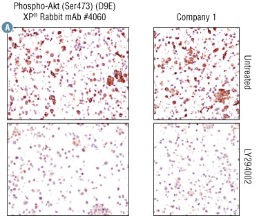
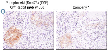
IHC was performed on LNCaP prostate cancer cell pellets, untreated or treated with LY294002 #9901 (a specific PI3 kinase inhibitor that inhibits Akt phosphorylation) (Figure A), as well as human breast carcinoma (Figure B).
The optimal dilutions for Phospho-Akt (Ser473) (D9E) XP® Rabbit mAb #4060 and competitor phospho-Akt antibody were first determined on cell pellets (Figure A).
This dilution was then used to compare the two antibodies on human breast cancer carcinoma tissue (Figure B). The target-specific staining of #4060 is significantly stronger and more distinct than that of the company 1 antibody.
While antibody staining can look quite similar when examined on cell pellets, tissue staining requires a higher level of antibody sensitivity so #4060 is the superior antibody for IHC on tissue sections.
Function: Akt is activated by phospholipid binding and by phosphorylation within the carboxy terminus at Ser473.
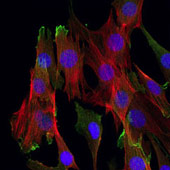
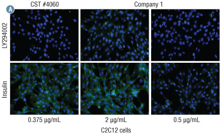
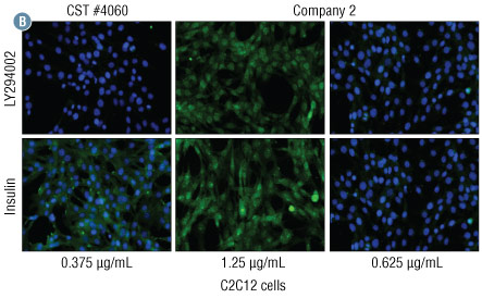
Samples: C2C12 mouse muscle progenitor cells were treated with LY294002 #9901 (a specific PI3 kinase inhibitor that inhibits Akt phosphorylation) or with insulin that induces Akt phosphorylation.
Results: The antibody concentrations used in the experiments are as follows:
This data shows that #4060 can give stronger signal with less antibody.
Results: The antibody concentrations used in the experiments are as follows:
This data shows that #4060 can give stronger signal with less antibody.
Function: Akt is activated by phospholipid binding and phosphorylation within the carboxy terminus at Ser473.
Samples: Jurkat cells were treated with either Calyculin A #9902, a potent phosphatase inhibitor, to preserve existing Akt phosphorylation levels or LY294002 #9901, a specific PI3 kinase inhibitor, to inhibit Akt phosphorylation via PI3 kinase.
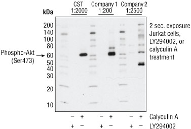
| CST #4060 | Competitor 1 | Competitor 2 | |
|---|---|---|---|
| Product | 4060 | ||
| Dilution | 1:2000 | 1:200* | 1:2500* |
| Conc. (μg/mL) | 0.043 | 0.5 | 0.2 |
*manufacturer’s recommended starting dilutions.