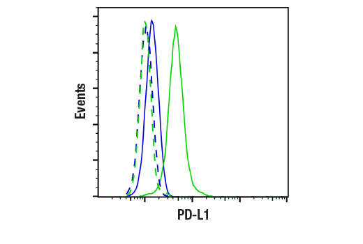
WB, IP, IHC-Bond, IHC-P, FC-FP
H
Endogenous
40-50
Rabbit IgG
#Q9NZQ7
29126
Product Information
Product Usage Information
| Application | Dilution |
|---|---|
| Western Blotting | 1:1000 |
| Immunoprecipitation | 1:50 |
| IHC Leica Bond | 1:200 - 1:800 |
| Immunohistochemistry (Paraffin) | 1:100 - 1:400 |
| Flow Cytometry (Fixed/Permeabilized) | 1:200 - 1:800 |
Storage
For a carrier free (BSA and azide free) version of this product see product #85164.
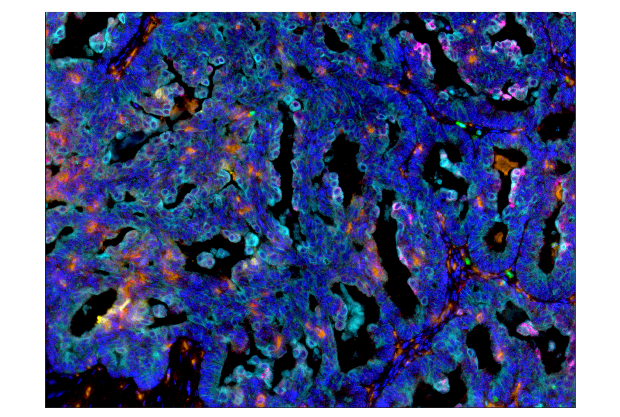
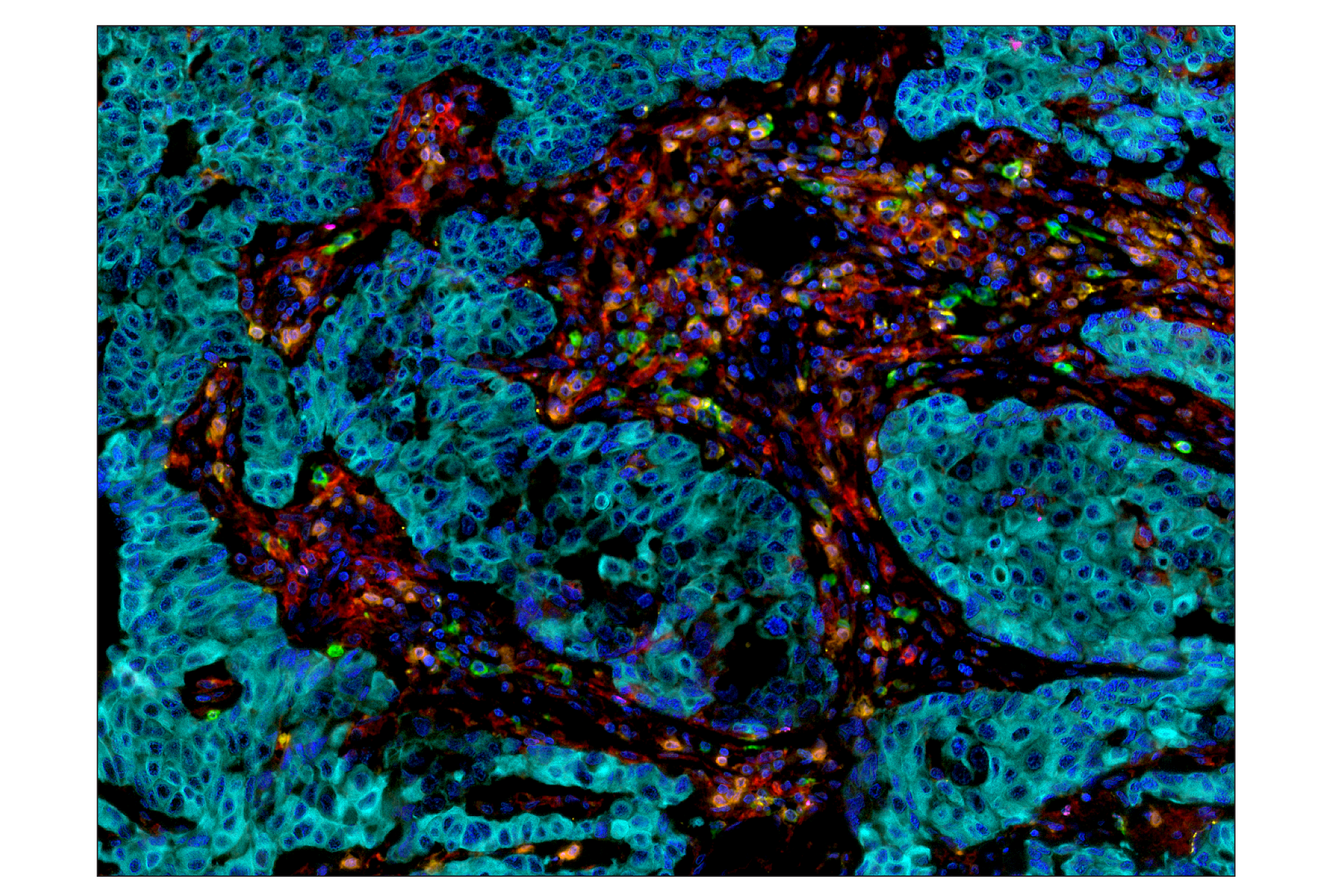
Specificity / Sensitivity
Species Reactivity:
Human
Source / Purification
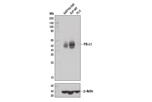
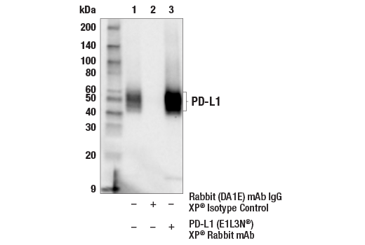
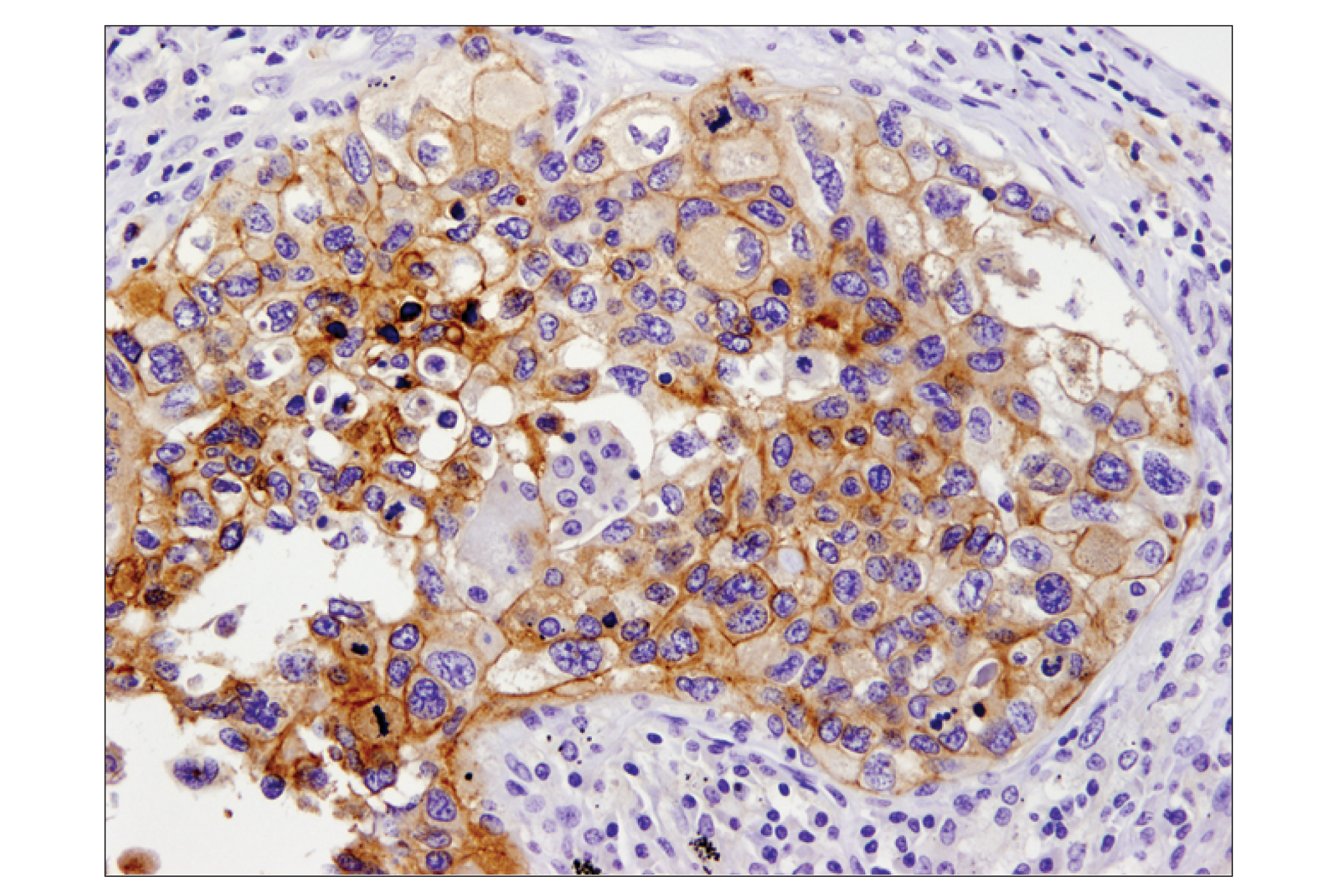
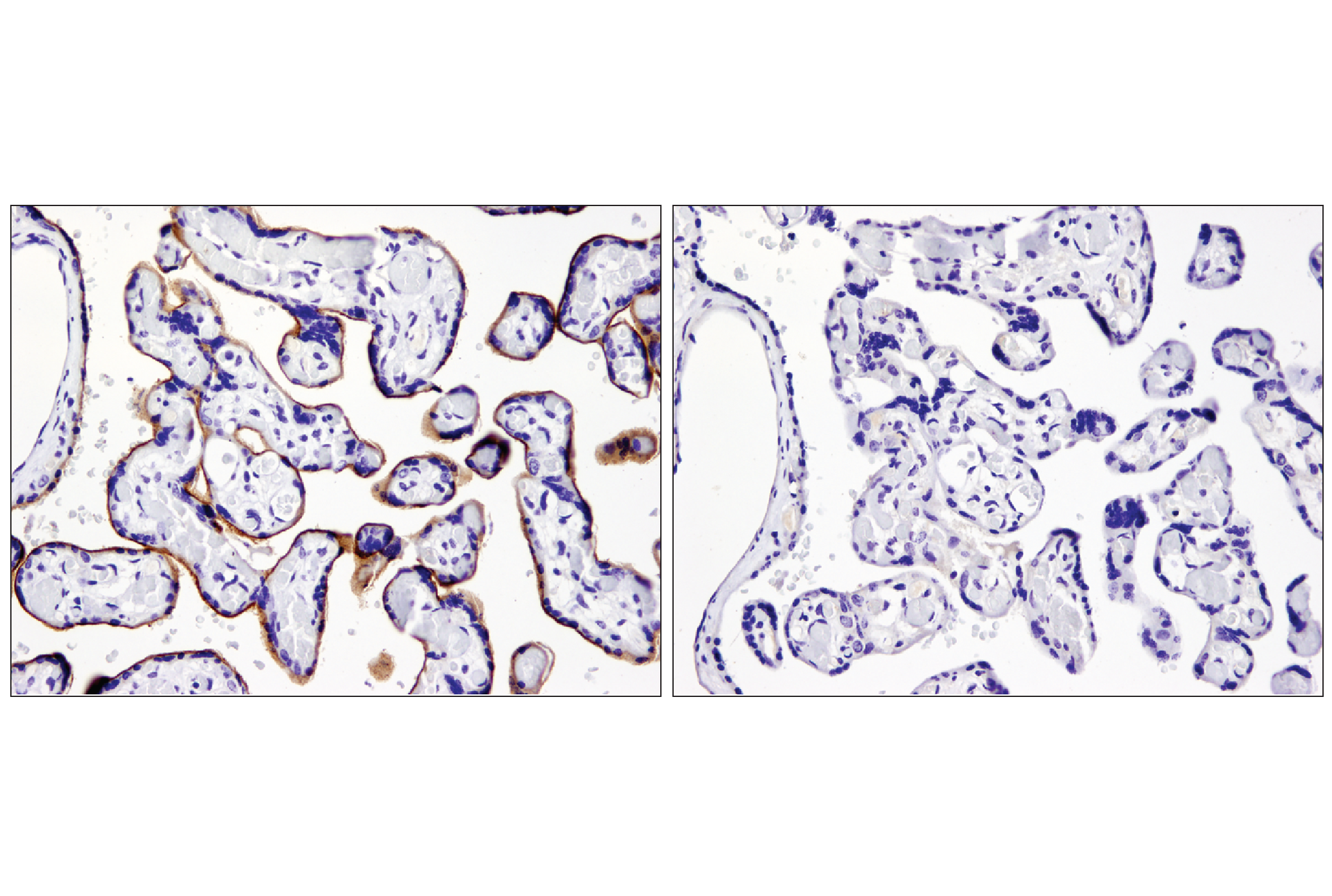
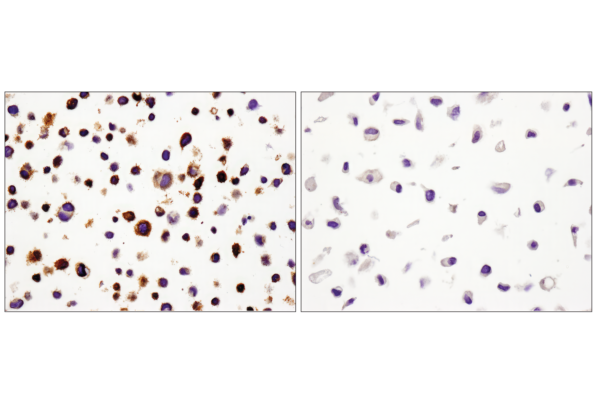
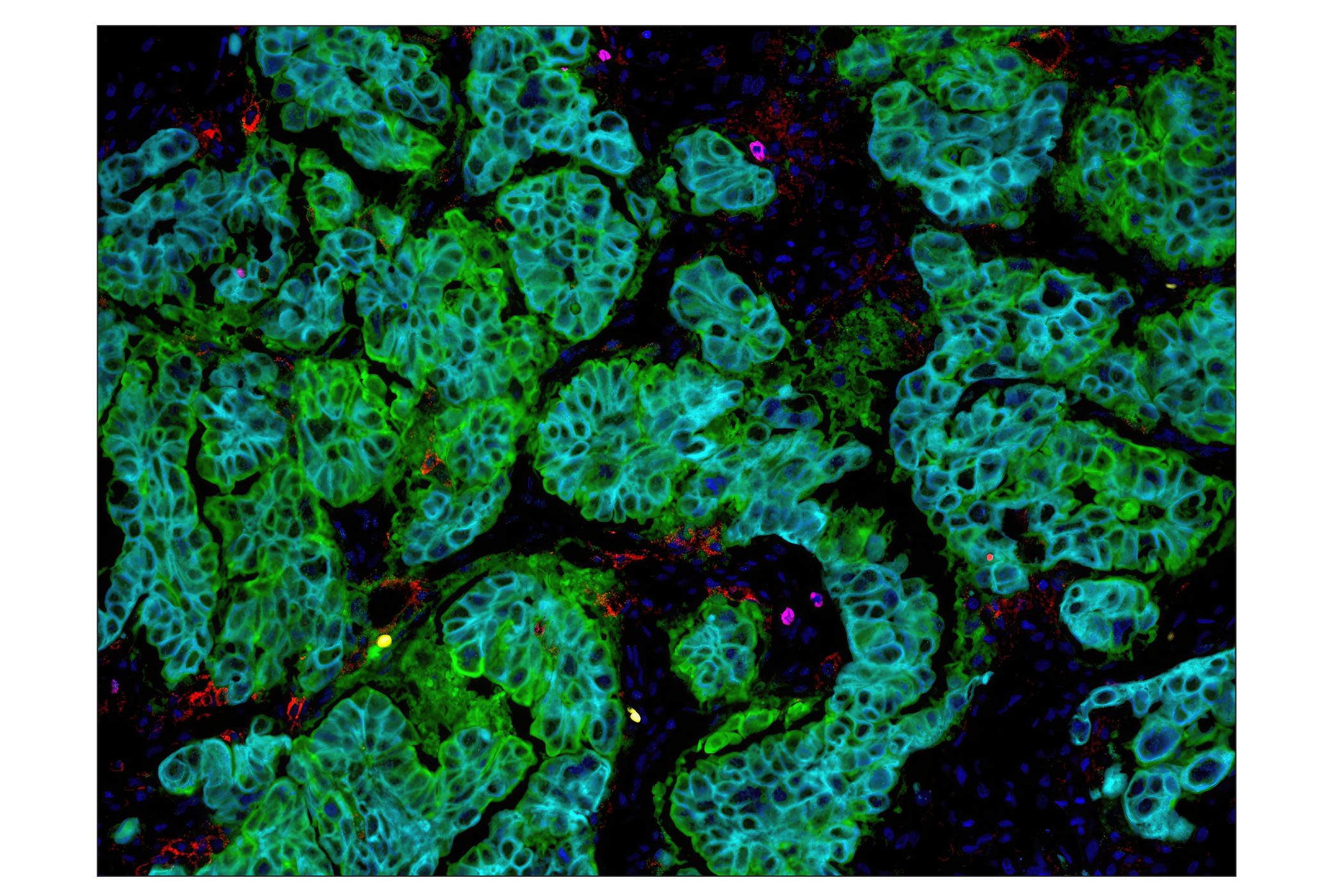
Monoclonal antibody is produced by immunizing animals with a synthetic peptide corresponding to residues near the carboxy terminus of human PD-L1 protein.
Background
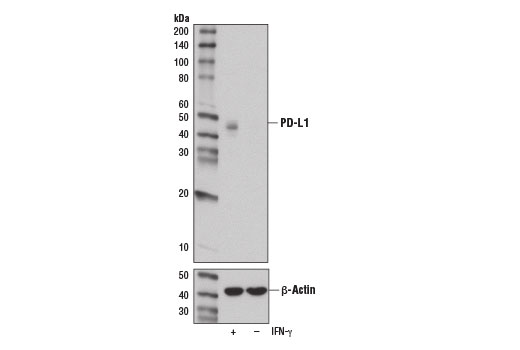
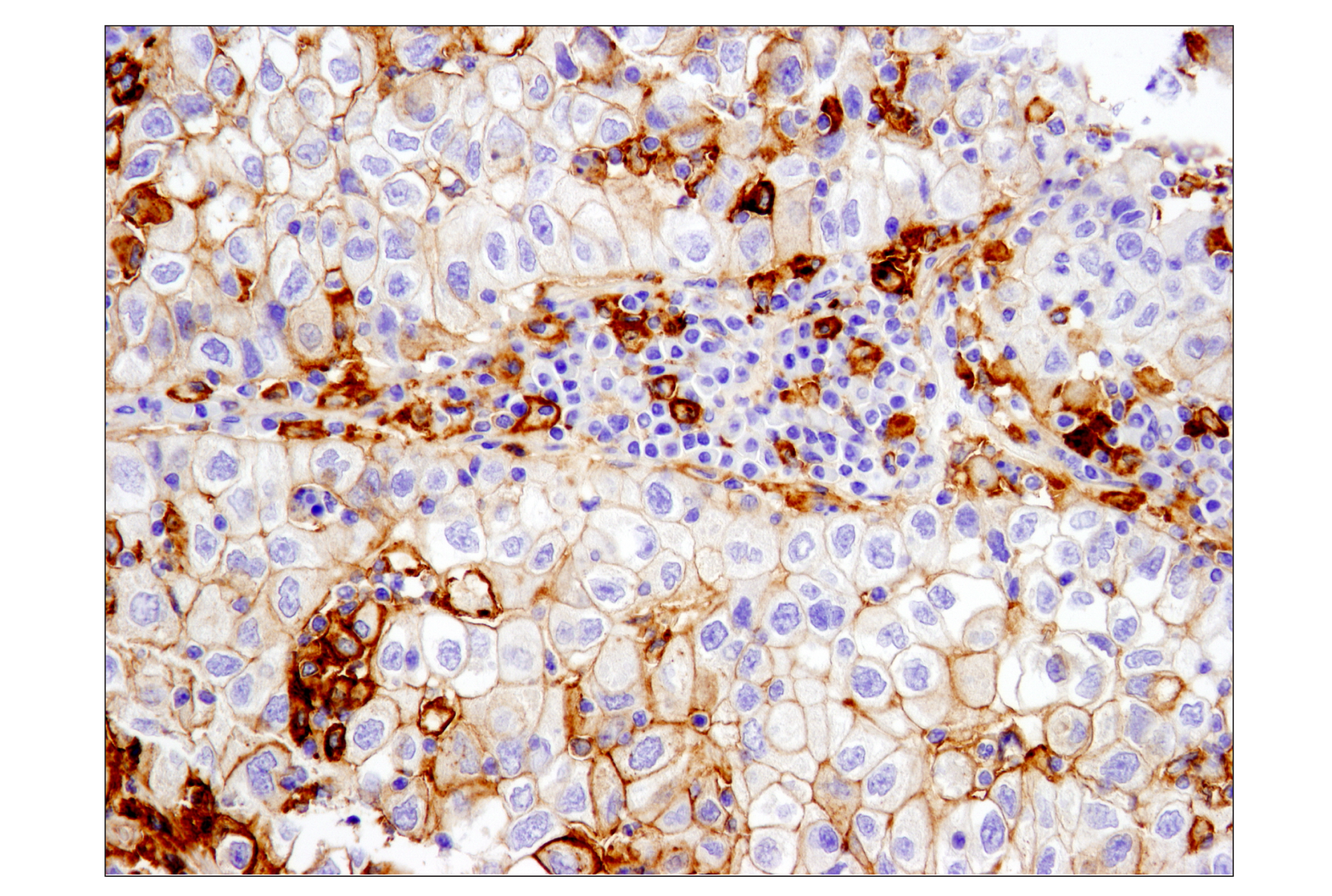
Programmed cell death 1 ligand 1 (PD-L1, B7-H1, CD274) is a member of the B7 family of cell surface ligands that regulate T cell activation and immune responses. The PD-L1 ligand binds the PD-1 transmembrane receptor and inhibits T cell activation. PD-L1 was discovered following a search for novel B7 protein homologs and was later shown to be expressed by antigen presenting cells, activated T cells, and tissues including placenta, heart, and lung (1-3). Similar in structure to related B7 family members, PD-L1 protein contains extracellular IgV and IgC domains and a short, cytoplasmic region. Research studies demonstrate that PD-L1 is expressed in several tumor types, including melanoma, ovary, colon, lung, breast, and renal cell carcinomas (4-6). Expression of PD-L1 in cancer is associated with tumor-infiltrating lymphocytes, which mediate PD-L1 expression through the release of interferon gamma (7). Additional research links PD-L1 expression to cancers associated with viral infections (8,9).
- Dong, H. et al. (1999) Nat Med 5, 1365-9.
- Freeman, G.J. et al. (2000) J Exp Med 192, 1027-34.
- Liang, S.C. et al. (2003) Eur J Immunol 33, 2706-16.
- Dong, H. et al. (2002) Nat Med 8, 793-800.
- Thompson, R.H. et al. (2006) Cancer Res 66, 3381-5.
- Pardoll, D.M. (2012) Nat Rev Cancer 12, 252-64.
- Taube, J.M. et al. (2012) Sci Transl Med 4, 127ra37.
- Lyford-Pike, S. et al. (2013) Cancer Res 73, 1733-41.
- Chen, B.J. et al. (2013) Clin Cancer Res 19, 3462-73.
- Wimberly, H. et al. (2014) Cancer Immunol Res , .
Species Reactivity
Species reactivity is determined by testing in at least one approved application (e.g., western blot).
Western Blot Buffer
IMPORTANT: For western blots, incubate membrane with diluted primary antibody in 5% w/v nonfat dry milk, 1X TBS, 0.1% Tween® 20 at 4°C with gentle shaking, overnight.
Applications Key
WB: Western Blotting IP: Immunoprecipitation IHC-Bond: IHC Leica Bond IHC-P: Immunohistochemistry (Paraffin) FC-FP: Flow Cytometry (Fixed/Permeabilized)
Cross-Reactivity Key
H: human M: mouse R: rat Hm: hamster Mk: monkey Vir: virus Mi: mink C: chicken Dm: D. melanogaster X: Xenopus Z: zebrafish B: bovine Dg: dog Pg: pig Sc: S. cerevisiae Ce: C. elegans Hr: horse GP: Guinea Pig Rab: rabbit All: all species expected
Trademarks and Patents
Limited Uses
Except as otherwise expressly agreed in a writing signed by a legally authorized representative of CST, the following terms apply to Products provided by CST, its affiliates or its distributors. Any Customer's terms and conditions that are in addition to, or different from, those contained herein, unless separately accepted in writing by a legally authorized representative of CST, are rejected and are of no force or effect.
Products are labeled with For Research Use Only or a similar labeling statement and have not been approved, cleared, or licensed by the FDA or other regulatory foreign or domestic entity, for any purpose. Customer shall not use any Product for any diagnostic or therapeutic purpose, or otherwise in any manner that conflicts with its labeling statement. Products sold or licensed by CST are provided for Customer as the end-user and solely for research and development uses. Any use of Product for diagnostic, prophylactic or therapeutic purposes, or any purchase of Product for resale (alone or as a component) or other commercial purpose, requires a separate license from CST. Customer shall (a) not sell, license, loan, donate or otherwise transfer or make available any Product to any third party, whether alone or in combination with other materials, or use the Products to manufacture any commercial products, (b) not copy, modify, reverse engineer, decompile, disassemble or otherwise attempt to discover the underlying structure or technology of the Products, or use the Products for the purpose of developing any products or services that would compete with CST products or services, (c) not alter or remove from the Products any trademarks, trade names, logos, patent or copyright notices or markings, (d) use the Products solely in accordance with CST Product Terms of Sale and any applicable documentation, and (e) comply with any license, terms of service or similar agreement with respect to any third party products or services used by Customer in connection with the Products.