WB, IP, IHC-Bond, IHC-P, FC-L
H
Endogenous
45-70
Rabbit IgG
#Q8TDQ0
84868
Product Information
Product Usage Information
| Application | Dilution |
|---|---|
| Western Blotting | 1:1000 |
| Immunoprecipitation | 1:50 |
| IHC Leica Bond | 1:50 - 1:200 |
| Immunohistochemistry (Paraffin) | 1:200 - 1:800 |
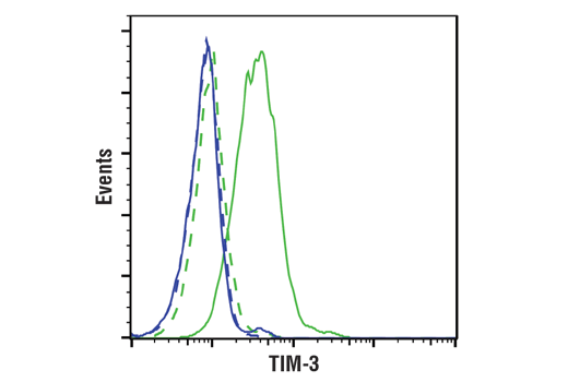
| Flow Cytometry (Live) | 1:50 |
Storage
For a carrier free (BSA and azide free) version of this product see product #81229.
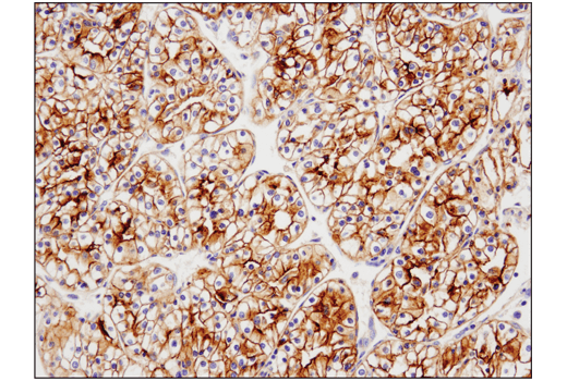
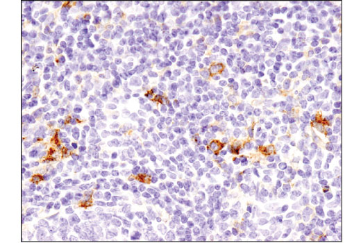
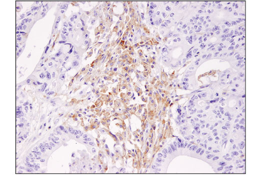
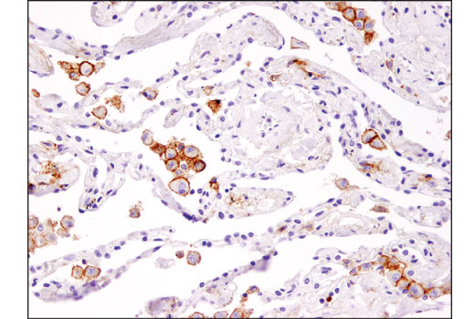
Specificity / Sensitivity
Species Reactivity:
Human
Source / Purification
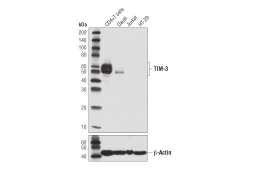
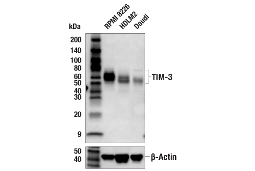
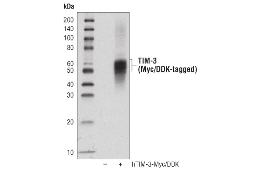
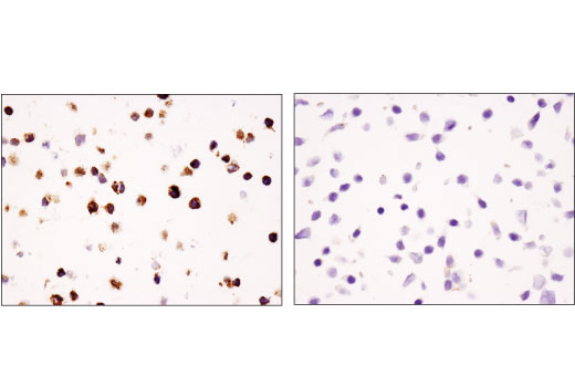
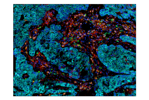
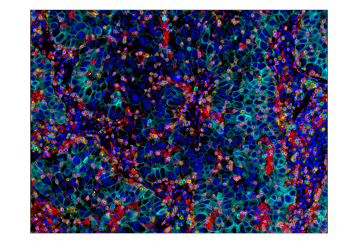
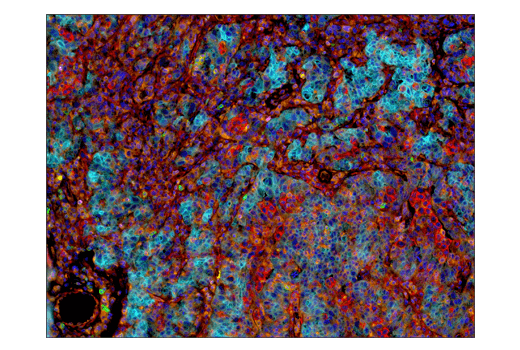
Monoclonal antibody is produced by immunizing animals with recombinant protein specific to the extracellular domain of human TIM-3 protein.
Background
T cell Ig- and mucin-domain-containing molecules (TIMs) are a family of transmembrane proteins expressed by various immune cells. TIM-3 is an inhibitory molecule that is induced following T cell activation (1-3 ). TIM-3 is expressed by exhausted T cells in the settings of chronic infection and cancer (4,5), and tumor-infiltrating T cells that coexpress PD-1 and TIM-3 exhibit the most severe exhausted phenotype (5). Tumor-infiltrating dendritic cells (DCs) also express TIM-3. TIM-3 expression on DCs was found to suppress innate immunity by reducing the immunogenicity of nucleic acids released by dying tumor cells (6). Research studies show that heterodimerization of TIM-3 with CEACAM-1 is critical for the inhibitory function of TIM-3, and co-blockade of TIM-3 and CEACAM-1 enhanced anti-tumor responses in a mouse model of colorectal cancer (7). In addition, blockade of TIM-3 in mouse models of autoimmunity enhanced the severity of disease (1). Finally, binding of Galectin-9 to TIM-3 expressed by Th1 cells induces T cell death (8).
- Monney, L. et al. (2002) Nature 415, 536-41.
- Sánchez-Fueyo, A. et al. (2003) Nat Immunol 4, 1093-101.
- Sabatos, C.A. et al. (2003) Nat Immunol 4, 1102-10.
- Jones, R.B. et al. (2008) J Exp Med 205, 2763-79.
- Sakuishi, K. et al. (2010) J Exp Med 207, 2187-94.
- Chiba, S. et al. (2012) Nat Immunol 13, 832-42.
- Huang, Y.H. et al. (2015) Nature 517, 386-90.
- Zhu, C. et al. (2005) Nat Immunol 6, 1245-52.
Species Reactivity
Species reactivity is determined by testing in at least one approved application (e.g., western blot).
Western Blot Buffer
IMPORTANT: For western blots, incubate membrane with diluted primary antibody in 5% w/v BSA, 1X TBS, 0.1% Tween® 20 at 4°C with gentle shaking, overnight.
Applications Key
WB: Western Blotting IP: Immunoprecipitation IHC-Bond: IHC Leica Bond IHC-P: Immunohistochemistry (Paraffin) FC-L: Flow Cytometry (Live)
Cross-Reactivity Key
H: human M: mouse R: rat Hm: hamster Mk: monkey Vir: virus Mi: mink C: chicken Dm: D. melanogaster X: Xenopus Z: zebrafish B: bovine Dg: dog Pg: pig Sc: S. cerevisiae Ce: C. elegans Hr: horse GP: Guinea Pig Rab: rabbit All: all species expected
Trademarks and Patents
Limited Uses
Except as otherwise expressly agreed in a writing signed by a legally authorized representative of CST, the following terms apply to Products provided by CST, its affiliates or its distributors. Any Customer's terms and conditions that are in addition to, or different from, those contained herein, unless separately accepted in writing by a legally authorized representative of CST, are rejected and are of no force or effect.
Products are labeled with For Research Use Only or a similar labeling statement and have not been approved, cleared, or licensed by the FDA or other regulatory foreign or domestic entity, for any purpose. Customer shall not use any Product for any diagnostic or therapeutic purpose, or otherwise in any manner that conflicts with its labeling statement. Products sold or licensed by CST are provided for Customer as the end-user and solely for research and development uses. Any use of Product for diagnostic, prophylactic or therapeutic purposes, or any purchase of Product for resale (alone or as a component) or other commercial purpose, requires a separate license from CST. Customer shall (a) not sell, license, loan, donate or otherwise transfer or make available any Product to any third party, whether alone or in combination with other materials, or use the Products to manufacture any commercial products, (b) not copy, modify, reverse engineer, decompile, disassemble or otherwise attempt to discover the underlying structure or technology of the Products, or use the Products for the purpose of developing any products or services that would compete with CST products or services, (c) not alter or remove from the Products any trademarks, trade names, logos, patent or copyright notices or markings, (d) use the Products solely in accordance with CST Product Terms of Sale and any applicable documentation, and (e) comply with any license, terms of service or similar agreement with respect to any third party products or services used by Customer in connection with the Products.