Proprietary antibody development technologies, along with our extensive validation and stringent quality control standards, deliver XP® monoclonal antibodies that perform exceptionally well in the widest range of research applications.
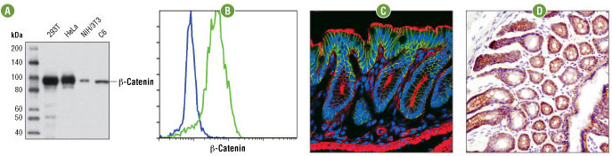
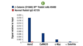
β-Catenin (D10A8) XP® Rabbit mAb #8480: Western blot analysis (A) of extracts from various cell lines using #8480. Flow cytometric analysis (B) of NCI-H28 (blue) or HeLa (green) cells using #8480. Confocal IF analysis (C) of mouse colon using #8480 (green). Actin filaments were labeled with DY-554 phalloidin (red). Blue pseudocolor = DRAQ5 #4084 (fluorescent DNA dye). IHC analysis (D) of frozen mouse colon using #8480. Chromatin immunoprecipitations (E) were performed with cross-linked chromatin from 4 x 10⁶ HCT 116 cells and either 20 μl of #8480 or 2 μl of Normal Rabbit IgG #2729 using SimpleChIP® Enzymatic Chromatin IP Kit (Magnetic Beads) #9003. The enriched DNA was quantified by real-time PCR using SimpleChIP® Human Axin2 Intron 1 Primers #8973, SimpleChIP® Human CaMK2D Intron 3 Primers #5111, human c-Myc promoter primers, and SimpleChIP® Human α Satellite Repeat Primers #4486. The amount of immunoprecipitated DNA in each sample is represented as signal relative to the total amount of input chromatin, which is equivalent to one.
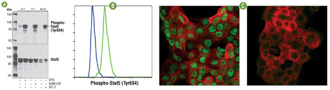
Phospho-Stat5 (Tyr694) (D47E7) XP® Rabbit mAb #4322: Western blot analysis (A) of extracts from UT-7 cells, untreated or treated with erythropoietin (EPO, 3 units/ml, 5 min), TF-1 cells, untreated or treated with Human Granulocyte Macrophage Colony Stimulating Factor (hGM-CSF) #8922 (100 ng/ml, 10 min), and NK-92 cells, untreated or treated with Human Interleukin-2 (hIL-2) #8907 (100 ng/ml, 10 min), using #4322 (upper) or a total protein Stat5 antibody (lower). Flow cytometric analysis (B) of TF-1 cells, untreated (blue) or GM-CSF-treated (green), using #4322. Confocal IF analysis (C) of A-431 cells, EGF-treated (left) or untreated (right), using #4322 (green) and Pan-Keratin (C11) Mouse mAb #4545 (red).
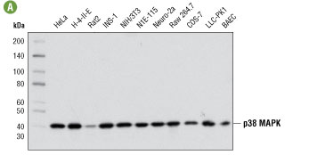
p38 MAPK (D13E1) XP® Rabbit mAb #8690: Western blot analysis (A) of extracts from various cell lines using #8690. Flow cytometric analysis (B) of HeLa cells using #8690 (blue) compared to concentration-matched Rabbit (DA1E) mAb IgG XP® Isotype Control #3900 (red). IHC analysis (C) of paraffin-embedded human breast carcinoma using #8690. Confocal IF analysis (D) of HeLa cells, untreated (left) or treated with UV (100 mJ/cm2, 30 min recovery; right), using #8690 (green). Actin filaments were labeled with DY-554 phalloidin (red).
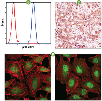
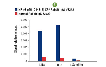
NF-κB p65 (D14E12) XP® Rabbit mAb #8242: Western blot analysis (A) of extracts from various cell lines using #8242. Flow cytometric analysis (B) of HeLa cells using #8242 (blue) compared to concentration matched Rabbit (DA1E) mAb IgG XP® Isotype Control #3900 (red). IHC analysis (C) of paraffin-embedded human chronic cholecystitis using #8242. Confocal IF analysis (D) of HT-1080 cells treated with hTNF-α #8902 (20 ng/ml, 20 min), using #8242 (green). Actin filaments were labeled with DY-554 phalloidin (red). Blue pseudocolor = DRAQ5® #4084 (fluorescent DNA dye). Chromatin immunoprecipitations (E) were performed with cross-linked chromatin from 4 x 10⁶ HeLa cells treated with hTNF-α #8902 (30 ng/ml, 1 hr) and either 5 μl of #8242 or 2 μl of Normal Rabbit IgG #2729 using SimpleChIP® Enzymatic Chromatin IP Kit (Magnetic Beads) #9003. The enriched DNA was quantified by real-time PCR using SimpleChIP® Human IκBα Promoter Primers #5552, human IL-8 promoter primers, and SimpleChIP® Human α Satellite Repeat Primers #4486. The amount of immunoprecipitated DNA in each sample is represented as signal relative to the total amount of input chromatin, which is equivalent to one.