Revision 1
#35981
Store at -20C
877-616-CELL (2355)
877-678-TECH (8324)
3 Trask Lane | Danvers | Massachusetts | 01923 | USA
For Research Use Only. Not for Use in Diagnostic Procedures.
Applications:
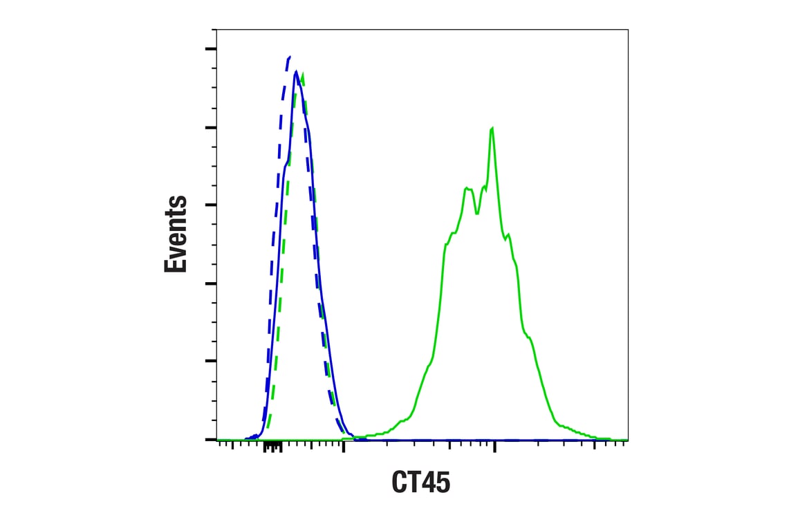
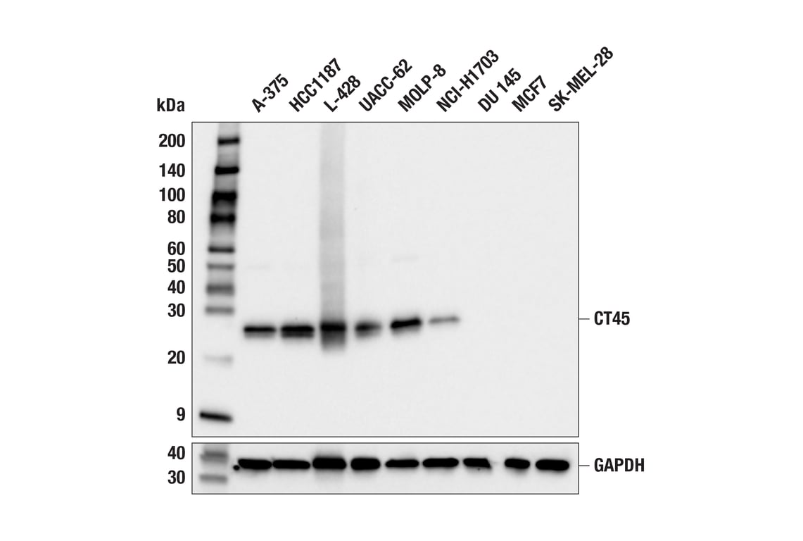
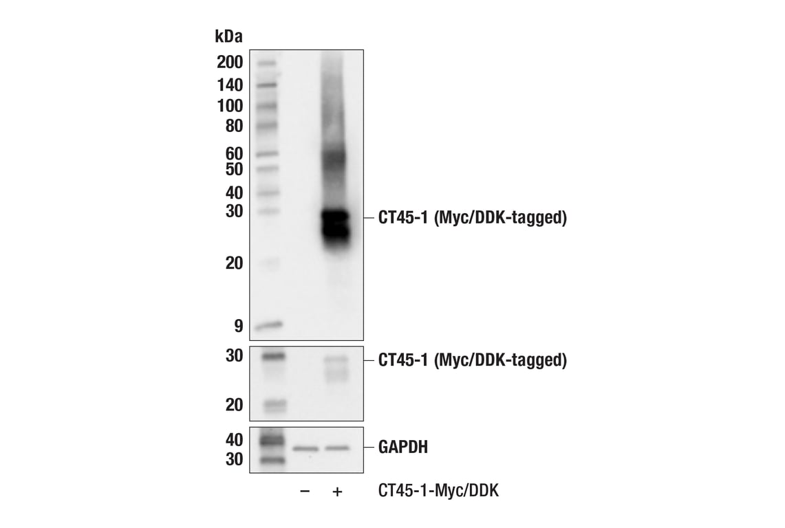
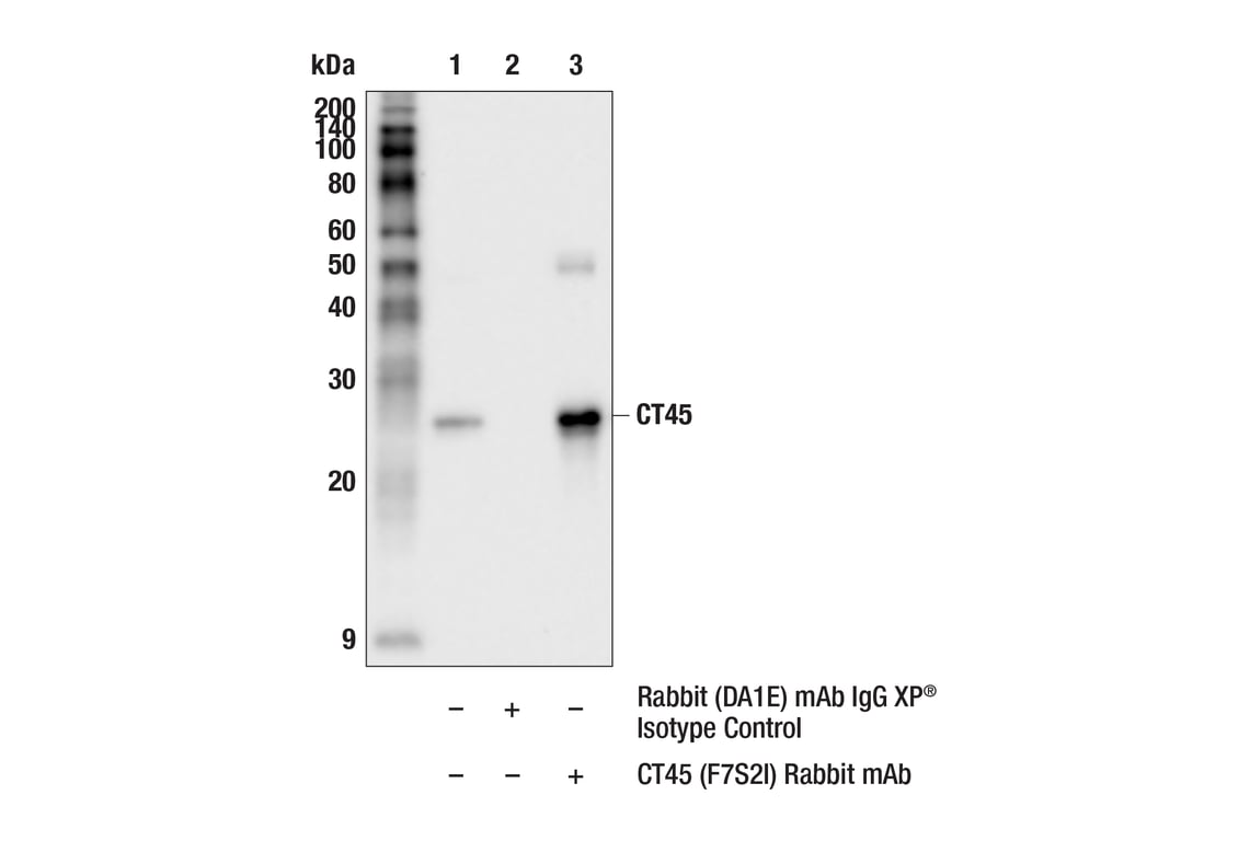
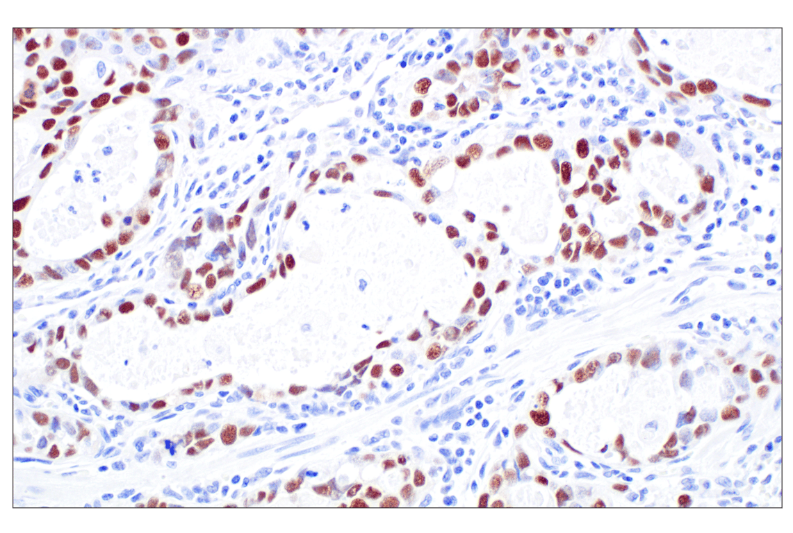
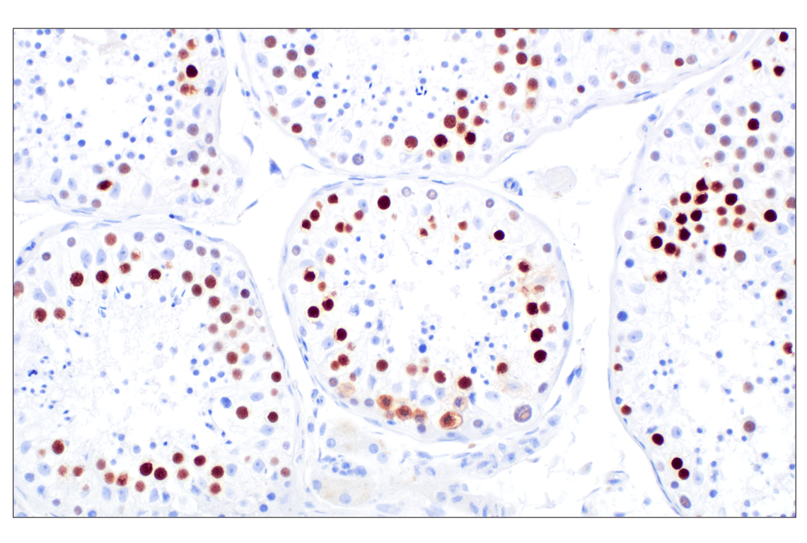
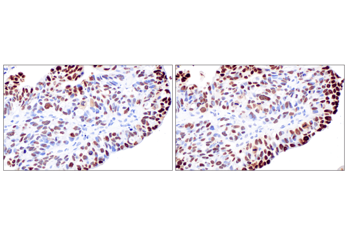
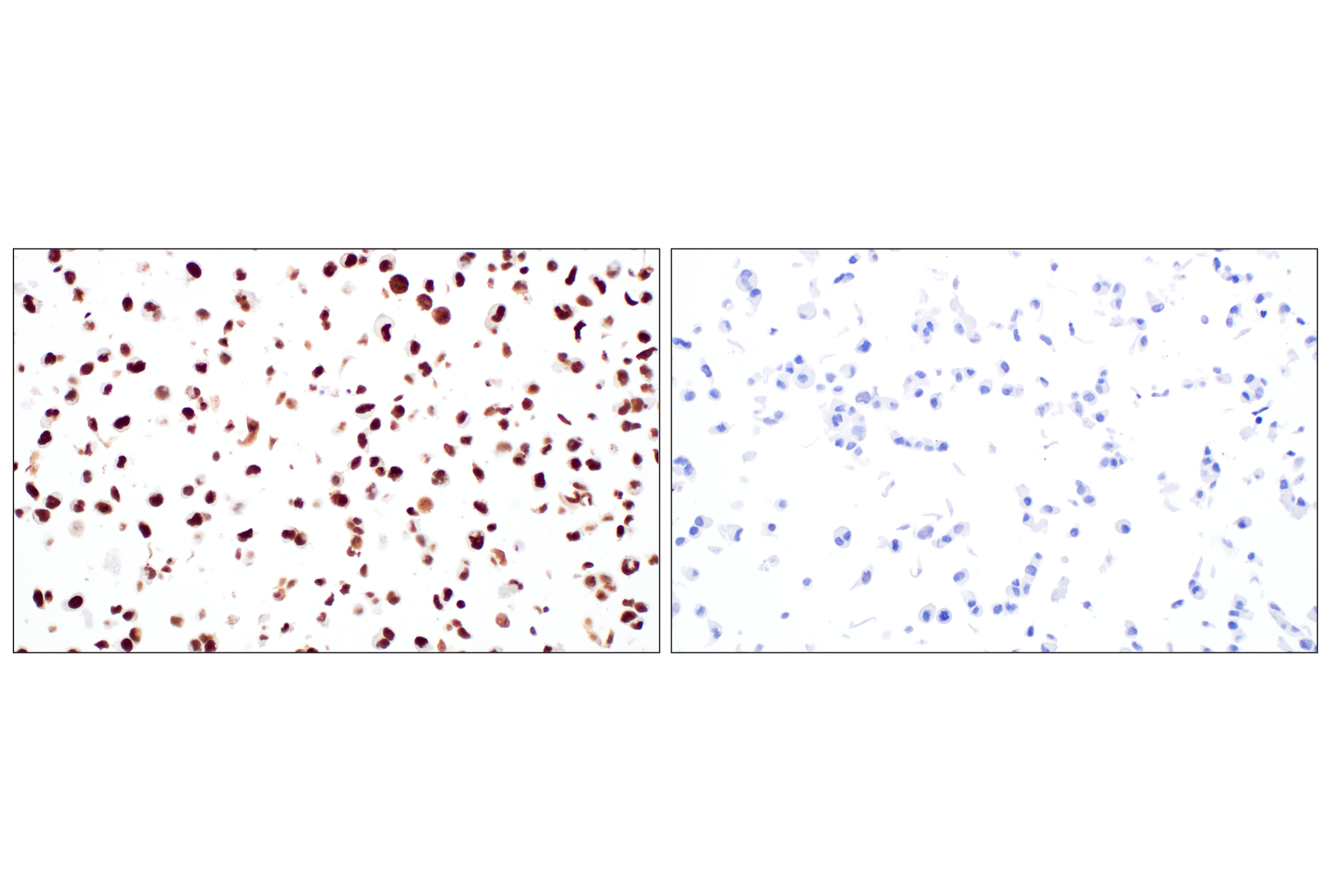
W, IP, IHC-P, FC-FP
Reactivity:
H
Sensitivity:
Endogenous
MW (kDa):
26
Source/Isotype:
Rabbit IgG
UniProt ID:
#Q5HYN5
Entrez-Gene Id:
541466
Product Usage Information
| Application | Dilution |
|---|---|
| Western Blotting | 1:1000 |
| Immunoprecipitation | 1:200 |
| Immunohistochemistry (Paraffin) | 1:4000 |
| Flow Cytometry (Fixed/Permeabilized) | 1:2400 - 1:9600 |
Storage
Specificity/Sensitivity
Source / Purification
Background
The CT45 family is a tandem cluster of highly similar genes that share many features with other classic CT genes, including Xq localization, multigene family, and identical or near-identical gene copies, indicating recent gene duplications (4). The nuclear protein antigen CT45 is often highly expressed in lung cancer and ovarian cancer, and the frequency and characteristics of CT45 expression are similar to those of other CT cancer vaccine targets currently in clinical trials, including NY-ESO-1 and MAGE-A (5). Overexpression of CT45A1 in breast cancer cells was found to upregulate various oncogenic and metastatic genes, promote epithelial-mesenchymal transition, and increase cell stemness, tumorigenesis, and metastasis (6). Additionally, research studies have shown that CT45A1 silencing suppresses cell proliferation and metastasis in breast and lung cancer cells by downregulating the ERK/CREB signaling pathway (6,7). Therefore, CT45A1 and other CT45 family members are excellent targets for anticancer drug discovery and targeted tumor therapy.
Background References
- Caballero, O.L. and Chen, Y.T. (2009) Cancer Sci 100, 2014-21.
- De Smet, C. et al. (1999) Mol Cell Biol 19, 7327-35.
- Gjerstorff, M.F. et al. (2015) Oncotarget 6, 15772-87.
- Chen, Y.T. et al. (2005) Proc Natl Acad Sci USA 102, 7940-5.
- Chen, Y.T. et al. (2009) Int J Cancer 124, 2893-8.
- Shang, B. et al. (2014) Cell Death Dis 5, e1285.
- Tang, F. et al. (2017) Mol Med Rep 16, 6708-14.
Species Reactivity
Species reactivity is determined by testing in at least one approved application (e.g., western blot).
Western Blot Buffer
IMPORTANT: For western blots, incubate membrane with diluted primary antibody in 5% w/v BSA, 1X TBS, 0.1% Tween® 20 at 4°C with gentle shaking, overnight.
Applications Key
W: Western Blotting IP: Immunoprecipitation IHC-P: Immunohistochemistry (Paraffin) FC-FP: Flow Cytometry (Fixed/Permeabilized)
Cross-Reactivity Key
H: Human
Trademarks and Patents
Cell Signaling Technology is a trademark of Cell Signaling Technology, Inc.
All other trademarks are the property of their respective owners. Visit cellsignal.com/trademarks for more information.
Limited Uses
Except as otherwise expressly agreed in a writing signed by a legally authorized representative of CST, the following terms apply to Products provided by CST, its affiliates or its distributors. Any Customer's terms and conditions that are in addition to, or different from, those contained herein, unless separately accepted in writing by a legally authorized representative of CST, are rejected and are of no force or effect.
Products are labeled with For Research Use Only or a similar labeling statement and have not been approved, cleared, or licensed by the FDA or other regulatory foreign or domestic entity, for any purpose. Customer shall not use any Product for any diagnostic or therapeutic purpose, or otherwise in any manner that conflicts with its labeling statement. Products sold or licensed by CST are provided for Customer as the end-user and solely for research and development uses. Any use of Product for diagnostic, prophylactic or therapeutic purposes, or any purchase of Product for resale (alone or as a component) or other commercial purpose, requires a separate license from CST. Customer shall (a) not sell, license, loan, donate or otherwise transfer or make available any Product to any third party, whether alone or in combination with other materials, or use the Products to manufacture any commercial products, (b) not copy, modify, reverse engineer, decompile, disassemble or otherwise attempt to discover the underlying structure or technology of the Products, or use the Products for the purpose of developing any products or services that would compete with CST products or services, (c) not alter or remove from the Products any trademarks, trade names, logos, patent or copyright notices or markings, (d) use the Products solely in accordance with CST Product Terms of Sale and any applicable documentation, and (e) comply with any license, terms of service or similar agreement with respect to any third party products or services used by Customer in connection with the Products.
Revision 1
Revision 1

Revision 1

Revision 1