Revision 1
#37690
Store at -20C
877-616-CELL (2355)
877-678-TECH (8324)
3 Trask Lane | Danvers | Massachusetts | 01923 | USA
For Research Use Only. Not for Use in Diagnostic Procedures.
Applications:
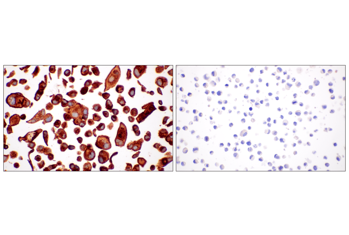
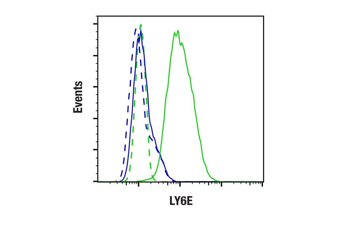
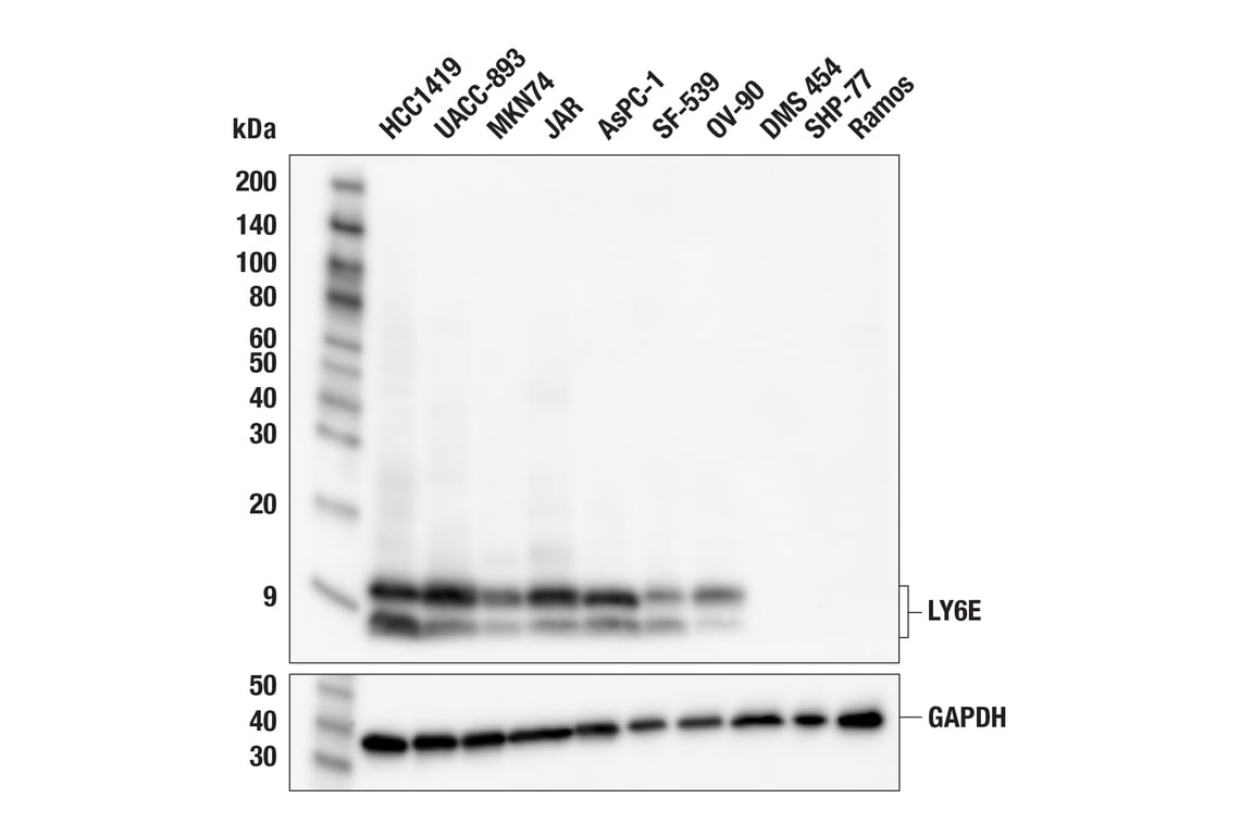
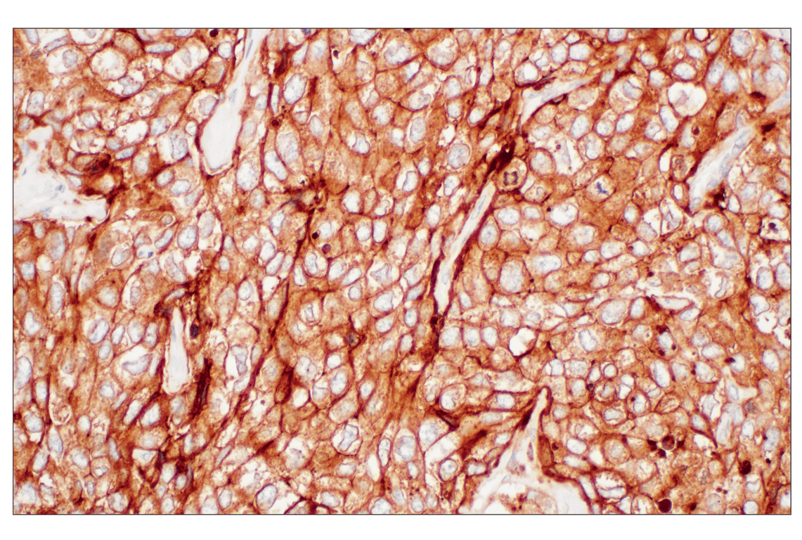
W, IHC-P, FC-FP
Reactivity:
H
Sensitivity:
Endogenous
MW (kDa):
8-13
Source/Isotype:
Human IgG
UniProt ID:
#Q16553
Entrez-Gene Id:
4061
Product Usage Information
| Application | Dilution |
|---|---|
| Western Blotting | 1:1000 |
| Immunohistochemistry (Paraffin) | 1:100 - 1:400 |
| Flow Cytometry (Fixed/Permeabilized) | 1:400 - 1:800 |
Storage
Specificity/Sensitivity
Source / Purification
Background
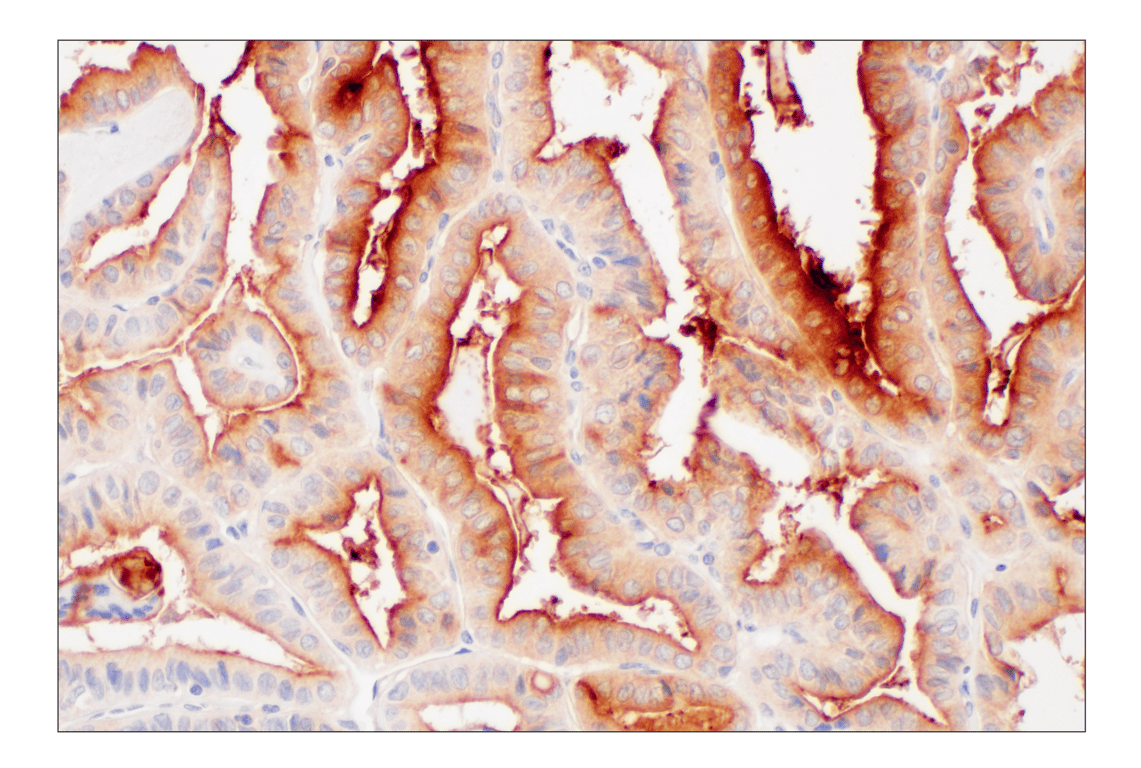
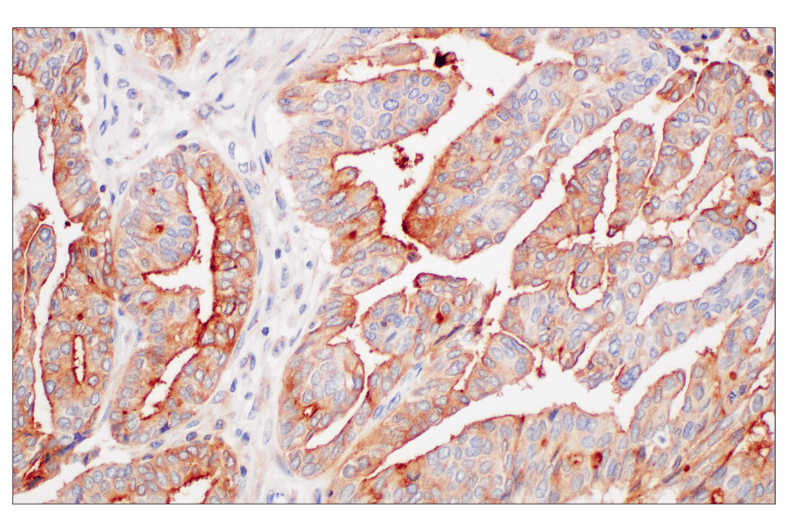
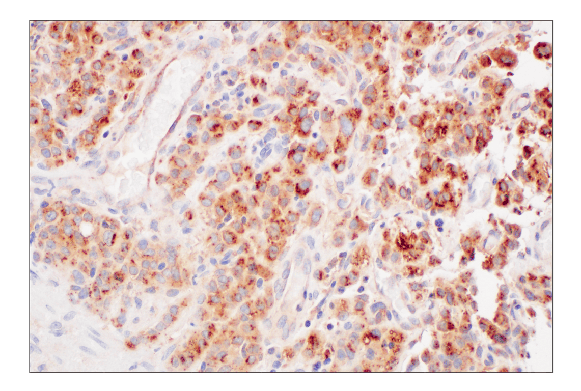
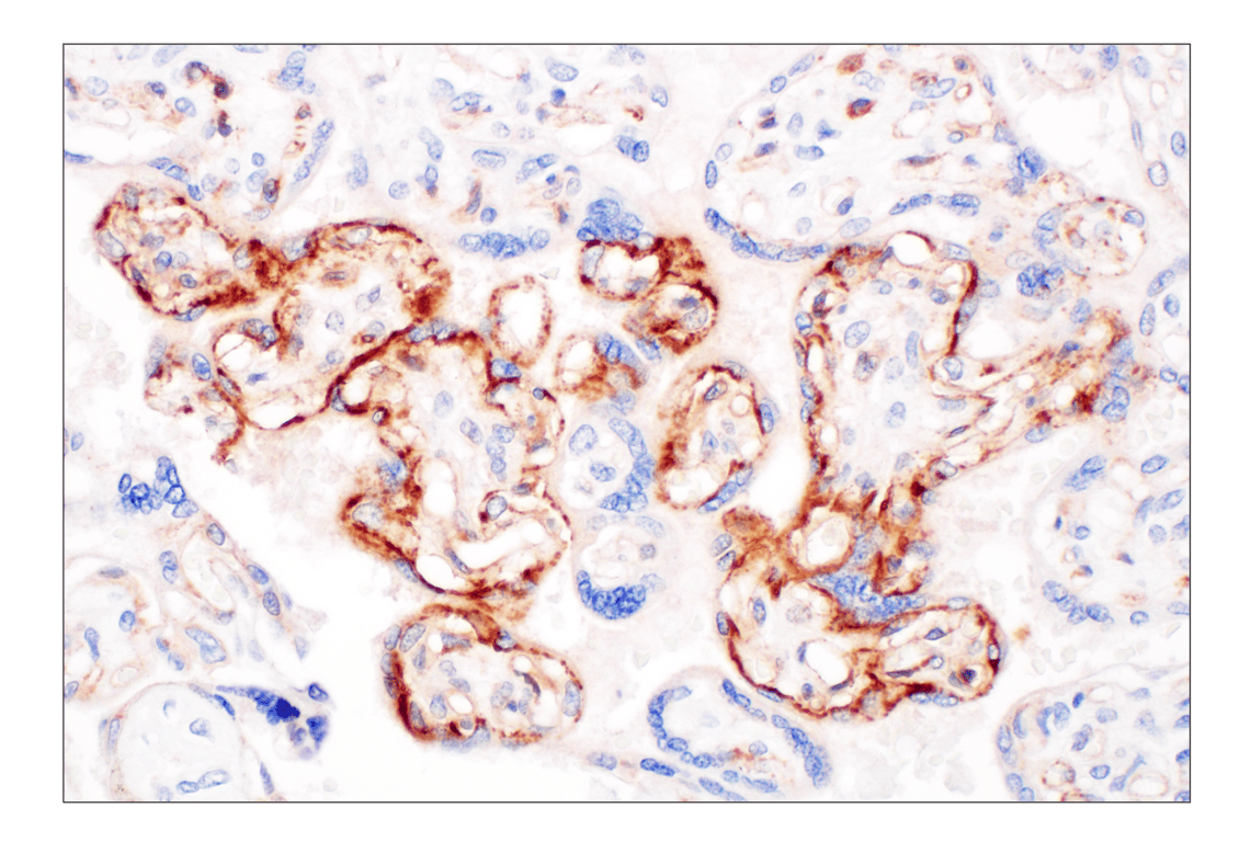
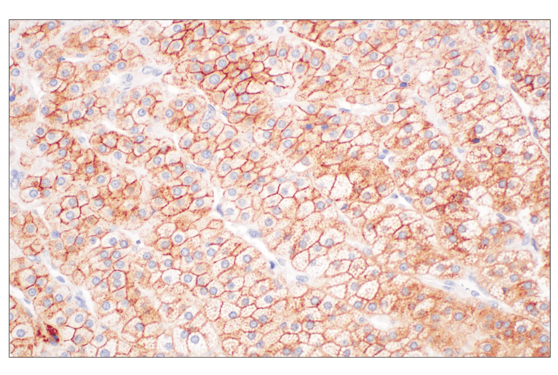
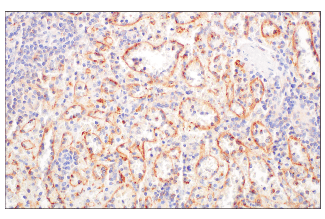
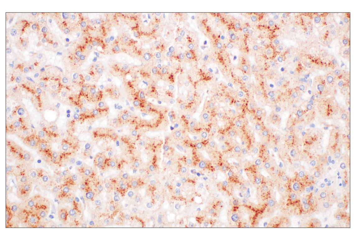
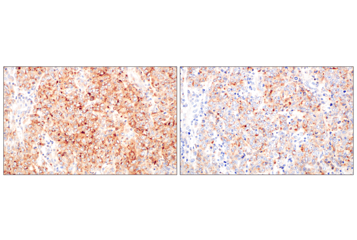
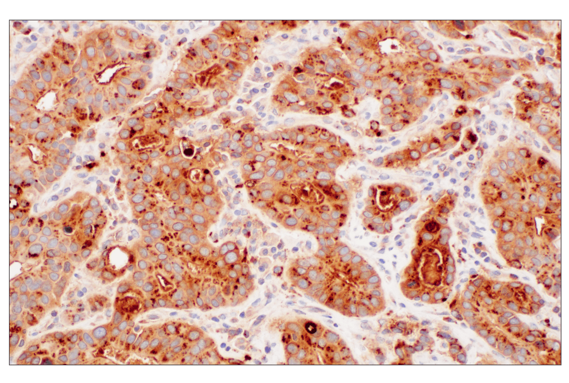
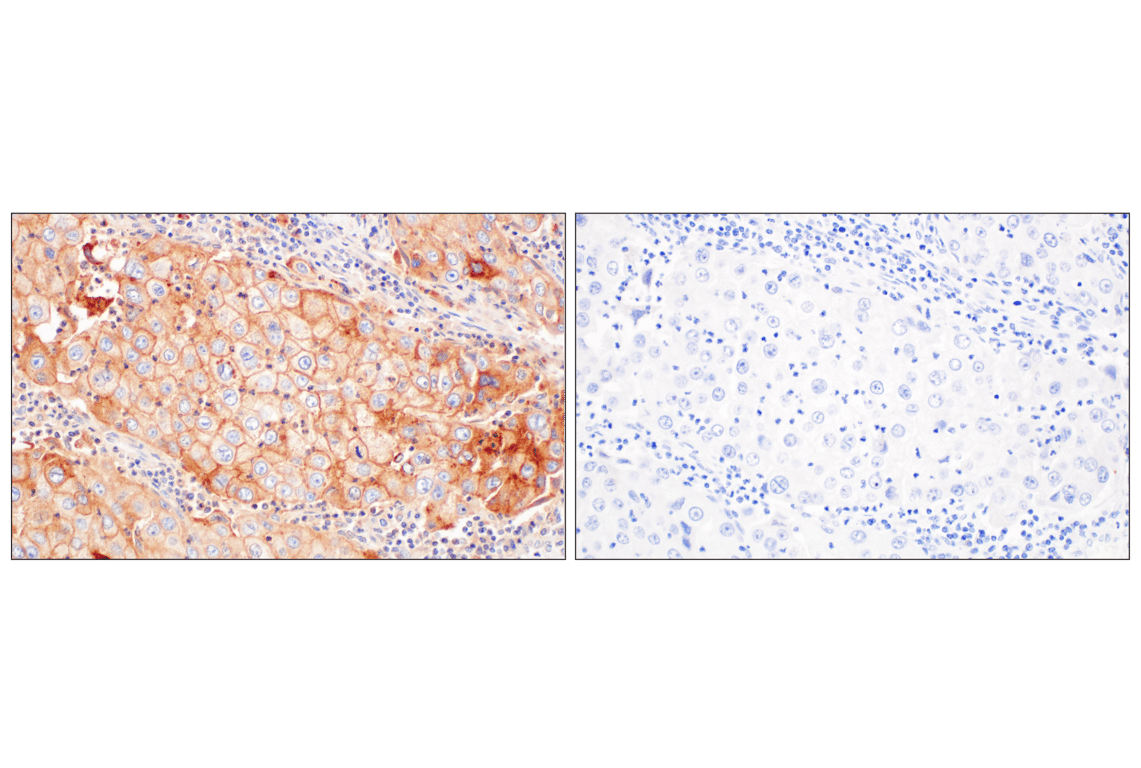
Lymphocyte antigen 6 family member E (LY6E), also known as stem cell antigen-2 (SCA-2), thymic shared antigen-1 (TSA-1), or retinoic acid-induced gene E (RIG-E), is widely expressed across various tissues, including the liver, spleen, lungs, and brain, as well as on several immune cell subsets. It plays critical roles in immune regulation, viral infection, and cancer progression, and is heavily induced by type I interferon (IFN). LY6E acts as a restriction factor against human coronaviruses by interfering with spike protein-mediated membrane fusion and blocking viral entry into host cells. Conversely, LY6E promotes the infection of other viruses, such as HIV-1, dengue, and Zika, by enhancing viral entry and fusion (7,8). LY6E is frequently overexpressed in several human malignancies, including breast and gastric cancers, where high expression correlates with poor survival outcomes (9). Recent research indicates that LY6E promotes tumor immune escape by facilitating the accumulation of M2 macrophages, driving the exclusion of CD8+ cytotoxic T cells from the tumor microenvironment (10). LY6E plays a complex role in regulating T cell activation and B cell development. It is heavily induced by IFN, acting as a classic interferon-stimulated gene (ISG). LY6E modulates T-lymphocyte proliferation and activation by interacting with CD3Z/CD247 (the zeta chain of the T-cell receptor complex) and can influence TCR sensitivity (10). The high expression of LY6E in tumors has established it as a promising target for pharmaceutical intervention, including antibody-drug conjugates (ADCs) (11). In the context of autoimmune disease, elevated LY6E levels are associated with systemic lupus erythematosus (SLE) activity and correlate with increased disease severity, likely due to the chronic interferon signature present in SLE patients (12).
Background References
- Bamezai, A. (2004) Arch Immunol Ther Exp (Warsz) 52, 255-66.
- Lee, P.Y. et al. (2013) J Leukoc Biol 94, 585-94.
- Fleming, T.J. et al. (1993) J Immunol 151, 2399-408.
- Tsetlin, V. (1999) Eur J Biochem 264, 281-6.
- Pflugh, D.L. et al. (2000) J Immunol 165, 313-21.
- Bronte, V. et al. (2016) Nat Commun 7, 12150.
- Yu, J. and Liu, S.L. (2019) Viruses 11, 1020. doi: 10.3390/v11111020.
- Yu, J. et al. (2017) J Biol Chem 292, 4674-4685.
- Yeom, C.J. et al. (2016) Oncotarget 7, 65837-65848.
- Hailin, L. et al. (2024) Cancer Immunol Immunother 74, 4.
- Dela Cruz Chuh, J. et al. (2021) MAbs 13, 1862452.
- Zheng, Y. et al. (2025) Front Immunol 16, 1476575.
Species Reactivity
Species reactivity is determined by testing in at least one approved application (e.g., western blot).
Western Blot Buffer
IMPORTANT: For western blots, incubate membrane with diluted primary antibody in 5% w/v BSA, 1X TBS, 0.1% Tween® 20 at 4°C with gentle shaking, overnight.
Applications Key
W: Western Blotting IHC-P: Immunohistochemistry (Paraffin) FC-FP: Flow Cytometry (Fixed/Permeabilized)
Cross-Reactivity Key
H: Human
Trademarks and Patents
Cell Signaling Technology is a trademark of Cell Signaling Technology, Inc.
All other trademarks are the property of their respective owners. Visit cellsignal.com/trademarks for more information.
Limited Uses
Except as otherwise expressly agreed in a writing signed by a legally authorized representative of CST, the following terms apply to Products provided by CST, its affiliates or its distributors. Any Customer's terms and conditions that are in addition to, or different from, those contained herein, unless separately accepted in writing by a legally authorized representative of CST, are rejected and are of no force or effect.
Products are labeled with For Research Use Only or a similar labeling statement and have not been approved, cleared, or licensed by the FDA or other regulatory foreign or domestic entity, for any purpose. Customer shall not use any Product for any diagnostic or therapeutic purpose, or otherwise in any manner that conflicts with its labeling statement. Products sold or licensed by CST are provided for Customer as the end-user and solely for research and development uses. Any use of Product for diagnostic, prophylactic or therapeutic purposes, or any purchase of Product for resale (alone or as a component) or other commercial purpose, requires a separate license from CST. Customer shall (a) not sell, license, loan, donate or otherwise transfer or make available any Product to any third party, whether alone or in combination with other materials, or use the Products to manufacture any commercial products, (b) not copy, modify, reverse engineer, decompile, disassemble or otherwise attempt to discover the underlying structure or technology of the Products, or use the Products for the purpose of developing any products or services that would compete with CST products or services, (c) not alter or remove from the Products any trademarks, trade names, logos, patent or copyright notices or markings, (d) use the Products solely in accordance with CST Product Terms of Sale and any applicable documentation, and (e) comply with any license, terms of service or similar agreement with respect to any third party products or services used by Customer in connection with the Products.
Revision 1
Revision 1
Revision 1
Revision 1
Revision 1