Revision 4

#38493

Store at -20C

CST Logo
Orders:

877-616-CELL (2355)

[email protected]

Support:

877-678-TECH (8324)

3 Trask Lane | Danvers | Massachusetts | 01923 | USA

For Research Use Only. Not for Use in Diagnostic Procedures.

Applications:
W, W-S, IP, IF-IC

Reactivity:
H M R

Sensitivity:
Endogenous

MW (kDa):
80

Source/Isotype:
Rabbit IgG

UniProt ID:
#O60488

Entrez-Gene Id:
2182

Product Usage Information

Application Dilution
Western Blotting 1:1000
Simple Western™ 1:10 - 1:50
Immunoprecipitation 1:50
Immunofluorescence (Immunocytochemistry) 1:200

Storage

Supplied in 10 mM sodium HEPES (pH 7.5), 150 mM NaCl, 100 µg/mL BSA, 50% glycerol, and less than 0.02% sodium azide. Store at –20°C. Do not aliquot the antibody.

Specificity/Sensitivity

ACSL4 (F6T3Z) Rabbit Monoclonal Antibody recognizes endogenous levels of total ACSL4 protein. Species reactivity for immunofluorescence is human only.

Source / Purification

Monoclonal antibody is produced by immunizing animals with a synthetic peptide corresponding to residues near the carboxy terminus of human ACSL4 protein.

Background

The long-chain fatty acyl-CoA synthetase (ACSL) family of enzymes convert free long-chain fatty acids into their active form, acyl-coenzyme A (acyl-CoA), and play dominant roles in both anabolic (fatty acid synthesis and lipogenesis) and catabolic (lipolysis and fatty acid β-oxidation) pathways (1). Acyl-CoA synthetase long-chain family member 4 (ACSL4) preferentially catalyzes the formation of arachidonoyl-coenzyme A (AA-CoA) by inserting coenzyme A (CoA) into arachidonic acid (AA) (1). ACSL4-catalyzed acyl-CoAs also participate in the regulation of steroidogenesis (2), eicosanoid biosynthesis (3), and phospholipid remodeling (4). ACSL4 is an essential enzyme for the conversion of two key ferroptosis-inducing signals, oxidized arachidonic and adrenic phosphatidylethanolamines (5,6). Genome-wide CRISPR/Cas9-based genetic screens have demonstrated that ACSL4 is an essential component for ferroptosis execution (6), and genetic or pharmacological inhibition of ACSL4 can initiate a specific anti-ferroptotic rescue pathway (5). ACSL4 expression is upregulated in ferroptosis-sensitive cancer cells compared with ferroptosis-resistant cells (7) and has been shown to predict sensitivity to ferroptosis in a panel of basal-like breast cancer cell lines (6). Moreover, high ACSL4 protein expression in hepatocellular carcinoma (HCC) clinical tissue specimens is associated with improved patient outcomes after sorafenib treatment and could serve as a predictive biomarker (8).

Species Reactivity

Species reactivity is determined by testing in at least one approved application (e.g., western blot).

Western Blot Buffer

IMPORTANT: For western blots, incubate membrane with diluted primary antibody in 5% w/v nonfat dry milk, 1X TBS, 0.1% Tween® 20 at 4°C with gentle shaking, overnight.

Applications Key

W: Western Blotting W-S: Simple Western™ IP: Immunoprecipitation IF-IC: Immunofluorescence (Immunocytochemistry)

Cross-Reactivity Key

H: Human M: Mouse R: Rat

Trademarks and Patents

Cell Signaling Technology is a trademark of Cell Signaling Technology, Inc.

All other trademarks are the property of their respective owners. Visit cellsignal.com/trademarks for more information.

Limited Uses

Except as otherwise expressly agreed in a writing signed by a legally authorized representative of CST, the following terms apply to Products provided by CST, its affiliates or its distributors. Any Customer's terms and conditions that are in addition to, or different from, those contained herein, unless separately accepted in writing by a legally authorized representative of CST, are rejected and are of no force or effect.

Products are labeled with For Research Use Only or a similar labeling statement and have not been approved, cleared, or licensed by the FDA or other regulatory foreign or domestic entity, for any purpose. Customer shall not use any Product for any diagnostic or therapeutic purpose, or otherwise in any manner that conflicts with its labeling statement. Products sold or licensed by CST are provided for Customer as the end-user and solely for research and development uses. Any use of Product for diagnostic, prophylactic or therapeutic purposes, or any purchase of Product for resale (alone or as a component) or other commercial purpose, requires a separate license from CST. Customer shall (a) not sell, license, loan, donate or otherwise transfer or make available any Product to any third party, whether alone or in combination with other materials, or use the Products to manufacture any commercial products, (b) not copy, modify, reverse engineer, decompile, disassemble or otherwise attempt to discover the underlying structure or technology of the Products, or use the Products for the purpose of developing any products or services that would compete with CST products or services, (c) not alter or remove from the Products any trademarks, trade names, logos, patent or copyright notices or markings, (d) use the Products solely in accordance with CST Product Terms of Sale and any applicable documentation, and (e) comply with any license, terms of service or similar agreement with respect to any third party products or services used by Customer in connection with the Products.

Orders: 877-616-CELL (2355) [email protected] Support: 877-678-TECH (8324) [email protected] Web: cellsignal.com
For Research Use Only. Not for Use in Diagnostic Procedures.

Revision 4

Cell Signaling Technology Logo
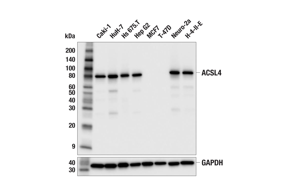
Western blot analysis of extracts from various cell lines using ACSL4 (F6T3Z) Rabbit mAb (upper) or GAPDH (D16H11) XP® Rabbit mAb #5174 (lower). Low expression of ACSL4 protein in MCF7 and T-47D cells is consistent with the predicted expression pattern.
Western Blotting Image 1: ACSL4 (F6T3Z) Rabbit Monoclonal Antibody
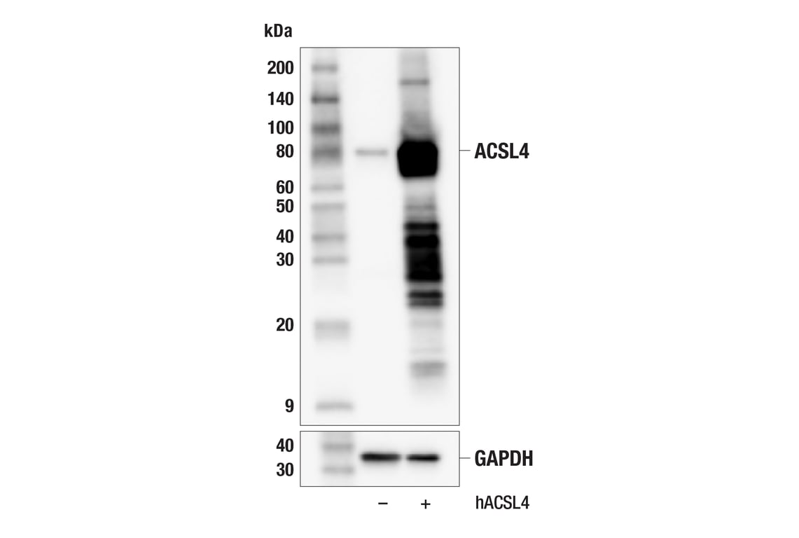
Western blot analysis of extracts from 293T cells, mock transfected (-) or transfected with a construct expressing full-length human ACSL4 protein (hACSL4; +), using ACSL4 (F6T3Z) Rabbit mAb (upper) or GAPDH (D16H11) XP® Rabbit mAb #5174 (lower).
Western Blotting Image 2: ACSL4 (F6T3Z) Rabbit Monoclonal Antibody
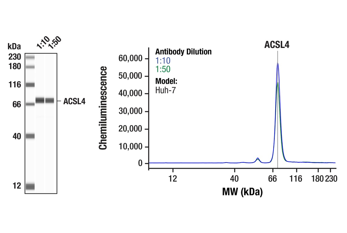
Simple Western™️ analysis of lysates (0.05mg/mL) from Huh-7 cells using ACSL4 (F6T3Z) Rabbit mAb #38493. The virtual lane view (left) shows the target band (as indicated) at 1:10 and 1:50 dilutions of primary antibody. The corresponding electropherogram view (right) plots chemiluminescence by molecular weight along the capillary at 1:10 (blue line) and 1:50 (green line) dilutions of primary antibody. This experiment was performed under reducing conditions on the Jess™️ Simple Western instrument from ProteinSimple, a BioTechne brand, using the 12 – 230 kDa separation module.
Western Blotting Image 1: ACSL4 (F6T3Z) Rabbit Monoclonal Antibody
Orders: 877-616-CELL (2355) [email protected] Support: 877-678-TECH (8324) [email protected] Web: cellsignal.com
For Research Use Only. Not for Use in Diagnostic Procedures.

Revision 4

Cell Signaling Technology Logo
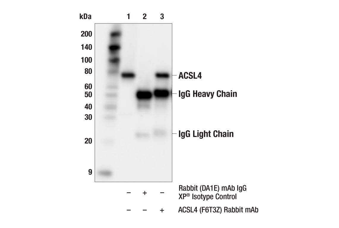
Immunoprecipitation of ACSL4 protein from HuH-7 cell extracts. Lane 1 is 10% input, lane 2 is Rabbit (DA1E) mAb IgG XP® Isotype Control #3900, and lane 3 is ACSL4 (F6T3Z) Rabbit mAb. Western blot analysis was performed using ACSL4 (F6T3Z) Rabbit mAb. Anti-rabbit IgG, HRP-linked Antibody #7074 was used as a secondary antibody.
Immunoprecipitation Image 1: ACSL4 (F6T3Z) Rabbit Monoclonal Antibody
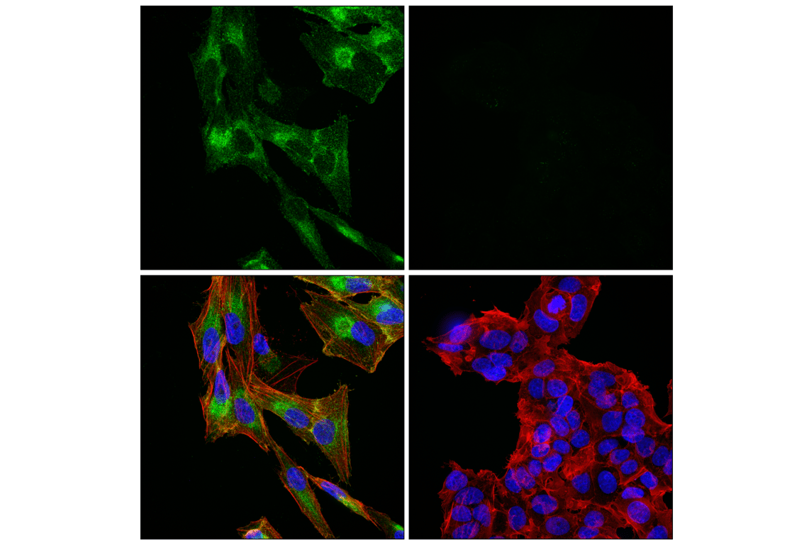
Confocal immunofluorescent analysis of Caki-1 cells (left, positive) and MCF7 cells (right, negative) using ACSL4 (F6T3Z) Rabbit mAb (green), DyLight 554 Phalloidin #13054 (red), and DAPI #4083 (blue).
Immunofluorescence Image 1: ACSL4 (F6T3Z) Rabbit Monoclonal Antibody
Orders: 877-616-CELL (2355) [email protected] Support: 877-678-TECH (8324) [email protected] Web: cellsignal.com
For Research Use Only. Not for Use in Diagnostic Procedures.