Revision 1
#52405
Store at -20C
877-616-CELL (2355)
877-678-TECH (8324)
3 Trask Lane | Danvers | Massachusetts | 01923 | USA
For Research Use Only. Not for Use in Diagnostic Procedures.
Applications:
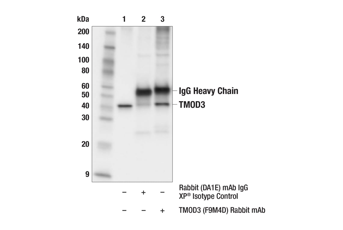
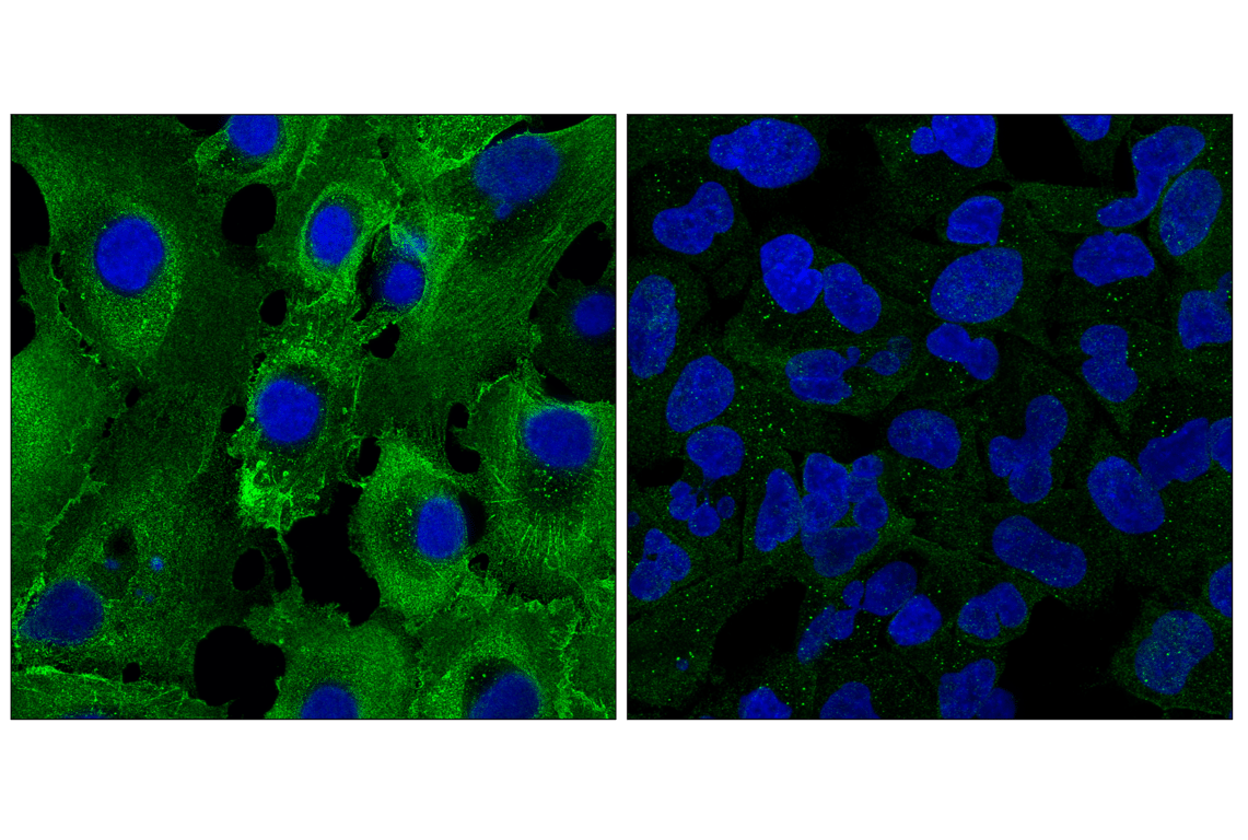
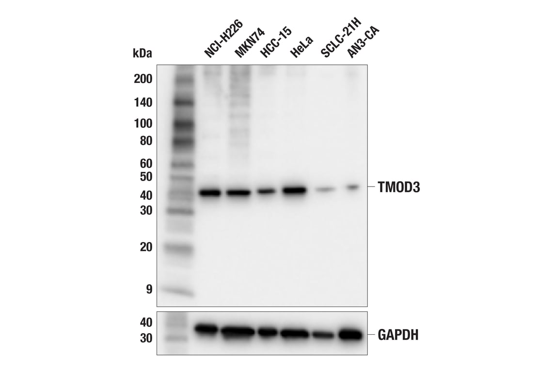
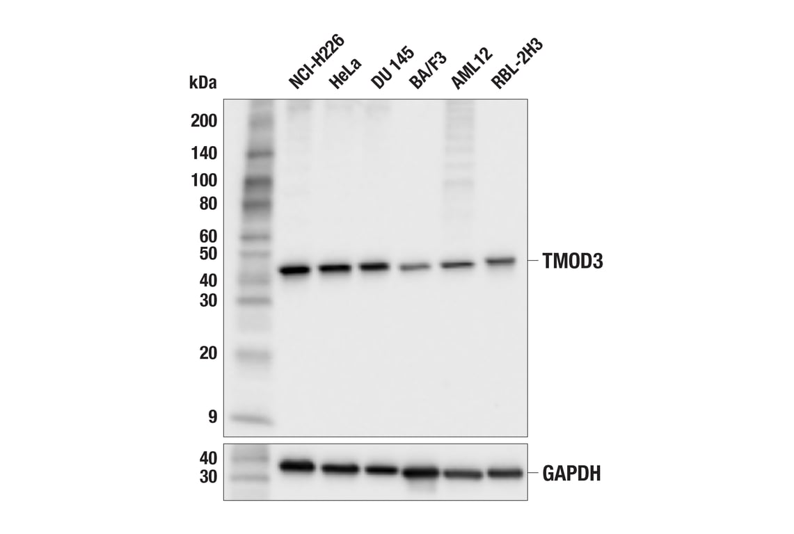
W, IP, IF-IC
Reactivity:
H M R
Sensitivity:
Endogenous
MW (kDa):
40
Source/Isotype:
Rabbit IgG
UniProt ID:
#Q9NYL9
Entrez-Gene Id:
29766
Product Usage Information
| Application | Dilution |
|---|---|
| Western Blotting | 1:1000 |
| Immunoprecipitation | 1:50 |
| Immunofluorescence (Immunocytochemistry) | 1:1600 - 1:6400 |
Storage
Specificity/Sensitivity
Source / Purification
Background
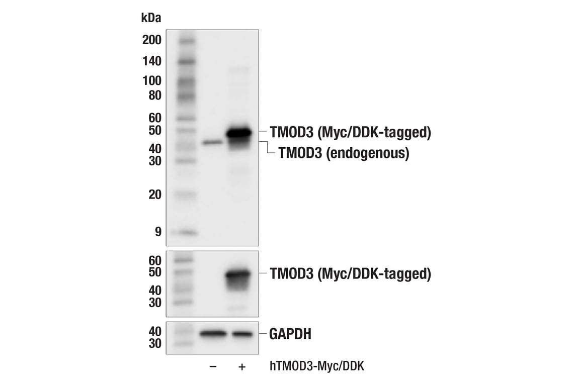
TMOD3 is widely expressed and, like other family members, binds tropomyosin and actin filaments (F-actin). This functions in the capping process (5). However, unlike other family members, TMOD3 is also able to bind to actin monomer (6). TMOD3 localizes to the lateral cell membrane in polarized epithelial cells and regulates cell shape (7). Loss of TMOD3 in mice leads to embryonic lethality with an impaired erythroid differential in the fetal liver (8). TMOD3 is uniquely phosphorylated by Akt2 upon insulin stimulation and regulates Glut4 trafficking through actin remodeling (9). Expression of TMOD3 may promote tumor progression, including liver cancer (10). Studies have also found a ferroptosis-related gene signature in lung adenocarcinoma that includes TMOD3 (11).
Background References
- Fischer, R.S. and Fowler, V.M. (2003) Trends Cell Biol 13, 593-601.
- Rao, J.N. et al. (2014) Science 345, 463-7.
- Ito-Kureha, T. et al. (2015) Cancer Res 75, 62-72.
- Agnelli, L. et al. (2012) Blood 120, 1274-81.
- Parreno, J. and Fowler, V.M. (2018) Biophys Rev 10, 1605-1615.
- Fischer, R.S. et al. (2006) J Biol Chem 281, 36454-65.
- Weber, K.L. et al. (2007) J Cell Sci 120, 3625-32.
- Sui, Z. et al. (2014) Blood 123, 758-67.
- Lim, C.Y. et al. (2015) Nat Commun 6, 5951.
- Jin, C. et al. (2019) Oncol Rep 41, 3060-3068.
- Tian, W. et al. (2024) J Gene Med 26, e3579.
Species Reactivity
Species reactivity is determined by testing in at least one approved application (e.g., western blot).
Western Blot Buffer
IMPORTANT: For western blots, incubate membrane with diluted primary antibody in 5% w/v nonfat dry milk, 1X TBS, 0.1% Tween® 20 at 4°C with gentle shaking, overnight.
Applications Key
W: Western Blotting IP: Immunoprecipitation IF-IC: Immunofluorescence (Immunocytochemistry)
Cross-Reactivity Key
H: Human M: Mouse R: Rat
Trademarks and Patents
Cell Signaling Technology is a trademark of Cell Signaling Technology, Inc.
All other trademarks are the property of their respective owners. Visit cellsignal.com/trademarks for more information.
Limited Uses
Except as otherwise expressly agreed in a writing signed by a legally authorized representative of CST, the following terms apply to Products provided by CST, its affiliates or its distributors. Any Customer's terms and conditions that are in addition to, or different from, those contained herein, unless separately accepted in writing by a legally authorized representative of CST, are rejected and are of no force or effect.
Products are labeled with For Research Use Only or a similar labeling statement and have not been approved, cleared, or licensed by the FDA or other regulatory foreign or domestic entity, for any purpose. Customer shall not use any Product for any diagnostic or therapeutic purpose, or otherwise in any manner that conflicts with its labeling statement. Products sold or licensed by CST are provided for Customer as the end-user and solely for research and development uses. Any use of Product for diagnostic, prophylactic or therapeutic purposes, or any purchase of Product for resale (alone or as a component) or other commercial purpose, requires a separate license from CST. Customer shall (a) not sell, license, loan, donate or otherwise transfer or make available any Product to any third party, whether alone or in combination with other materials, or use the Products to manufacture any commercial products, (b) not copy, modify, reverse engineer, decompile, disassemble or otherwise attempt to discover the underlying structure or technology of the Products, or use the Products for the purpose of developing any products or services that would compete with CST products or services, (c) not alter or remove from the Products any trademarks, trade names, logos, patent or copyright notices or markings, (d) use the Products solely in accordance with CST Product Terms of Sale and any applicable documentation, and (e) comply with any license, terms of service or similar agreement with respect to any third party products or services used by Customer in connection with the Products.
Revision 1
Revision 1