Revision 2

#53721

Store at +4C

CST Logo
Orders:

877-616-CELL (2355)

[email protected]

Support:

877-678-TECH (8324)

3 Trask Lane | Danvers | Massachusetts | 01923 | USA

For Research Use Only. Not for Use in Diagnostic Procedures.

Product Includes Product # Quantity (Petite)
Anti-rabbit IgG (H+L), F(ab')2 Fragment (Alexa Fluor® 488 Conjugate)44121 x 50 µl
VersicanG1-Fc (Rabbit IgG)159331 x 25 µl

Description

The Hyaluronan Complete Tissue Staining Kit (Alexa Fluor® 488) can be used to visualize hyaluronan (HA) levels in paraffin-embedded tissue sections. The kit includes an HA binding fusion protein, VersicanG1-Fc (Rabbit IgG) #15933, which specifically binds to HA in tissues, and Anti-rabbit IgG (H+L), F(ab')2 Fragment (Alexa Fluor® 488 Conjugate) #4412 for detection of the Fc portion of VersicanG1-Fc (Rabbit IgG).

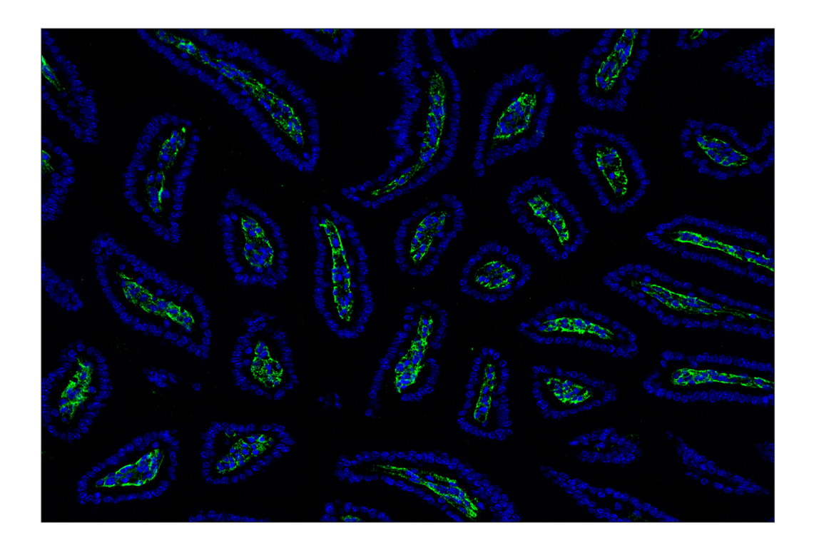
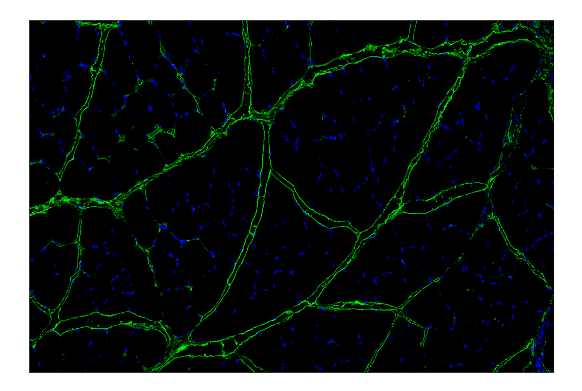
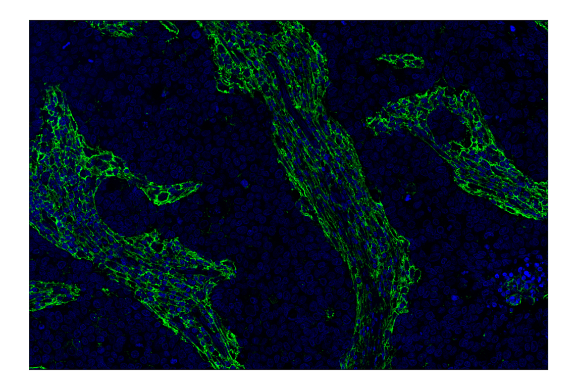
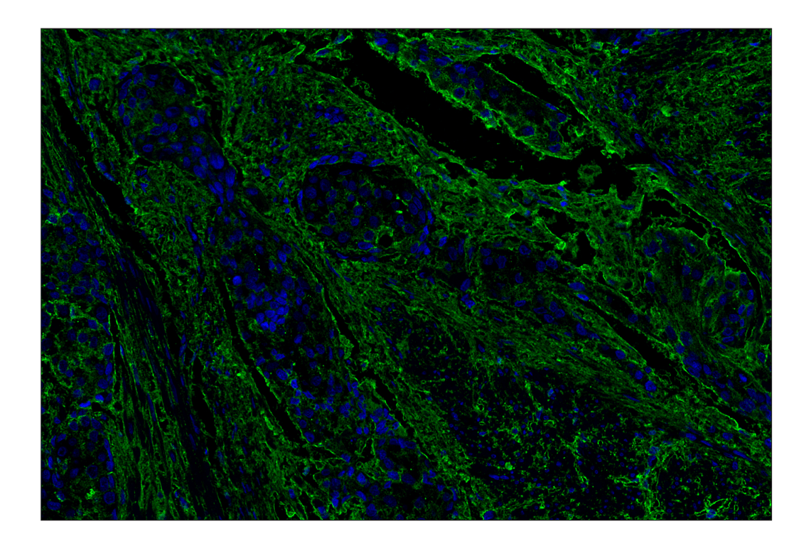
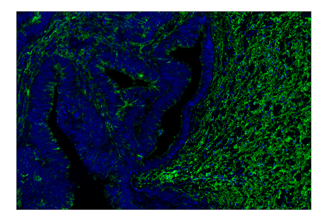
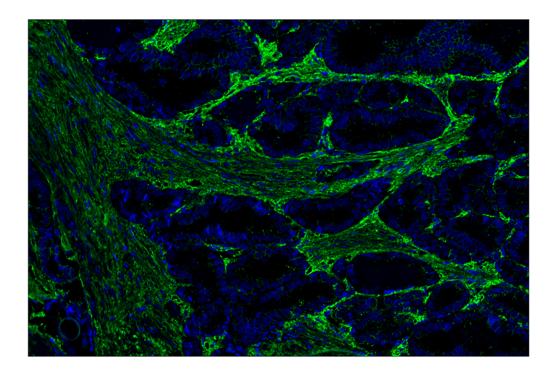
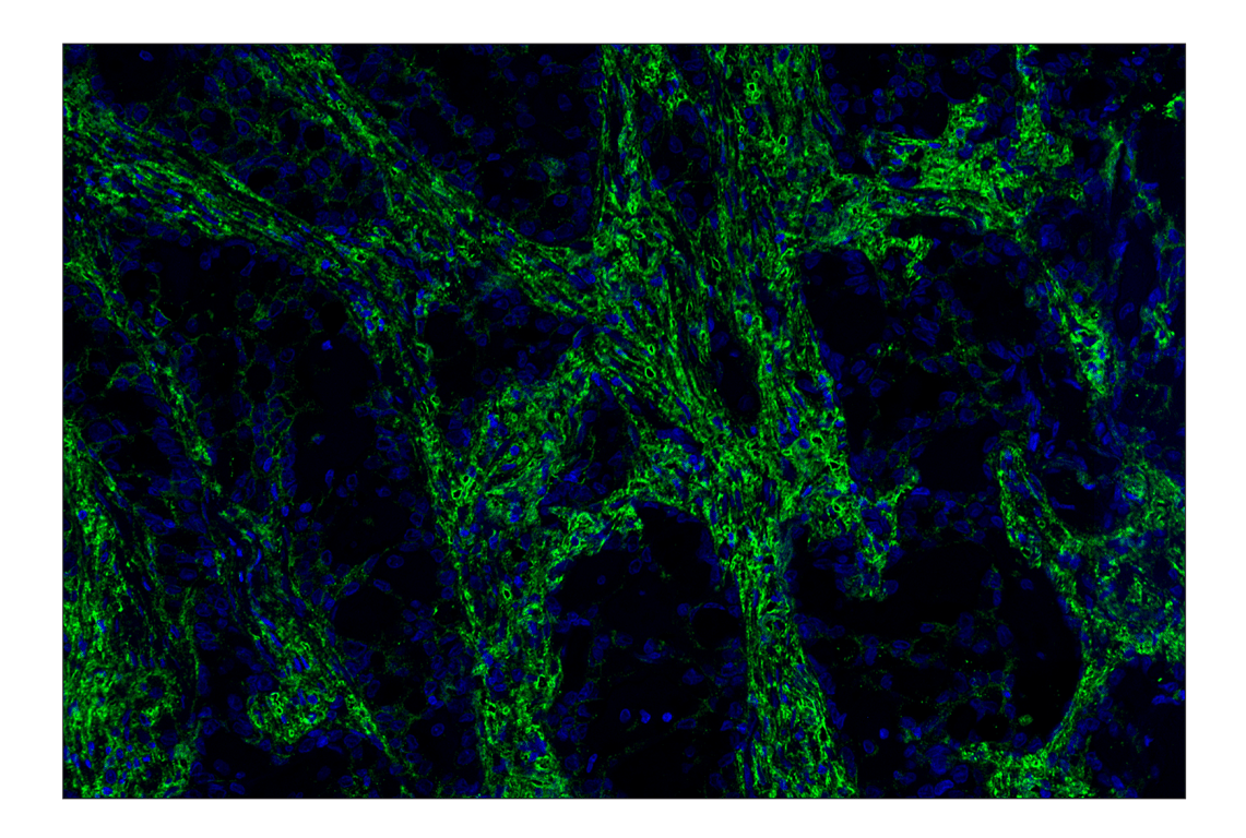
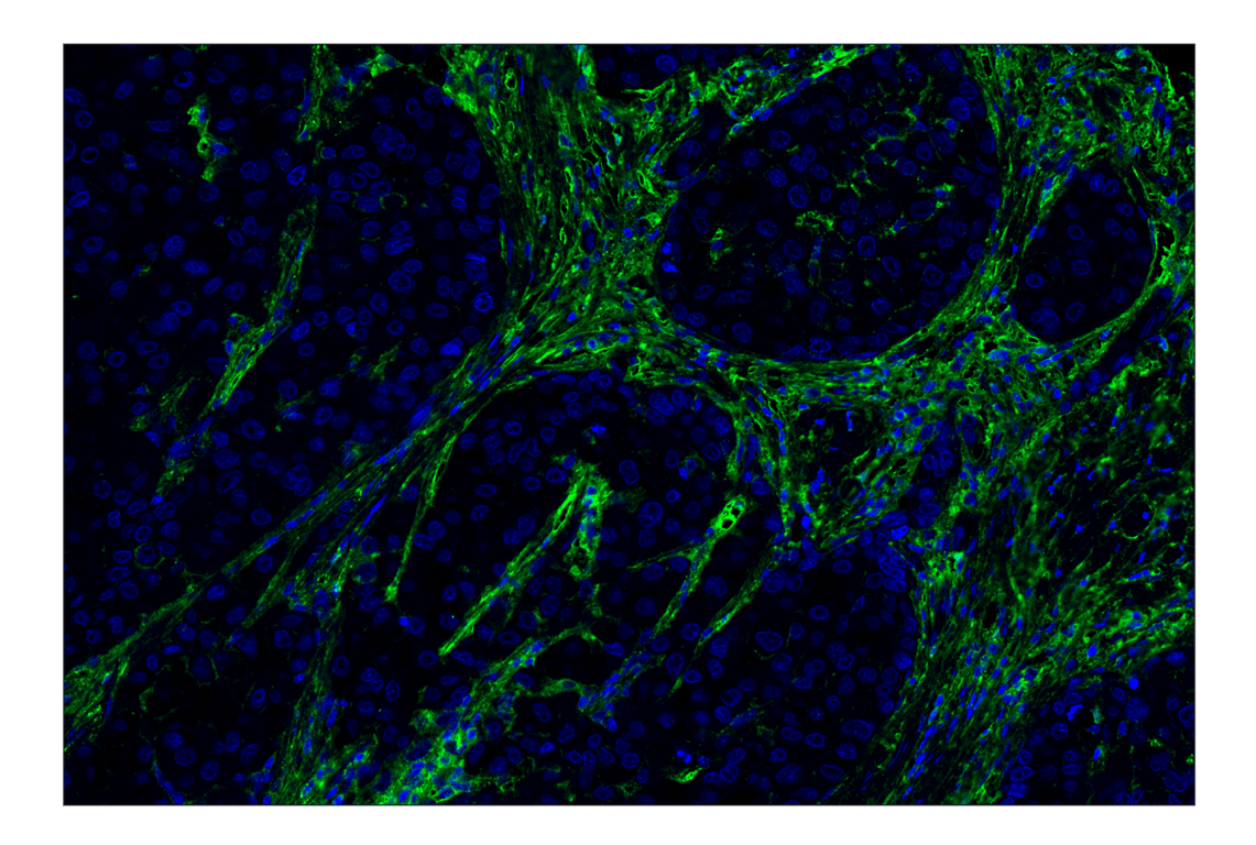
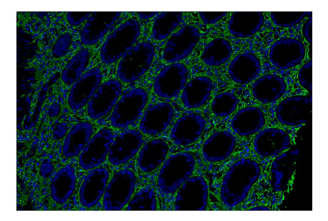
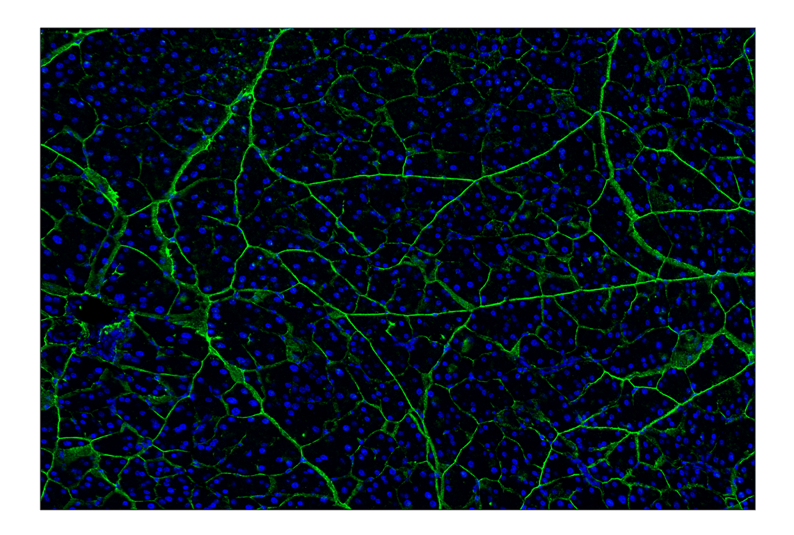
This kit detects endogenous levels of HA in human, mouse, rat, and monkey tissues; it may also be used for HA staining of tissues from other animal species.

Storage

All components in this kit are stable in accordance with the date printed on the outer packaging label when stored at the recommended temperature.

Background

Hyaluronan (HA) comprises a major component of the extracellular matrix (ECM) in vertebrate connective tissues and is abundant in the cartilage, skin, brain, vitreous body, umbilical cord, and synovial fluid (1). The HA polysaccharide is a non-sulfated linear glycosaminoglycan composed of repeating disaccharide units of (1,3)-β-D-N-acetyl-glucosamine (GlcNAc)-β (1,4)-D-glucuronic acid (GlcA)-β (1). HA levels are significantly increased during inflammatory conditions such as myocardial infarction, arthritis, and during transplant, as well as cancer development (2). Two forms of HA have been classified based on size: low molecular weight (LMW HA, < 200 kDa) and high molecular weight (HMW HA > 1 million Da). Despite being composed of similar subunits, LMW HA and HMW HA exhibit different biological activities (3,4). The HMW HA molecule provides tissue hygroscopicity and viscoelasticity, modulates tissue hydration and osmotic balance, and possesses anti-inflammatory, anti-proliferative, anti-angiogenic, and immunosuppressive properties. The LMW HA molecule stimulates the expression of proinflammatory chemokines to regulate tissue wound healing and promotes cancer metastasis (3,4).

Trademarks and Patents

Cell Signaling Technology is a trademark of Cell Signaling Technology, Inc.

Alexa Fluor is a registered trademark of Life Technologies Corporation.

This product is provided under an intellectual property license from Life Technologies Corporation. The transfer of this product is conditioned on the buyer using the purchased product solely in research conducted by the buyer, excluding contract research or any fee for service research, and the buyer must not (1) use this product or its components for (a) diagnostic, therapeutic or prophylactic purposes; (b) testing, analysis or screening services, or information in return for compensation on a per-test basis; or (c) manufacturing or quality assurance or quality control, and/or (2) sell or transfer this product or its components for resale, whether or not resold for use in research. For information on purchasing a license to this product for purposes other than as described above, contact Life Technologies Corporation, 5791 Van Allen Way, Carlsbad, CA 92008 USA or [email protected].

All other trademarks are the property of their respective owners. Visit cellsignal.com/trademarks for more information.

Limited Uses

Except as otherwise expressly agreed in a writing signed by a legally authorized representative of CST, the following terms apply to Products provided by CST, its affiliates or its distributors. Any Customer's terms and conditions that are in addition to, or different from, those contained herein, unless separately accepted in writing by a legally authorized representative of CST, are rejected and are of no force or effect.

Products are labeled with For Research Use Only or a similar labeling statement and have not been approved, cleared, or licensed by the FDA or other regulatory foreign or domestic entity, for any purpose. Customer shall not use any Product for any diagnostic or therapeutic purpose, or otherwise in any manner that conflicts with its labeling statement. Products sold or licensed by CST are provided for Customer as the end-user and solely for research and development uses. Any use of Product for diagnostic, prophylactic or therapeutic purposes, or any purchase of Product for resale (alone or as a component) or other commercial purpose, requires a separate license from CST. Customer shall (a) not sell, license, loan, donate or otherwise transfer or make available any Product to any third party, whether alone or in combination with other materials, or use the Products to manufacture any commercial products, (b) not copy, modify, reverse engineer, decompile, disassemble or otherwise attempt to discover the underlying structure or technology of the Products, or use the Products for the purpose of developing any products or services that would compete with CST products or services, (c) not alter or remove from the Products any trademarks, trade names, logos, patent or copyright notices or markings, (d) use the Products solely in accordance with CST Product Terms of Sale and any applicable documentation, and (e) comply with any license, terms of service or similar agreement with respect to any third party products or services used by Customer in connection with the Products.

Orders: 877-616-CELL (2355) [email protected] Support: 877-678-TECH (8324) [email protected] Web: cellsignal.com
For Research Use Only. Not for Use in Diagnostic Procedures.

Revision 2

Cell Signaling Technology Logo
Immunohistochemical analysis of paraffin-embedded human ductal breast carcinoma using Hyaluronan Complete Tissue Staining Kit (Alexa Fluor® 488) (green) and DAPI #4083 (blue).
Immunohistochemistry Image 1: Hyaluronan Complete Tissue Staining Kit (Alexa Fluor<sup>®</sup> 488)
Immunohistochemical analysis of paraffin-embedded human adenoid cystic carcinoma of the salivary gland using Hyaluronan Complete Tissue Staining Kit (Alexa Fluor® 488) (green) and DAPI #4083 (blue).
Immunohistochemistry Image 2: Hyaluronan Complete Tissue Staining Kit (Alexa Fluor<sup>®</sup> 488)

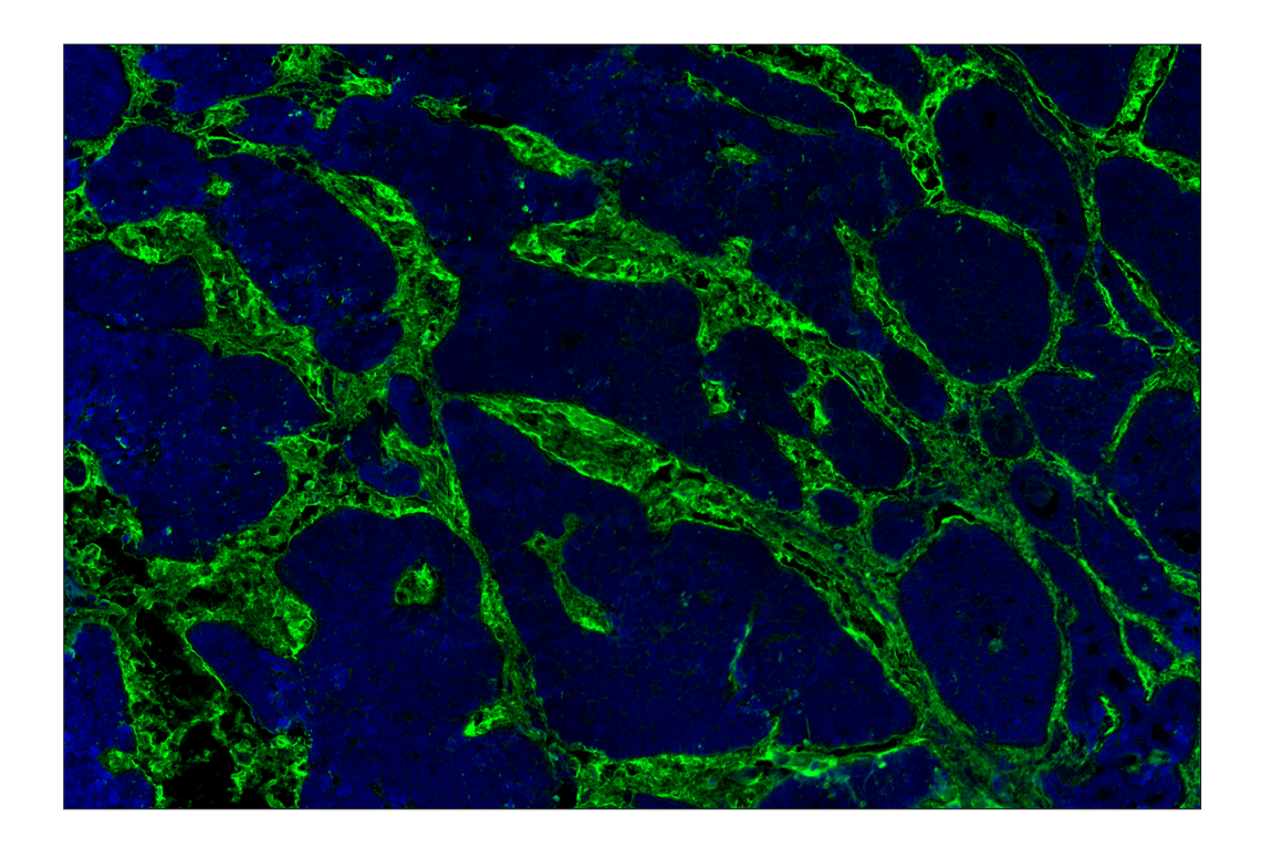
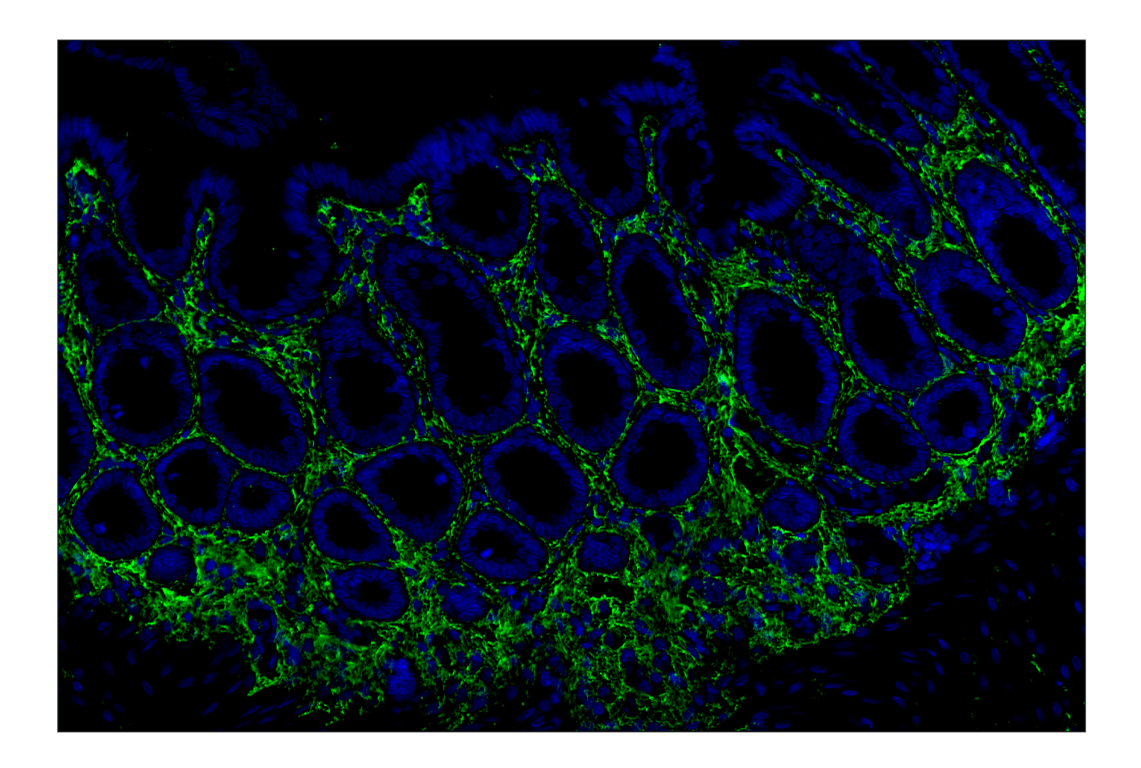
Immunohistochemical analysis of paraffin-embedded human urothelial carcinoma using Hyaluronan Complete Tissue Staining Kit (Alexa Fluor® 488) (green) and DAPI #4083 (blue).

Immunohistochemistry Image 3: Hyaluronan Complete Tissue Staining Kit (Alexa Fluor<sup>®</sup> 488)
Orders: 877-616-CELL (2355) [email protected] Support: 877-678-TECH (8324) [email protected] Web: cellsignal.com
For Research Use Only. Not for Use in Diagnostic Procedures.

Revision 2

Cell Signaling Technology Logo
Immunohistochemical analysis of paraffin-embedded human colon adenocarcinoma using Hyaluronan Complete Tissue Staining Kit (Alexa Fluor® 488) (green) and DAPI #4083 (blue).
Immunohistochemistry Image 4: Hyaluronan Complete Tissue Staining Kit (Alexa Fluor<sup>®</sup> 488)
Immunohistochemical analysis of paraffin-embedded human papillary thyroid carcinoma using Hyaluronan Complete Tissue Staining Kit (Alexa Fluor® 488) (green) and DAPI #4083 (blue).
Immunohistochemistry Image 5: Hyaluronan Complete Tissue Staining Kit (Alexa Fluor<sup>®</sup> 488)
Immunohistochemical analysis of paraffin-embedded human lung adenocarcinoma using Hyaluronan Complete Tissue Staining Kit (Alexa Fluor® 488) (green) and DAPI #4083 (blue).
Immunohistochemistry Image 6: Hyaluronan Complete Tissue Staining Kit (Alexa Fluor<sup>®</sup> 488)
Orders: 877-616-CELL (2355) [email protected] Support: 877-678-TECH (8324) [email protected] Web: cellsignal.com
For Research Use Only. Not for Use in Diagnostic Procedures.

Revision 2

Cell Signaling Technology Logo
Immunohistochemical analysis of paraffin-embedded human hepatocellular carcinoma using Hyaluronan Complete Tissue Staining Kit (Alexa Fluor® 488) (green) and DAPI #4083 (blue).
Immunohistochemistry Image 7: Hyaluronan Complete Tissue Staining Kit (Alexa Fluor<sup>®</sup> 488)
Immunohistochemical analysis of paraffin-embedded rhesus monkey colon using Hyaluronan Complete Tissue Staining Kit (Alexa Fluor® 488) (green) and DAPI #4083 (blue).
Immunohistochemistry Image 8: Hyaluronan Complete Tissue Staining Kit (Alexa Fluor<sup>®</sup> 488)
Immunohistochemical analysis of paraffin-embedded mouse pancreas using Hyaluronan Complete Tissue Staining Kit (Alexa Fluor® 488) (green) and DAPI #4083 (blue).
Immunohistochemistry Image 9: Hyaluronan Complete Tissue Staining Kit (Alexa Fluor<sup>®</sup> 488)
Orders: 877-616-CELL (2355) [email protected] Support: 877-678-TECH (8324) [email protected] Web: cellsignal.com
For Research Use Only. Not for Use in Diagnostic Procedures.

Revision 2

Cell Signaling Technology Logo
Immunohistochemical analysis of paraffin-embedded mouse small intestine using Hyaluronan Complete Tissue Staining Kit (Alexa Fluor® 488) (green) and DAPI #4083 (blue).
Immunohistochemistry Image 10: Hyaluronan Complete Tissue Staining Kit (Alexa Fluor<sup>®</sup> 488)
Immunohistochemical analysis of paraffin-embedded rat colon using Hyaluronan Complete Tissue Staining Kit (Alexa Fluor® 488) (green) and DAPI #4083 (blue).
Immunohistochemistry Image 11: Hyaluronan Complete Tissue Staining Kit (Alexa Fluor<sup>®</sup> 488)
Immunohistochemical analysis of paraffin-embedded rat skeletal muscle using Hyaluronan Complete Tissue Staining Kit (Alexa Fluor® 488) (green) and DAPI #4083 (blue).
Immunohistochemistry Image 12: Hyaluronan Complete Tissue Staining Kit (Alexa Fluor<sup>®</sup> 488)
Orders: 877-616-CELL (2355) [email protected] Support: 877-678-TECH (8324) [email protected] Web: cellsignal.com
For Research Use Only. Not for Use in Diagnostic Procedures.