Revision 1
#62696
Store at -20C
877-616-CELL (2355)
877-678-TECH (8324)
3 Trask Lane | Danvers | Massachusetts | 01923 | USA
For Research Use Only. Not for Use in Diagnostic Procedures.
Applications:
W, IP, IF-IC
Reactivity:
H
Sensitivity:
Endogenous
MW (kDa):
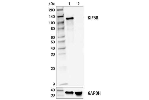
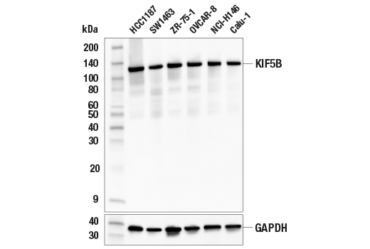
110
Source/Isotype:
Rabbit IgG
UniProt ID:
#P33176
Entrez-Gene Id:
3799
Product Usage Information
| Application | Dilution |
|---|---|
| Western Blotting | 1:1000 |
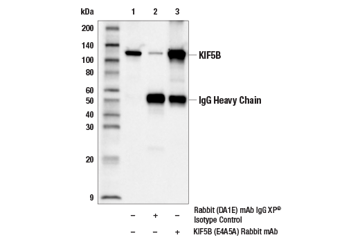
| Immunoprecipitation | 1:100 |
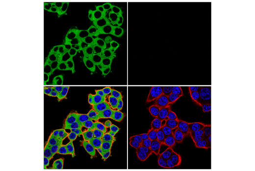
| Immunofluorescence (Immunocytochemistry) | 1:200 - 1:800 |
Storage
Specificity/Sensitivity
Source / Purification
Background
KIF5 consists of three family members referred to as KIF5A, KIF5B, and KIF5C (7). KIF5A and KIF5C are specifically expressed in neurons whereas KIF5B is expressed ubiquitously (7-10). Targeted disruption of the kif5B gene resulted in embryonic lethality (11). These and other studies demonstrate that KIF5B plays an important role in the localization and distribution of major organelles, including the mitochondria and lysosome, and can contribute to autophagy (11-13). In addition, gene rearrangements involving KIF5B fusions to ALK and RET have been identified as drivers for lung cancer and other malignancies (14-18).
Background References
- Hirokawa, N. et al. (2009) Nat Rev Mol Cell Biol 10, 682-96.
- Yu, Y. and Feng, Y.M. (2010) Cancer 116, 5150-60.
- Park, J.J. et al. (2008) Mol Endocrinol 22, 989-1005.
- Hirokawa, N. et al. (2010) Neuron 68, 610-38.
- Yoshimura, Y. et al. (2010) Mol Cell Biol 30, 2206-19.
- Hirokawa, N. and Noda, Y. (2008) Physiol Rev 88, 1089-118.
- Nakagawa, T. et al. (1997) Proc Natl Acad Sci U S A 94, 9654-9.
- Aizawa, H. et al. (1992) J Cell Biol 119, 1287-96.
- Kanai, Y. et al. (2000) J Neurosci 20, 6374-84.
- Meng, Y.X. et al. (1997) Endocrinology 138, 1979-87.
- Tanaka, Y. et al. (1998) Cell 93, 1147-58.
- Cardoso, C.M. et al. (2009) PLoS One 4, e4424.
- Du, W. et al. (2016) Dev Cell 37, 326-336.
- Takeuchi, K. et al. (2009) Clin Cancer Res 15, 3143-9.
- Wong, D.W. et al. (2011) Cancer 117, 2709-18.
- Takeuchi, K. et al. (2012) Nat Med 18, 378-81.
- Ju, Y.S. et al. (2012) Genome Res 22, 436-45.
- Kohno, T. et al. (2012) Nat Med 18, 375-7.
Species Reactivity
Species reactivity is determined by testing in at least one approved application (e.g., western blot).
Western Blot Buffer
IMPORTANT: For western blots, incubate membrane with diluted primary antibody in 5% w/v BSA, 1X TBS, 0.1% Tween® 20 at 4°C with gentle shaking, overnight.
Applications Key
W: Western Blotting IP: Immunoprecipitation IF-IC: Immunofluorescence (Immunocytochemistry)
Cross-Reactivity Key
H: Human
Trademarks and Patents
Cell Signaling Technology is a trademark of Cell Signaling Technology, Inc.
All other trademarks are the property of their respective owners. Visit cellsignal.com/trademarks for more information.
Limited Uses
Except as otherwise expressly agreed in a writing signed by a legally authorized representative of CST, the following terms apply to Products provided by CST, its affiliates or its distributors. Any Customer's terms and conditions that are in addition to, or different from, those contained herein, unless separately accepted in writing by a legally authorized representative of CST, are rejected and are of no force or effect.
Products are labeled with For Research Use Only or a similar labeling statement and have not been approved, cleared, or licensed by the FDA or other regulatory foreign or domestic entity, for any purpose. Customer shall not use any Product for any diagnostic or therapeutic purpose, or otherwise in any manner that conflicts with its labeling statement. Products sold or licensed by CST are provided for Customer as the end-user and solely for research and development uses. Any use of Product for diagnostic, prophylactic or therapeutic purposes, or any purchase of Product for resale (alone or as a component) or other commercial purpose, requires a separate license from CST. Customer shall (a) not sell, license, loan, donate or otherwise transfer or make available any Product to any third party, whether alone or in combination with other materials, or use the Products to manufacture any commercial products, (b) not copy, modify, reverse engineer, decompile, disassemble or otherwise attempt to discover the underlying structure or technology of the Products, or use the Products for the purpose of developing any products or services that would compete with CST products or services, (c) not alter or remove from the Products any trademarks, trade names, logos, patent or copyright notices or markings, (d) use the Products solely in accordance with CST Product Terms of Sale and any applicable documentation, and (e) comply with any license, terms of service or similar agreement with respect to any third party products or services used by Customer in connection with the Products.
Revision 1
Revision 1