Revision 1
#66212
Store at -20C
877-616-CELL (2355)
877-678-TECH (8324)
3 Trask Lane | Danvers | Massachusetts | 01923 | USA
For Research Use Only. Not for Use in Diagnostic Procedures.
| Product Includes | Product # | Quantity | Mol. Wt | Isotype/Source |
|---|---|---|---|---|
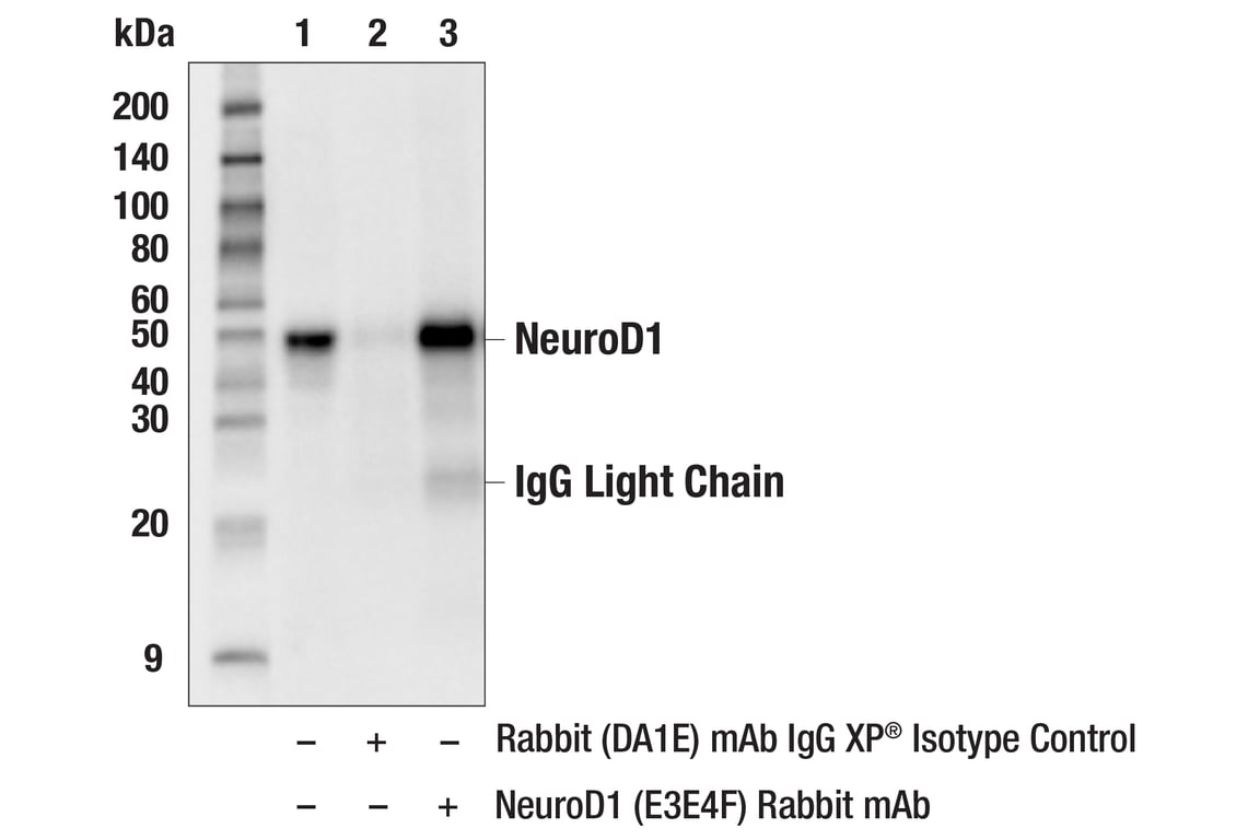
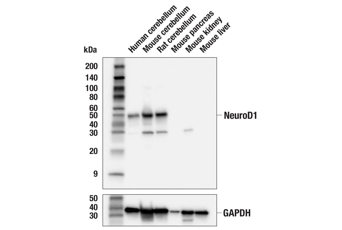
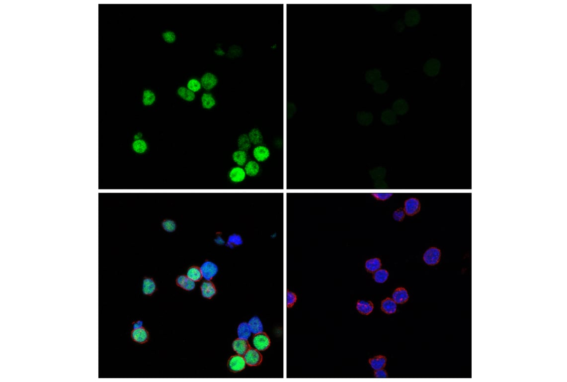
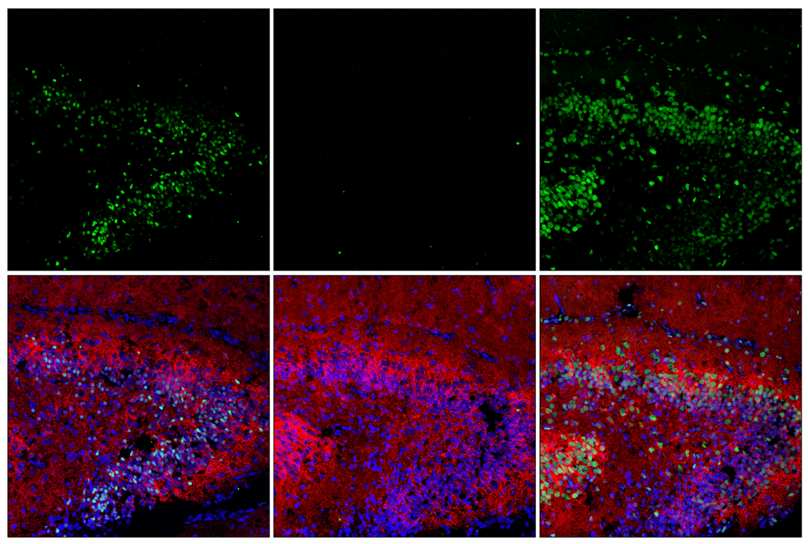
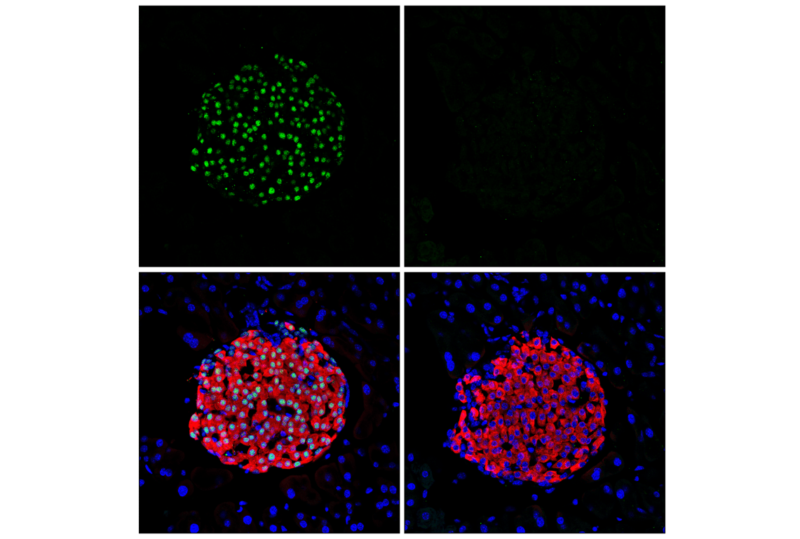
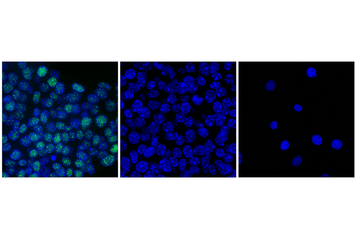
| NeuroD1 (E3E4F) Rabbit Monoclonal Antibody | 62953 | 100 µl | 49 kDa | Rabbit IgG |
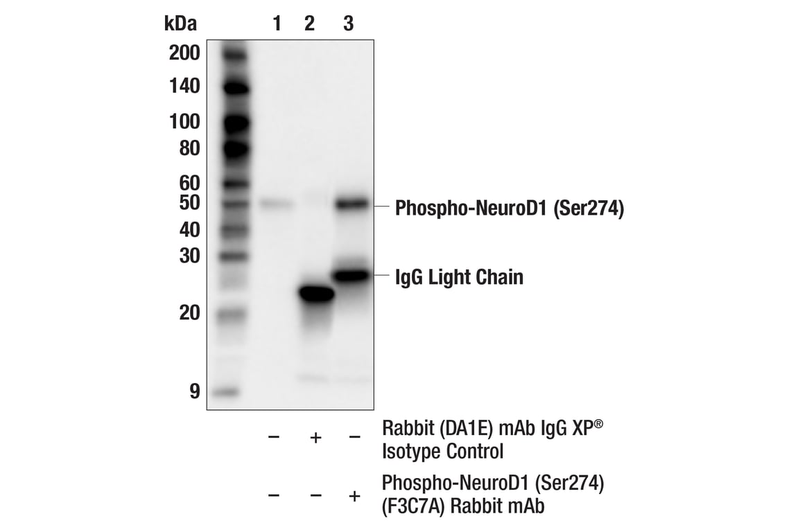
| Phospho-NeuroD1 (Ser274) (F3C7A) Rabbit Monoclonal Antibody | 92657 | 100 µl | 49 kDa | Rabbit IgG |
Please visit cellsignal.com for individual component applications, species cross-reactivity, dilutions, protocols, and additional product information.
UniProt ID:
#Q13562
Entrez-Gene Id:
4760
Description
Storage
Background
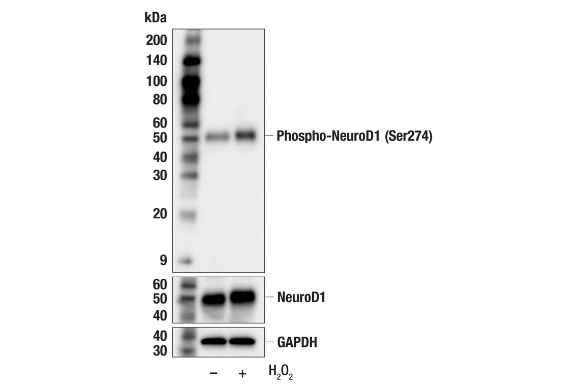
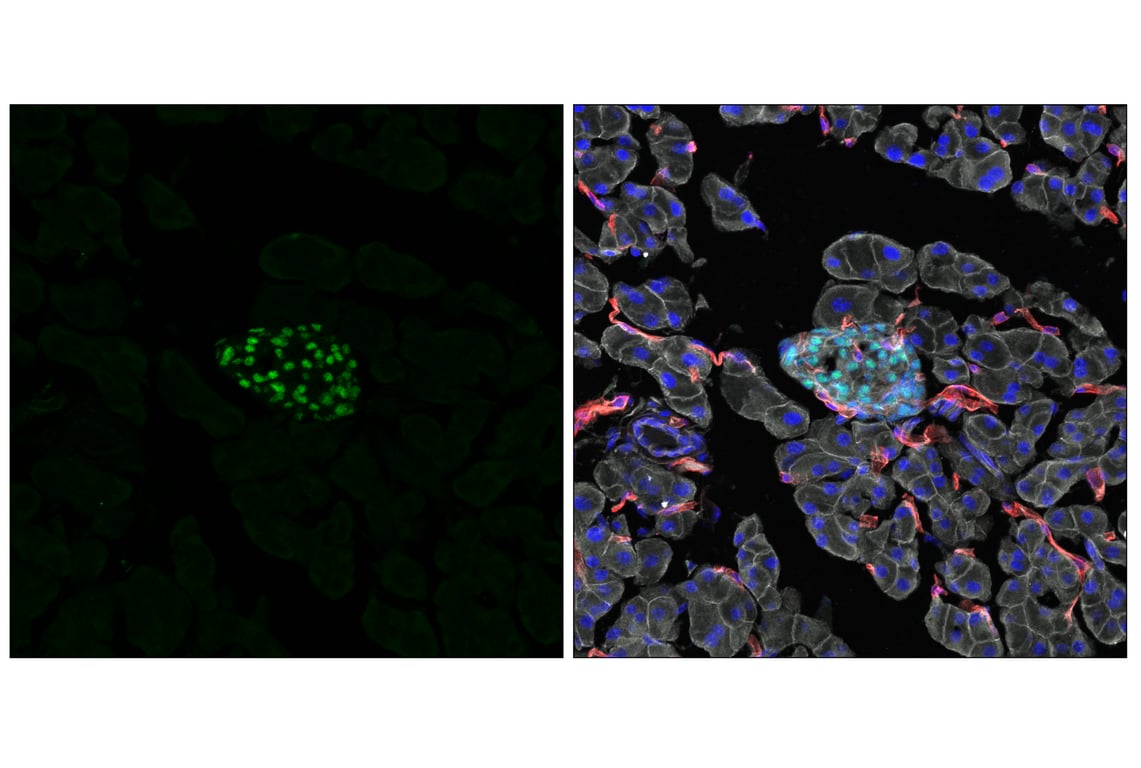
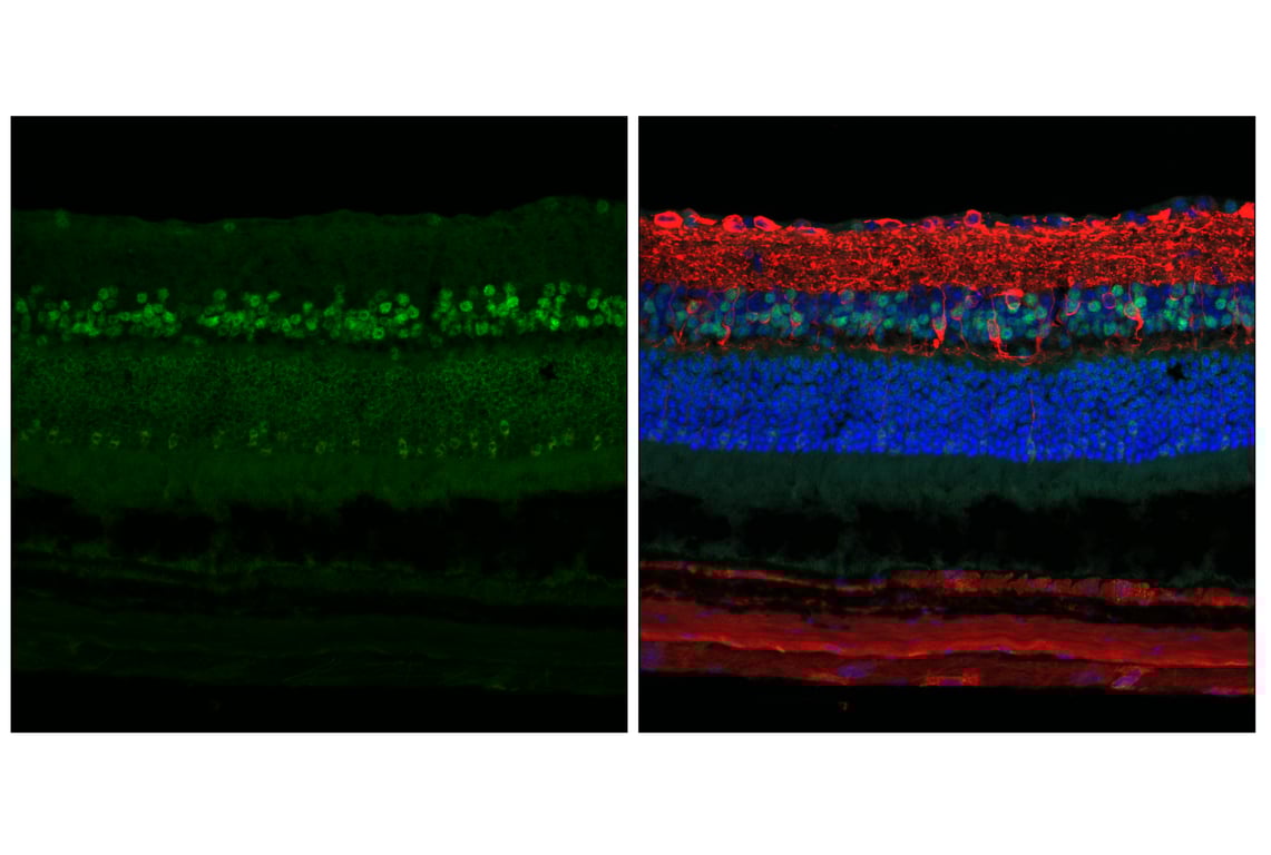
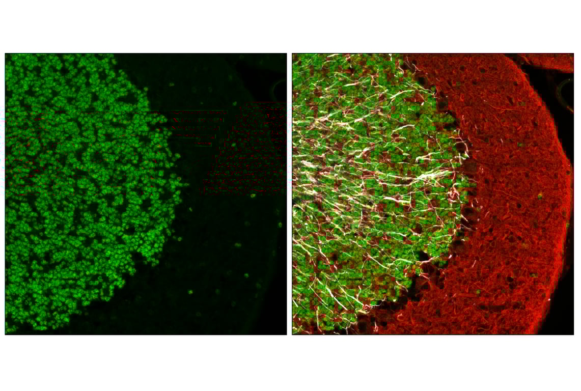
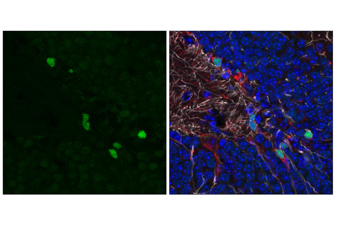
Erk1/2 kinase activity is stimulated by glucose and calcium influx in pancreatic beta cells, and the subsequent phosphorylation of NeuroD1 at Ser274 promotes insulin gene transcription (7,8). Phosphorylated NeuroD1 in the pancreas is increasingly translocated to the nucleus, where it directly interacts with the insulin promoter (9). Active, nuclear NeuroD1 acts in synergy with Pdx1 to activate insulin, NKX2.2, and islet amyloid polypeptide (IAPP) transcription while suppressing Pdx1-driven transcription of somatostatin (10,11). In developing rat pineal gland, phosphorylation of NeuroD1 at Ser274 and Ser336 is tightly correlated with NeuroD1 nuclear localization, and cytoplasmic to nuclear shuttling of the protein is under cyclical day and night adrenergic control (12). In 293T cells, transfection of a nonphosphorylatable Ser274Ala mutant blocked NeuroD1 ubiquitination and proteasomal degradation (13). The localization of phosphorylated NeuroD1 is reversed in these cells compared to pancreatic beta cells, with a Ser274Glu phospho-mimic localized primarily to the cytoplasm in the former, further evidence of the context-specific nature of this modification. (13).
Background References
- Schonhoff, S.E. et al. (2004) Endocrinology 145, 2639-2644.
- Sharma, A. et al. (1999) Mol. Cell Biol. 19, 704-713.
- Chae, J.H. et al. (2004) Mol. Cells 18, 271-288.
- Gaudillière, B. et al. (2004) Neuron 41, 229-241.
- Miyata, T. et al. (1999) Genes Dev. 13, 1647-1652.
- Naya, F.J. et al. (1997) Genes Dev. 11, 2323-2334.
- Khoo, S. and Cobb, M.H. (1997) Proc Natl Acad Sci U S A 94, 5599-604.
- Khoo, S. et al. (2003) J Biol Chem 278, 32969-77.
- Petersen, H.V. et al. (2002) FEBS Lett 528, 241-5.
- Itkin-Ansari, P. et al. (2005) Dev Dyn 233, 946-53.
- Babu, D.A. et al. (2008) J Biol Chem 283, 8164-72.
- Castro, A.E. et al. (2015) J Pineal Res 58, 439-51.
- Lee, T.Y. et al. (2020) Exp Neurobiol 29, 189-206.
Trademarks and Patents
Cell Signaling Technology is a trademark of Cell Signaling Technology, Inc.
PhosphoPlus is a registered trademark of Cell Signaling Technology, Inc.
All other trademarks are the property of their respective owners. Visit cellsignal.com/trademarks for more information.
Limited Uses
Except as otherwise expressly agreed in a writing signed by a legally authorized representative of CST, the following terms apply to Products provided by CST, its affiliates or its distributors. Any Customer's terms and conditions that are in addition to, or different from, those contained herein, unless separately accepted in writing by a legally authorized representative of CST, are rejected and are of no force or effect.
Products are labeled with For Research Use Only or a similar labeling statement and have not been approved, cleared, or licensed by the FDA or other regulatory foreign or domestic entity, for any purpose. Customer shall not use any Product for any diagnostic or therapeutic purpose, or otherwise in any manner that conflicts with its labeling statement. Products sold or licensed by CST are provided for Customer as the end-user and solely for research and development uses. Any use of Product for diagnostic, prophylactic or therapeutic purposes, or any purchase of Product for resale (alone or as a component) or other commercial purpose, requires a separate license from CST. Customer shall (a) not sell, license, loan, donate or otherwise transfer or make available any Product to any third party, whether alone or in combination with other materials, or use the Products to manufacture any commercial products, (b) not copy, modify, reverse engineer, decompile, disassemble or otherwise attempt to discover the underlying structure or technology of the Products, or use the Products for the purpose of developing any products or services that would compete with CST products or services, (c) not alter or remove from the Products any trademarks, trade names, logos, patent or copyright notices or markings, (d) use the Products solely in accordance with CST Product Terms of Sale and any applicable documentation, and (e) comply with any license, terms of service or similar agreement with respect to any third party products or services used by Customer in connection with the Products.
Revision 1
Revision 1
Revision 1
Revision 1
Revision 1
Revision 1