Revision 3
#88034
Store at -20C
877-616-CELL (2355)
877-678-TECH (8324)
3 Trask Lane | Danvers | Massachusetts | 01923 | USA
For Research Use Only. Not for Use in Diagnostic Procedures.
Applications:
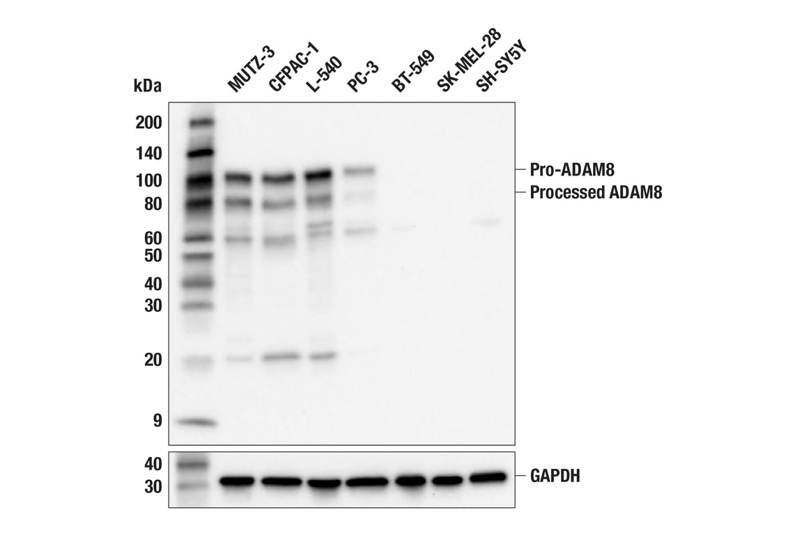
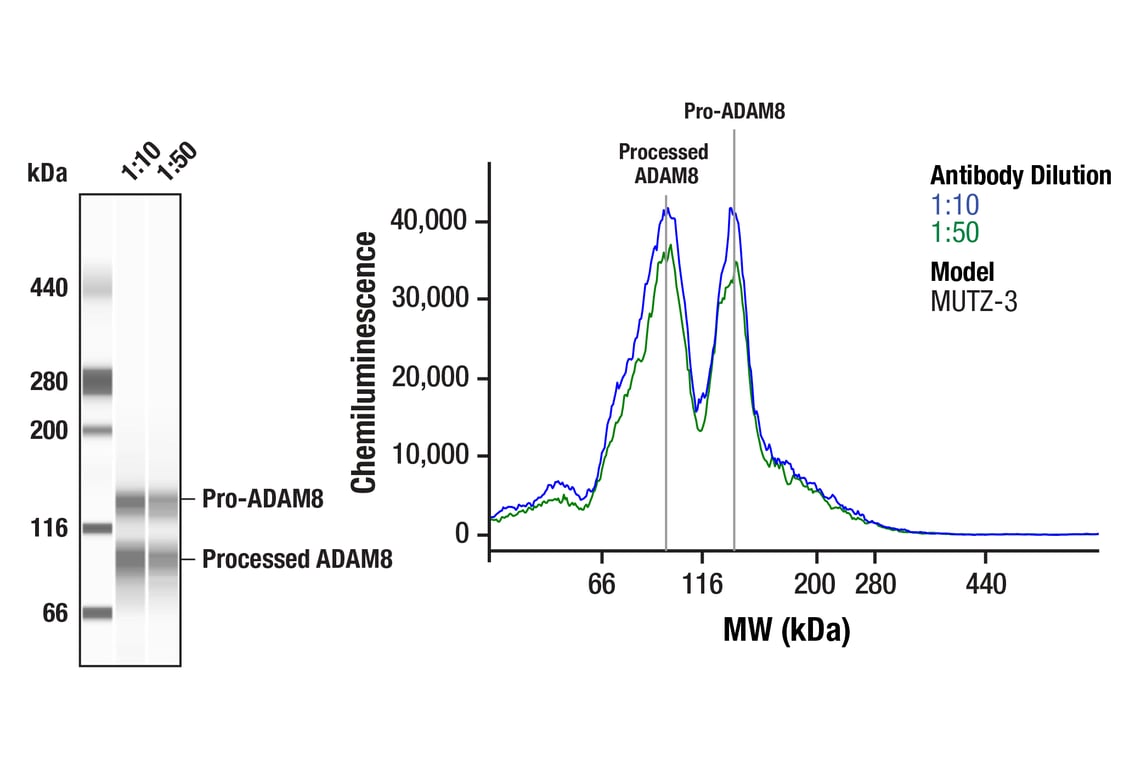
W, W-S
Reactivity:
H
Sensitivity:
Endogenous
MW (kDa):
100, 80
Source/Isotype:
Rabbit IgG
UniProt ID:
#P78325
Entrez-Gene Id:
101
Product Usage Information
| Application | Dilution |
|---|---|
| Western Blotting | 1:1000 |
| Simple Western™ | 1:10 - 1:50 |
Storage
Specificity/Sensitivity
Source / Purification
Background
ADAM8 is unique among family members in its relatively broad expression across various immune cells, including monocytes, macrophages, neutrophils, eosinophils, dendritic cells, and lymphocytes, as well as in epithelial cells in various tissues. ADAM8 is generally expressed at low levels in normal adult tissues but is significantly upregulated in response to inflammatory stimuli and various pathological conditions. ADAM8 is aberrantly overexpressed in numerous cancer types and is increasingly recognized as a prognostic biomarker and therapeutic target. High expression of ADAM8 in breast cancer, particularly in triple-negative breast cancer (TNBC), has been shown to correlate with aggressive phenotype, tumor growth, dissemination, and metastasis, and poor patient outcome. In this tumor type, it is thought to influence cell motility and interaction with the extracellular matrix. ADAM8 is highly expressed in glioblastoma (GBM) tumors, specifically within the tumor environment where it is thought to affect intracellular kinase (e.g., STAT3, MAPK, PI3K/Akt) signaling, and promote angiogenesis, thereby enhancing chemoresistance. Upregulation of ADAM8 is also associated with increased migration and invasiveness of cancer cells in pancreatic tumors, and correlates with reduced survival. ADAM8 expression is found in hepatocellular carcinoma and is associated with tumor size, differentiation, recurrence, metastasis, and poor prognosis. It can promote proliferation, migration, and invasion of hepatoma cells. Upregulation of ADAM8 is also observed in colon cancer, where it is thought to promote invasion by inducing epithelial-mesenchymal transition (EMT) via the TGF-β/Smad2/3 signaling pathway. Lastly, overexpression of ADAM8 is associated with lung cancer progression (3-7).
Background References
- Giebeler, N. and Zigrino, P. (2016) Toxins (Basel) 8, 122.
- Schlöndorff, J. and Blobel, C.P. (1999) J Cell Sci 112 ( Pt 21), 3603-17.
- Mierke, C.T. (2023) Front Cell Dev Biol 11, 1130823.
- Romagnoli, M. et al. (2014) EMBO Mol Med 6, 278-94.
- Schlomann, U. et al. (2015) Nat Commun 6, 6175.
- Li, Y. et al. (2021) Biol Chem 402, 195-206.
- Ishikawa, N. et al. (2004) Clin Cancer Res 10, 8363-70.
Species Reactivity
Species reactivity is determined by testing in at least one approved application (e.g., western blot).
Western Blot Buffer
IMPORTANT: For western blots, incubate membrane with diluted primary antibody in 5% w/v BSA, 1X TBS, 0.1% Tween® 20 at 4°C with gentle shaking, overnight.
Applications Key
W: Western Blotting W-S: Simple Western™
Cross-Reactivity Key
H: Human
Trademarks and Patents
Cell Signaling Technology is a trademark of Cell Signaling Technology, Inc.
All other trademarks are the property of their respective owners. Visit cellsignal.com/trademarks for more information.
Limited Uses
Except as otherwise expressly agreed in a writing signed by a legally authorized representative of CST, the following terms apply to Products provided by CST, its affiliates or its distributors. Any Customer's terms and conditions that are in addition to, or different from, those contained herein, unless separately accepted in writing by a legally authorized representative of CST, are rejected and are of no force or effect.
Products are labeled with For Research Use Only or a similar labeling statement and have not been approved, cleared, or licensed by the FDA or other regulatory foreign or domestic entity, for any purpose. Customer shall not use any Product for any diagnostic or therapeutic purpose, or otherwise in any manner that conflicts with its labeling statement. Products sold or licensed by CST are provided for Customer as the end-user and solely for research and development uses. Any use of Product for diagnostic, prophylactic or therapeutic purposes, or any purchase of Product for resale (alone or as a component) or other commercial purpose, requires a separate license from CST. Customer shall (a) not sell, license, loan, donate or otherwise transfer or make available any Product to any third party, whether alone or in combination with other materials, or use the Products to manufacture any commercial products, (b) not copy, modify, reverse engineer, decompile, disassemble or otherwise attempt to discover the underlying structure or technology of the Products, or use the Products for the purpose of developing any products or services that would compete with CST products or services, (c) not alter or remove from the Products any trademarks, trade names, logos, patent or copyright notices or markings, (d) use the Products solely in accordance with CST Product Terms of Sale and any applicable documentation, and (e) comply with any license, terms of service or similar agreement with respect to any third party products or services used by Customer in connection with the Products.
Revision 3