Revision 3
#89348
Store at -20C
877-616-CELL (2355)
877-678-TECH (8324)
3 Trask Lane | Danvers | Massachusetts | 01923 | USA
For Research Use Only. Not for Use in Diagnostic Procedures.
Applications:
W, W-S, IHC-P
Reactivity:
H
Sensitivity:
Endogenous
MW (kDa):
51-60
Source/Isotype:
Rabbit IgG
UniProt ID:
#P19544
Entrez-Gene Id:
7490
Product Usage Information
| Application | Dilution |
|---|---|
| Western Blotting | 1:1000 |
| Simple Western™ | 1:50 - 1:200 |
| Immunohistochemistry (Paraffin) | 1:400 - 1:1600 |
Storage
Specificity/Sensitivity
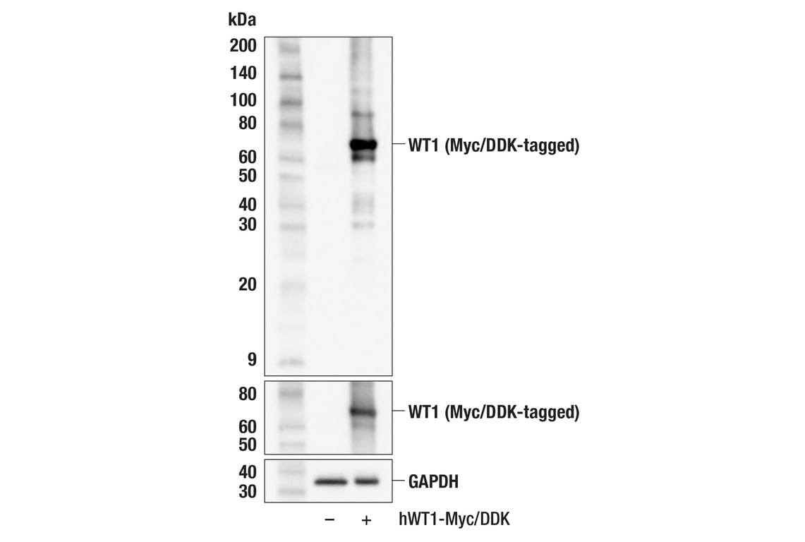
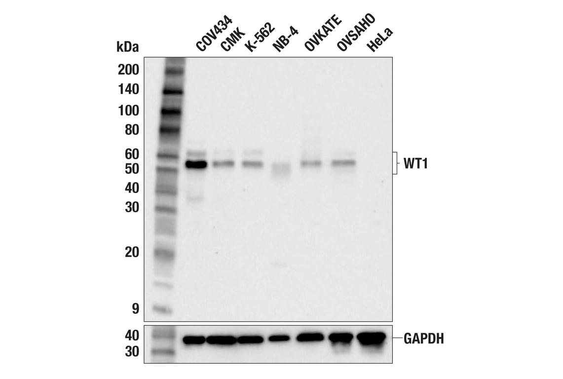
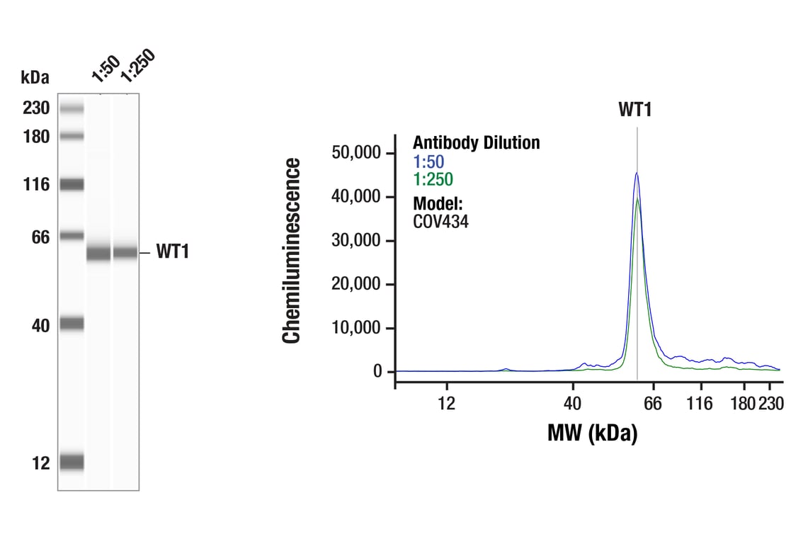
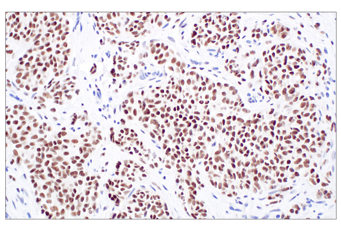
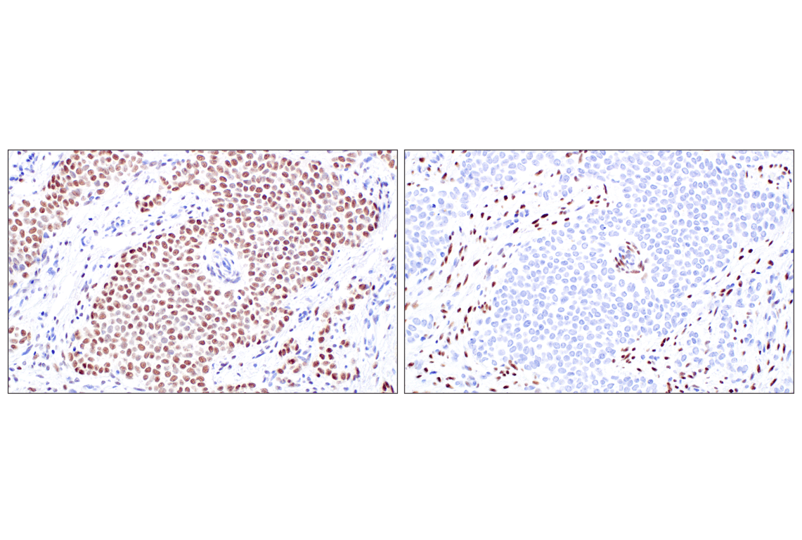
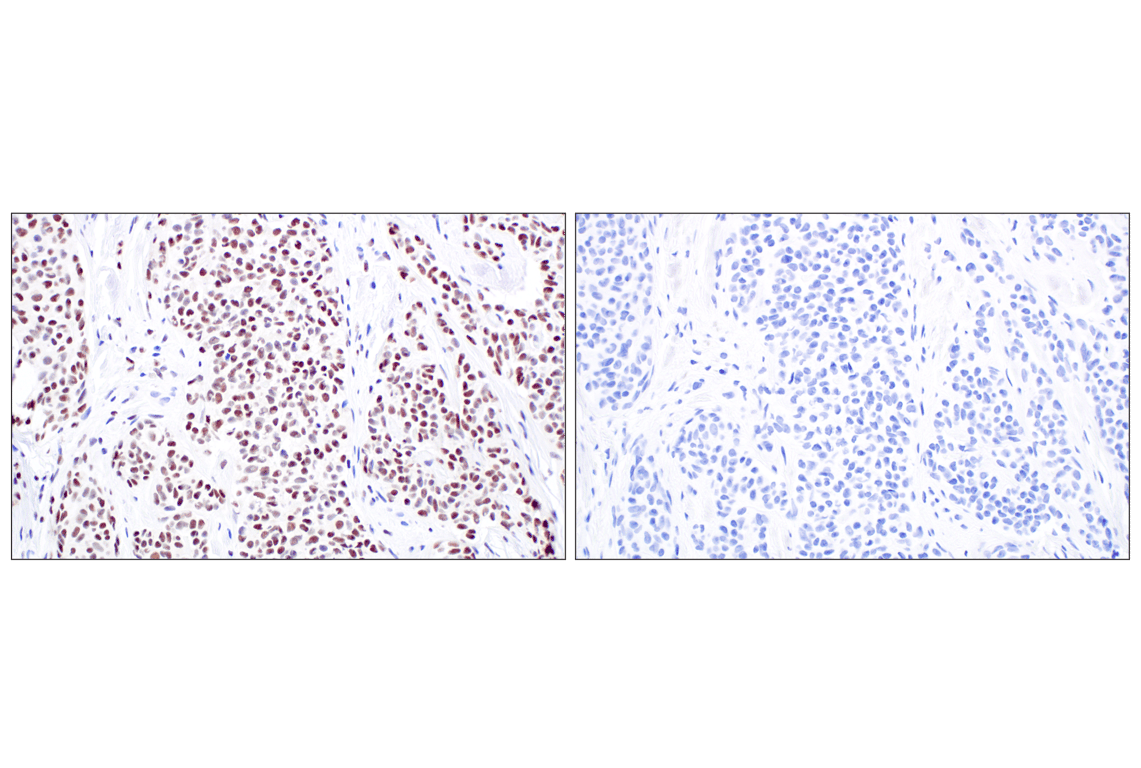
WT1 (F3T7B) Rabbit mAb (Carboxy-terminal Antigen) recognizes endogenous levels of total WT1 protein.
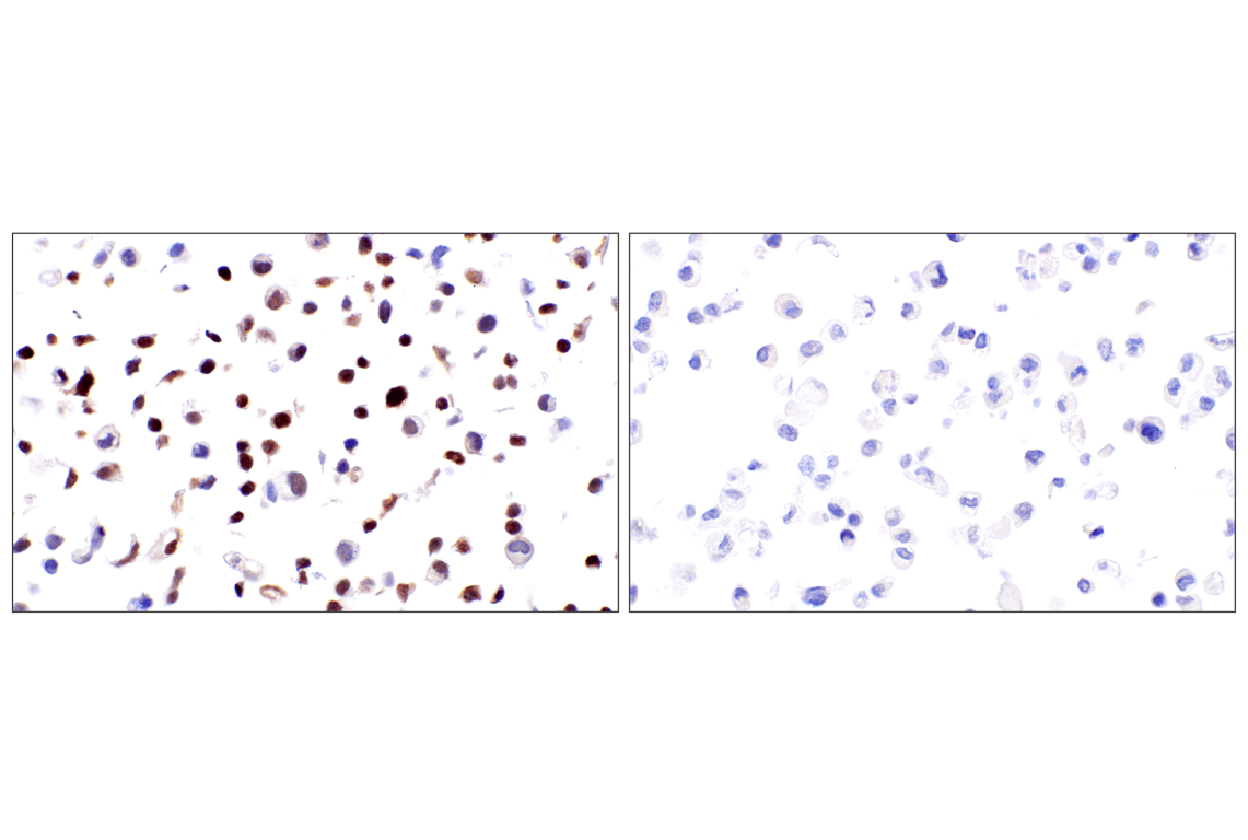
This antibody is recommended for the detection of fusion proteins containing the C-terminus region of WT1 found in desmoplastic small round cell tumors by immunohistochemistry. For the detection of wild-type WT1, it is recommended to use WT1 (D8I7F) XP® Rabbit mAb #83535.
Non-specific staining was observed in kidney, liver, and small intestine by immunohistochemistry.
Species predicted to react based on 100% sequence homology
Source / Purification
Background
WT1 has a myriad of biological functions and a host of interacting partners and target genes (6). It can behave as a transcriptional activator, or a repressor, and can act as an oncogene or a tumor suppressor (7). It exerts influence over the epigenetic landscape, and also has post-translational influence of gene expression through RNA interactions (8). The diverse biological roles of WT1 have been attributed to the existence of multiple isoforms and post-translational modifications of the protein (9).
Background References
- Royer-Pokora, B. et al. (2004) Am J Med Genet A 127A, 249-57.
- Kohsaka, T. et al. (1999) Hum Mutat 14, 466-70.
- Little, M. et al. (2000) Hum Mutat 15, 389.
- Takata, A. et al. (2000) J Med Genet 37, 698-701.
- Suri, M. et al. (2007) Am J Med Genet A 143A, 2312-20.
- Toska, E. and Roberts, S.G. (2014) Biochem J 461, 15-32.
- Yang, L. et al. (2007) Leukemia 21, 868-76.
- Weiss, T.C. and Romaniuk, P.J. (2009) Biochemistry 48, 148-55.
- Haber, D.A. et al. (1991) Proc Natl Acad Sci USA 88, 9618-22.
Species Reactivity
Species reactivity is determined by testing in at least one approved application (e.g., western blot).
Western Blot Buffer
IMPORTANT: For western blots, incubate membrane with diluted primary antibody in 5% w/v BSA, 1X TBS, 0.1% Tween® 20 at 4°C with gentle shaking, overnight.
Applications Key
W: Western Blotting W-S: Simple Western™ IHC-P: Immunohistochemistry (Paraffin)
Cross-Reactivity Key
H: Human
Trademarks and Patents
Cell Signaling Technology is a trademark of Cell Signaling Technology, Inc.
All other trademarks are the property of their respective owners. Visit cellsignal.com/trademarks for more information.
Limited Uses
Except as otherwise expressly agreed in a writing signed by a legally authorized representative of CST, the following terms apply to Products provided by CST, its affiliates or its distributors. Any Customer's terms and conditions that are in addition to, or different from, those contained herein, unless separately accepted in writing by a legally authorized representative of CST, are rejected and are of no force or effect.
Products are labeled with For Research Use Only or a similar labeling statement and have not been approved, cleared, or licensed by the FDA or other regulatory foreign or domestic entity, for any purpose. Customer shall not use any Product for any diagnostic or therapeutic purpose, or otherwise in any manner that conflicts with its labeling statement. Products sold or licensed by CST are provided for Customer as the end-user and solely for research and development uses. Any use of Product for diagnostic, prophylactic or therapeutic purposes, or any purchase of Product for resale (alone or as a component) or other commercial purpose, requires a separate license from CST. Customer shall (a) not sell, license, loan, donate or otherwise transfer or make available any Product to any third party, whether alone or in combination with other materials, or use the Products to manufacture any commercial products, (b) not copy, modify, reverse engineer, decompile, disassemble or otherwise attempt to discover the underlying structure or technology of the Products, or use the Products for the purpose of developing any products or services that would compete with CST products or services, (c) not alter or remove from the Products any trademarks, trade names, logos, patent or copyright notices or markings, (d) use the Products solely in accordance with CST Product Terms of Sale and any applicable documentation, and (e) comply with any license, terms of service or similar agreement with respect to any third party products or services used by Customer in connection with the Products.
Revision 3
Revision 3
(metastatic site) using WT1 (F3T7B) Rabbit mAb.
Revision 3