Revision 4
#99618
Store at -20C
877-616-CELL (2355)
877-678-TECH (8324)
3 Trask Lane | Danvers | Massachusetts | 01923 | USA
For Research Use Only. Not for Use in Diagnostic Procedures.
Applications:
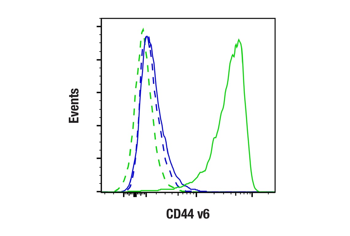
W, IHC-P, FC-L
Reactivity:
H
Sensitivity:
Endogenous
MW (kDa):
200-220
Source/Isotype:
Mouse IgG1 kappa
UniProt ID:
#P16070
Entrez-Gene Id:
960
Product Usage Information
| Application | Dilution |
|---|---|
| Western Blotting | 1:1000 |
| Immunohistochemistry (Paraffin) | 1:50 - 1:200 |
| Flow Cytometry (Live) | 1:100 - 1:400 |
Storage
Specificity/Sensitivity
Source / Purification
Background
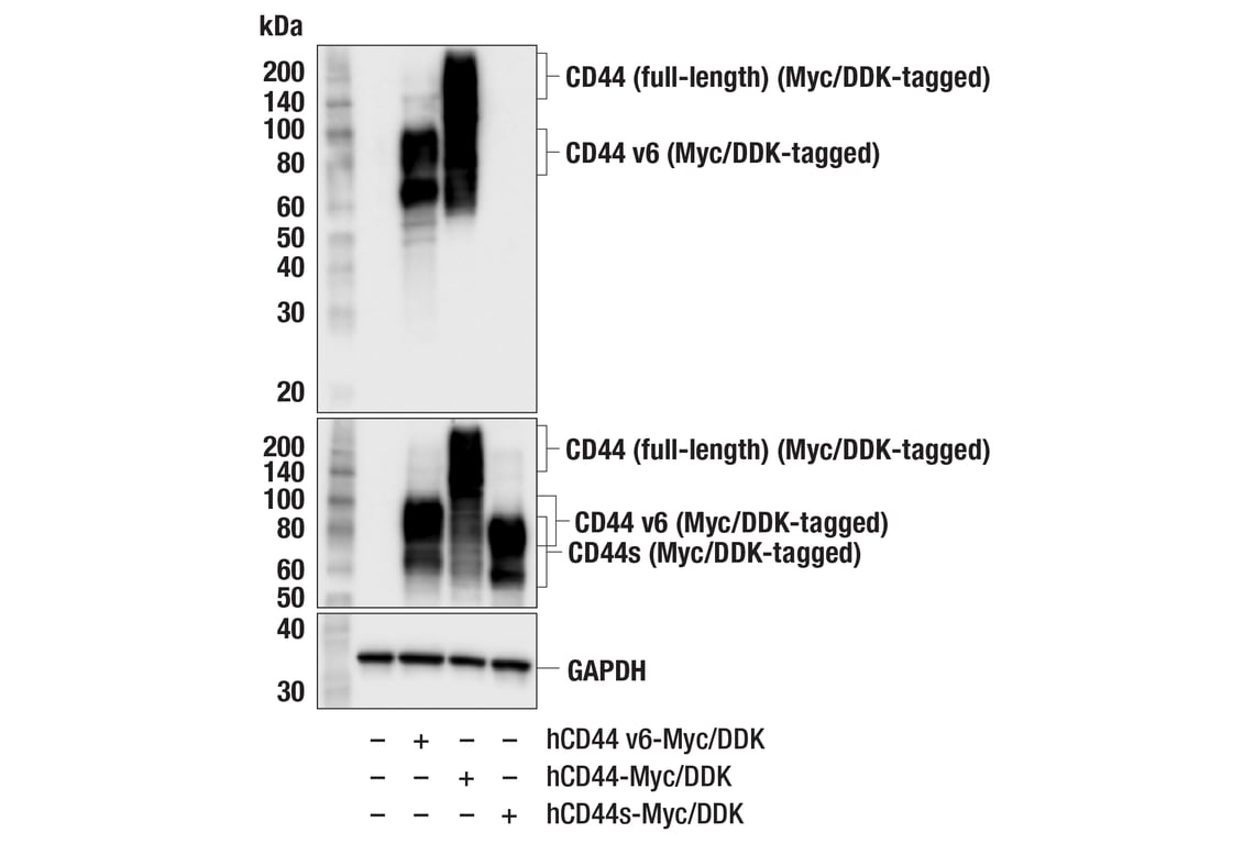
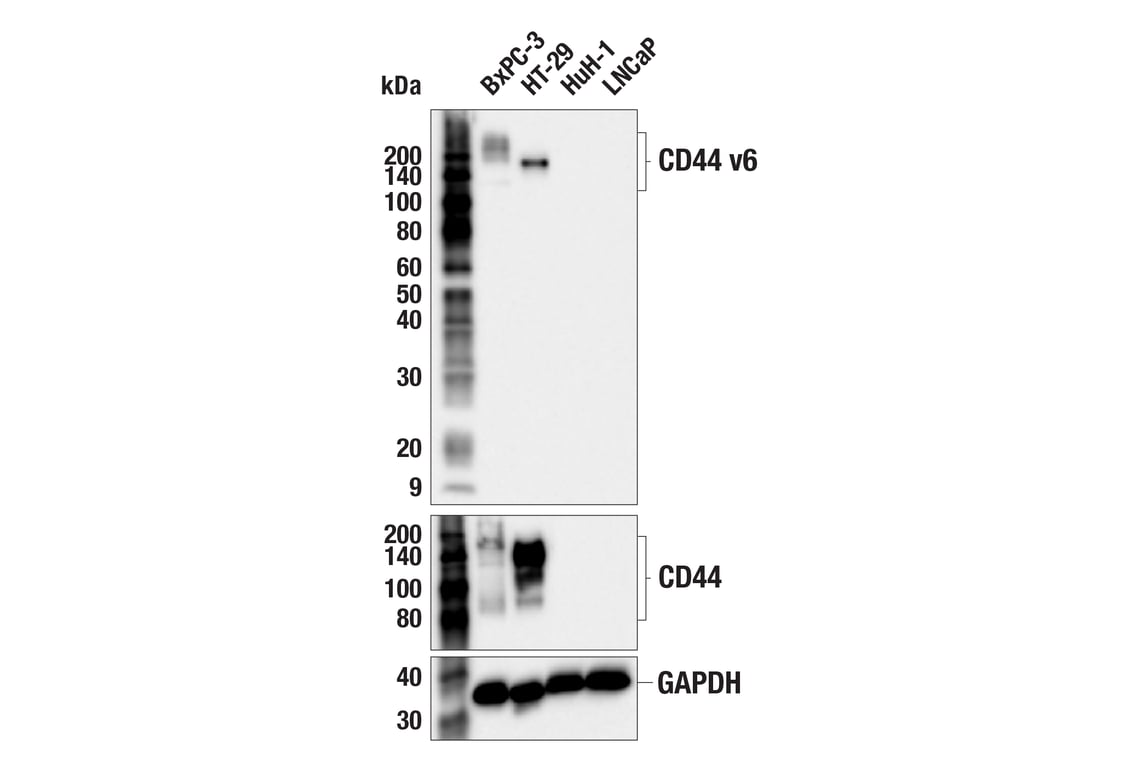
Human CD44 consists of 19 exons, of which 10 are expressed in the standard isoform (CD44s) and all other isoforms. The nine variant exons (v2-v10) inserted between the constant regions via alternative splicing are the source of CD44 heterogeneity, and can dramatically alter the cell-adhesion properties of CD44-expressing cells (7-10). Expression of CD44 isoforms containing exon v6 is associated with metastasis and poor clinical outcomes in colorectal cancer, osteosarcoma, breast cancer, head and neck squamous cell carcinoma, endometriosis, and pancreatic carcinoma (11-16).
Among pancreatic ductal adenocarcinomas (PDAC) cell lines, those that highly express CD44v, including CD44 v6, exhibit an epithelial or MET phenotype, express E-cadherin, and have an increased growth rate (9). Conversely, PDAC cells that highly express CD44s exhibit a mesenchymal phenotype, have high gemcitabine resistance, and co-express proteins associated with EMT transition, including vimentin and ZEB-1 (9). In vivo, PDAC cells have the ability to switch between expression of these CD44 isoforms in response to chemotherapy, demonstrating the importance of CD44-targeted therapies for treatment of some cancers (9).
Background References
- Goodison, S. et al. (1999) Mol. Pathol. 52, 189-196.
- Cichy, J. and Puré, E. (2003) J. Cell Biol. 161, 839-843.
- Bourguignon, L.Y. et al. (1997) J. Biol. Chem. 272, 27913-27918.
- Legg, J.W. et al. (2002) Nat. Cell Biol. 4, 399-407.
- Yonemura, S. et al. (1998) J. Cell Biol. 140, 885-895.
- Tsukita, S. et al. (1994) J. Cell Biol. 126, 391-401.
- Ejima, Ryo, et al. (2023) Int J Mol Sci. 24(4):4007
- Chen, C. et al. (2018) J Hematol Oncol 11, 64.
- Zhao, S. et al. (2016) Clin Cancer Res 22, 5592-5604.
- Rudzki, Z. and Jothy, S. (1997) Mol Pathol 50, 57-71.
- Ma, L. et al. (2019) Cell Death Dis 10, 30.
- Liang, S. et al. (2024) Future Oncol 20, 1799-1806.
- Kaufmann, M. et al. (1995) Lancet 345, 615-9.
- Athanassiou-Papaefthymiou, M. et al. (2014) Int J Immunopathol Pharmacol 27, 337-49.
- Knudtson, J.F. et al. (2020) F S Sci 1, 188-194.
- Li, Z. et al. (2014) Diagn Pathol 9, 79.
Species Reactivity
Species reactivity is determined by testing in at least one approved application (e.g., western blot).
Western Blot Buffer
IMPORTANT: For western blots, incubate membrane with diluted primary antibody in 5% w/v nonfat dry milk, 1X TBS, 0.1% Tween® 20 at 4°C with gentle shaking, overnight.
Applications Key
W: Western Blotting IHC-P: Immunohistochemistry (Paraffin) FC-L: Flow Cytometry (Live)
Cross-Reactivity Key
H: Human
Trademarks and Patents
Cell Signaling Technology is a trademark of Cell Signaling Technology, Inc.
All other trademarks are the property of their respective owners. Visit cellsignal.com/trademarks for more information.
Limited Uses
Except as otherwise expressly agreed in a writing signed by a legally authorized representative of CST, the following terms apply to Products provided by CST, its affiliates or its distributors. Any Customer's terms and conditions that are in addition to, or different from, those contained herein, unless separately accepted in writing by a legally authorized representative of CST, are rejected and are of no force or effect.
Products are labeled with For Research Use Only or a similar labeling statement and have not been approved, cleared, or licensed by the FDA or other regulatory foreign or domestic entity, for any purpose. Customer shall not use any Product for any diagnostic or therapeutic purpose, or otherwise in any manner that conflicts with its labeling statement. Products sold or licensed by CST are provided for Customer as the end-user and solely for research and development uses. Any use of Product for diagnostic, prophylactic or therapeutic purposes, or any purchase of Product for resale (alone or as a component) or other commercial purpose, requires a separate license from CST. Customer shall (a) not sell, license, loan, donate or otherwise transfer or make available any Product to any third party, whether alone or in combination with other materials, or use the Products to manufacture any commercial products, (b) not copy, modify, reverse engineer, decompile, disassemble or otherwise attempt to discover the underlying structure or technology of the Products, or use the Products for the purpose of developing any products or services that would compete with CST products or services, (c) not alter or remove from the Products any trademarks, trade names, logos, patent or copyright notices or markings, (d) use the Products solely in accordance with CST Product Terms of Sale and any applicable documentation, and (e) comply with any license, terms of service or similar agreement with respect to any third party products or services used by Customer in connection with the Products.
Revision 4
Revision 4
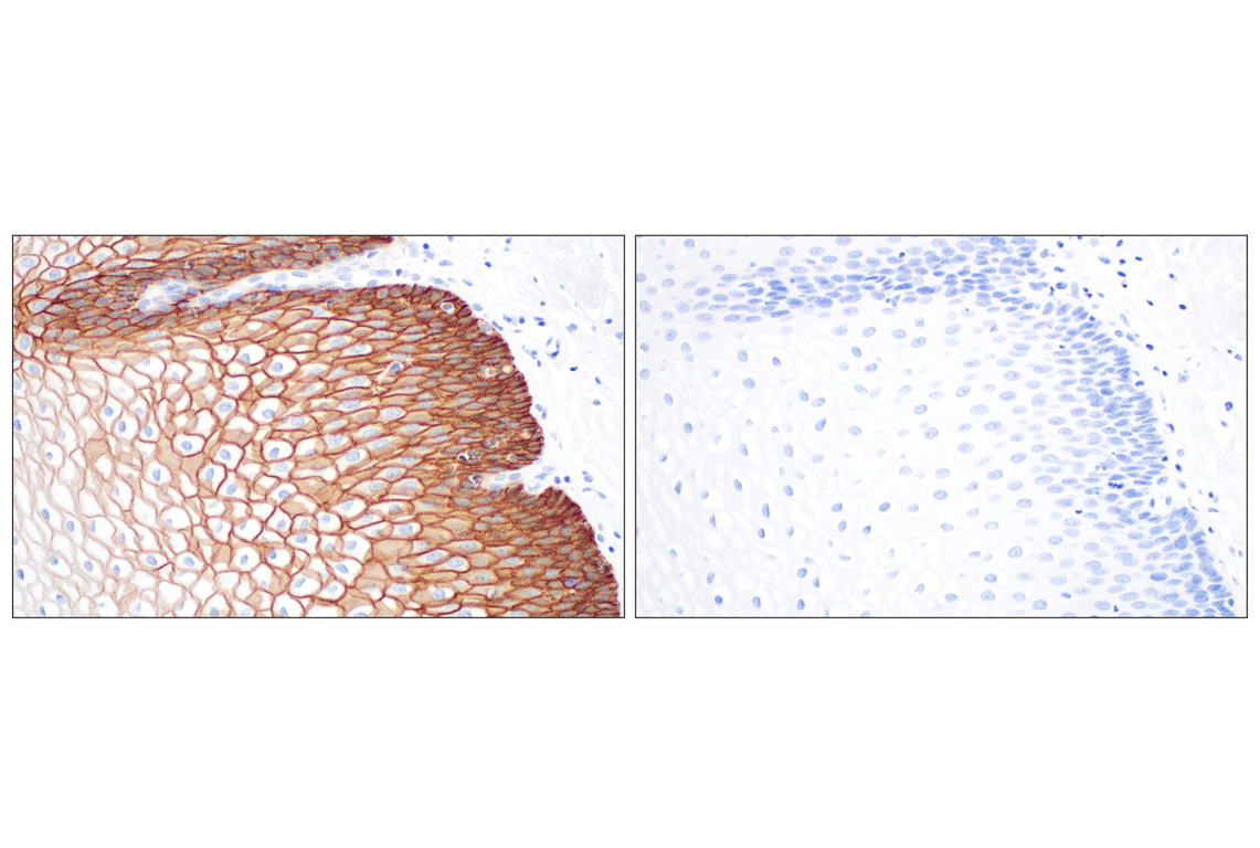
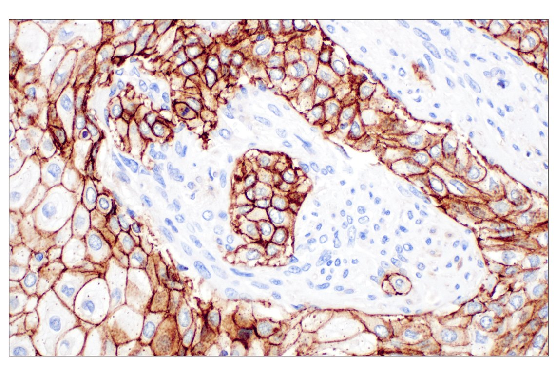
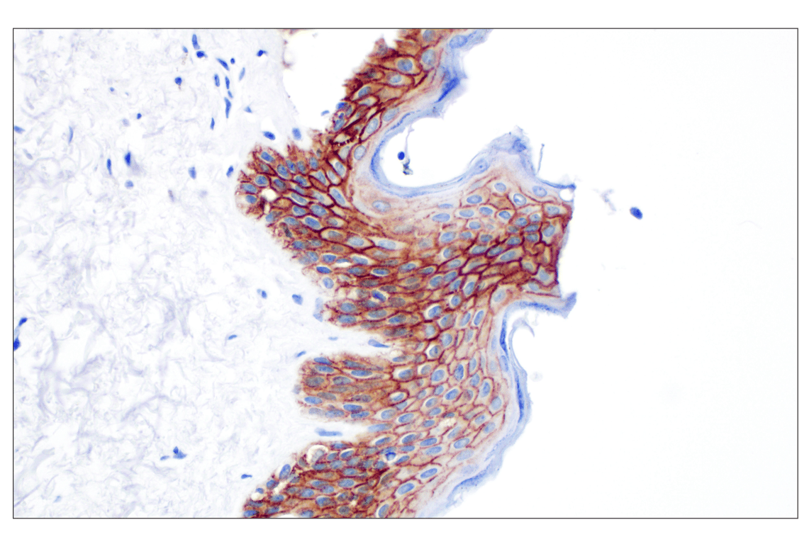
Immunohistochemical analysis of paraffin-embedded normal human skin using CD44 v6 (C44Mab-9) Mouse mAb.
Revision 4
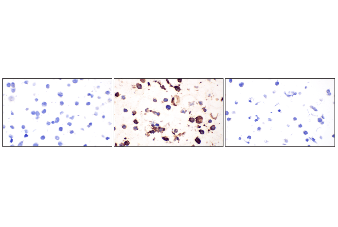
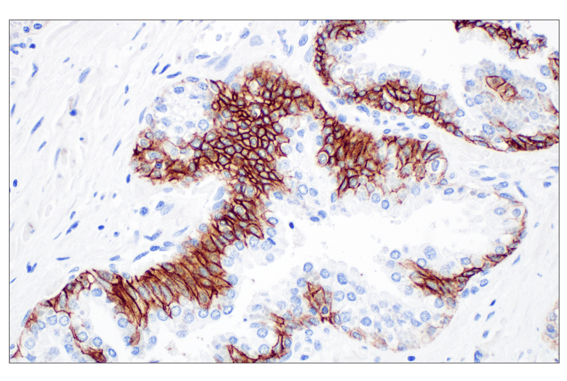
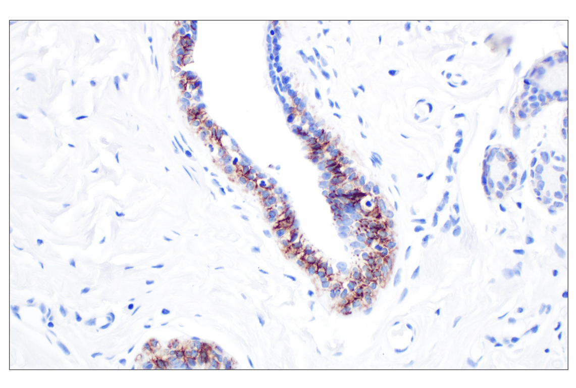
Immunohistochemical analysis of paraffin-embedded normal human breast using CD44 v6 (C44Mab-9) Mouse mAb.
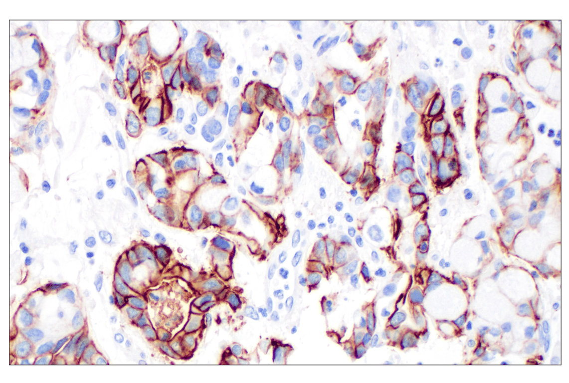
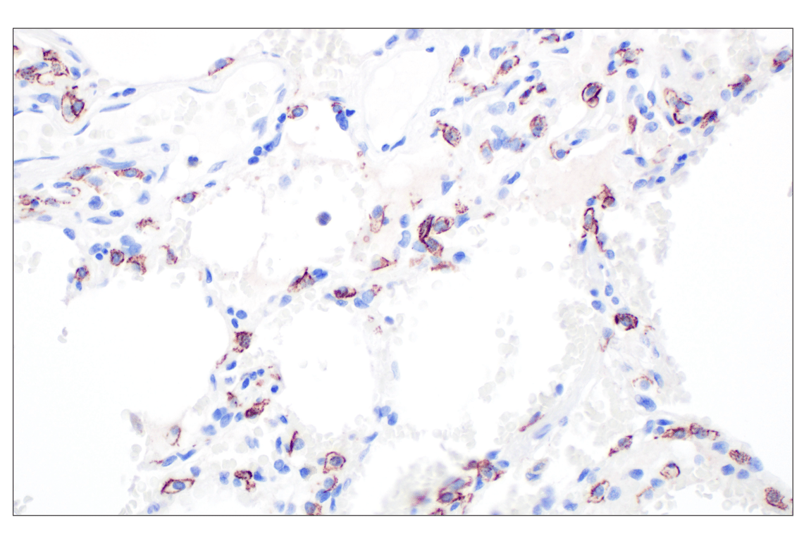
Immunohistochemical analysis of paraffin-embedded normal human lung using CD44 v6 (C44Mab-9) Mouse mAb.
Revision 4