Product Information
Chloroquine is supplied as a lyophilized powder. For a 50 mM stock, reconstitute the 150 mg in 5.82 ml sterile dH2O. First add 1 ml dH2O to the tube containing the chemical, vortex, and dispense into a new, larger tube. Repeat this action two or three more times to transfer any residual material. Add additional dH2O to the new tube to bring the volume up to 5.82 ml. Filter sterilize into sterile tube. Utilize a syringe and 0.2 μm syringe filter to minimize sample loss.
Working concentrations and length of treatment can vary depending on the desired effect, but it is typically used at 25-100 μM for 12-48 hr.
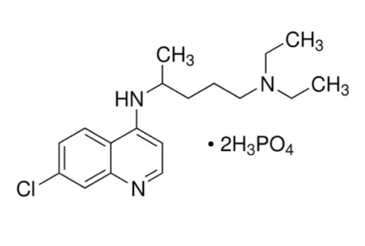
| Molecular Weight | 515.9 g/mol |
| Purity | >98% |
| Molecular Formula | C18H26ClN3•2H3PO4 |
| CAS | 50-63-5 |
| Solubility | Soluble in H2O at 25mg/ml. |
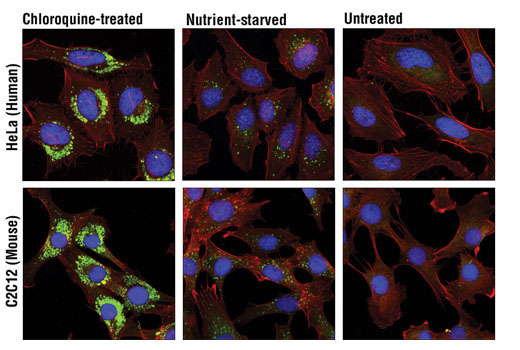
Chloroquine (CQ) is a lysosomotropic agent with an extensive range of biological effects (1). Historically known for its anti-malarial activity, chloroquine is a widely used biological research tool for studying autophagy inhibition. Research studies demonstrate that chloroquine accumulates in acidic lysosomes and increases the lysosomal pH. This inhibits lysosomal hydrolases and prevents autophagosomal fusion and degradation, which can result in apoptotic or necrotic cell death (1-4). Inhibition of chloroquine-induced apoptosis with the V-ATPase inhibitor bafilomycin A1 has been observed in several cell types (4). Chloroquine also enhances the anti-neoplastic effects of the histone deacetylase inhibitor vorinostat (SAHA) (5).
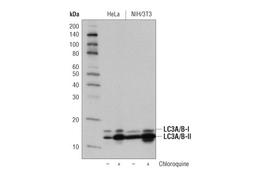
Chloroquine treatment of cells leads to accumulation of light chain 3-II (LC3-II) (1-3). This autophagy marker resides within autophagosomal membranes during the autophagic process and is degraded upon fusion with lysosomes. Chloroquine inhibition of these fusion events effectively blocks LC3-II degradation.
This product has applications to SARS-CoV-2 research into the mechanisms of the Novel Coronavirus, which has caused the COVID-19 pandemic.
Except as otherwise expressly agreed in a writing signed by a legally authorized representative of CST, the following terms apply to Products provided by CST, its affiliates or its distributors. Any Customer's terms and conditions that are in addition to, or different from, those contained herein, unless separately accepted in writing by a legally authorized representative of CST, are rejected and are of no force or effect.
Products are labeled with For Research Use Only or a similar labeling statement and have not been approved, cleared, or licensed by the FDA or other regulatory foreign or domestic entity, for any purpose. Customer shall not use any Product for any diagnostic or therapeutic purpose, or otherwise in any manner that conflicts with its labeling statement. Products sold or licensed by CST are provided for Customer as the end-user and solely for research and development uses. Any use of Product for diagnostic, prophylactic or therapeutic purposes, or any purchase of Product for resale (alone or as a component) or other commercial purpose, requires a separate license from CST. Customer shall (a) not sell, license, loan, donate or otherwise transfer or make available any Product to any third party, whether alone or in combination with other materials, or use the Products to manufacture any commercial products, (b) not copy, modify, reverse engineer, decompile, disassemble or otherwise attempt to discover the underlying structure or technology of the Products, or use the Products for the purpose of developing any products or services that would compete with CST products or services, (c) not alter or remove from the Products any trademarks, trade names, logos, patent or copyright notices or markings, (d) use the Products solely in accordance with CST Product Terms of Sale and any applicable documentation, and (e) comply with any license, terms of service or similar agreement with respect to any third party products or services used by Customer in connection with the Products.
| Cat. # | Size | Qty. | Price |
|---|---|---|---|
| 14774S | 150 mg |
|