| # | Product Name | Applications | Reactivity | |
|---|---|---|---|---|
| 4873 | HSP70 (6B3) Rat mAb |
|
H Mk |
| REACTIVITY | H M R Mk |
| SENSITIVITY | Endogenous |
| MW (kDa) | |
| SOURCE | Rabbit |
Product Information
| Application | Dilution |
|---|---|
| Flow Cytometry (Fixed/Permeabilized) | 1:50 |
All reagents required for this protocol may be efficiently purchased together in our Intracellular Flow Cytometry Kit (Methanol) #13593, or individually using the catalog numbers listed below.
NOTE: Prepare solutions with reverse osmosis deionized (RODI) or equivalent grade water.
NOTE: When including fluorescent cellular dyes in your experiment (including viability dyes, DNA dyes, etc.), please refer to the dye product page for the recommended protocol. Visit www.cellsignal.com for a full listing of cellular dyes validated for use in flow cytometry.
NOTE: Adherent cells or tissue should be dissociated and in single-cell suspension prior to fixation.
NOTE: Optimal centrifugation conditions will vary depending upon cell type and reagent volume. Generally, 150-300g for 1-5 minutes will be sufficient to pellet the cells.
NOTE: If using whole blood, lyse red blood cells and wash by centrifugation prior to fixation.
NOTE: Antibodies targeting CD markers or other extracellular proteins may be added prior to fixation if the epitope is disrupted by formaldehyde and/or methanol. The antibodies will remain bound to the target of interest during the fixation and permeabilization process. However, note that some fluorophores (including PE and APC) are damaged by methanol and thus should not be added prior to permeabilization. Conduct a small-scale experiment if you are unsure.
NOTE: Count cells using a hemocytometer or alternative method.
posted July 2009
revised June 2020
Protocol Id: 407
Human, Mouse, Rat, Monkey
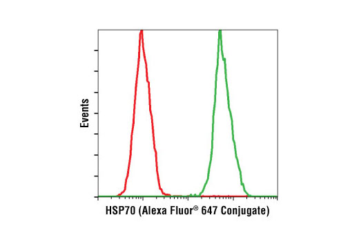
Polyclonal antibodies are produced by immunizing animals with a synthetic peptide corresponding to residues surrounding Asp69 of human HSP70. Antibodies are purified by protein A and peptide affinity chromatography. This antibody was conjugated to Alexa Fluor® 647 under optimal conditions with an F/P ratio of 2-6. The Alexa Fluor® 647 dye is maximally excited by red light (e.g. 633 nm He-Ne laser). Antibody conjugates of the Alexa Fluor® 647 dye produce bright far-red-fluorescence emission, with a peak at 665 nm.
HSP70 and HSP90 are molecular chaperones expressed constitutively under normal conditions to maintain protein homeostasis and are induced upon environmental stress (1). Both HSP70 and HSP90 are able to interact with unfolded proteins to prevent irreversible aggregation and catalyze the refolding of their substrates in an ATP- and co-chaperone-dependent manner (1). HSP70 has a broad range of substrates including newly synthesized and denatured proteins, while HSP90 tends to have a more limited subset of substrates, most of which are signaling molecules. HSP70 and HSP90 often function collaboratively in a multi-chaperone system, which requires a minimal set of co-chaperones: HSP40, Hop, and p23 (2,3). The co-chaperones either regulate the intrinsic ATPase activity of the chaperones or recruit chaperones to specific substrates or subcellular compartments (1,4). When the ubiquitin ligase CHIP associates with the HSP70/HSP90 complex as a cofactor, the unfolded substrates are subjected to degradation by the proteasome (4). The biological functions of HSP70/HSP90 extend beyond their chaperone activity. They are essential for the maturation and inactivation of nuclear hormones and other signaling molecules (1,3). They also play a role in vesicle formation and protein trafficking (2).
Explore pathways related to this product.
STRING - Known and Predicted Protein-Protein Interactions.
Except as otherwise expressly agreed in a writing signed by a legally authorized representative of CST, the following terms apply to Products provided by CST, its affiliates or its distributors. Any Customer's terms and conditions that are in addition to, or different from, those contained herein, unless separately accepted in writing by a legally authorized representative of CST, are rejected and are of no force or effect.
Products are labeled with For Research Use Only or a similar labeling statement and have not been approved, cleared, or licensed by the FDA or other regulatory foreign or domestic entity, for any purpose. Customer shall not use any Product for any diagnostic or therapeutic purpose, or otherwise in any manner that conflicts with its labeling statement. Products sold or licensed by CST are provided for Customer as the end-user and solely for research and development uses. Any use of Product for diagnostic, prophylactic or therapeutic purposes, or any purchase of Product for resale (alone or as a component) or other commercial purpose, requires a separate license from CST. Customer shall (a) not sell, license, loan, donate or otherwise transfer or make available any Product to any third party, whether alone or in combination with other materials, or use the Products to manufacture any commercial products, (b) not copy, modify, reverse engineer, decompile, disassemble or otherwise attempt to discover the underlying structure or technology of the Products, or use the Products for the purpose of developing any products or services that would compete with CST products or services, (c) not alter or remove from the Products any trademarks, trade names, logos, patent or copyright notices or markings, (d) use the Products solely in accordance with CST Product Terms of Sale and any applicable documentation, and (e) comply with any license, terms of service or similar agreement with respect to any third party products or services used by Customer in connection with the Products.