View Featured Offers >>
8663
Cellular Localization Alexa Fluor® 647 Conjugated Antibody Sampler Kit
Antibody Conjugates
Antibody Sampler Kit

Cellular Localization Alexa Fluor® 647 Conjugated Antibody Sampler Kit #8663

Citations (0)
Confocal immunofluorescent analysis of C2C12 cells using GAPDH (14C10) Rabbit mAb (Alexa Fluor® 647 Conjugate) #3907 (pseudocolor blue, left) compared to concentration matched Rabbit (DA1E) mAb IgG XP® Isotype Control (Alexa Fluor® 647 Conjugate (pseudocolor blue, right). Red = Propidium Iodide (PI)/RNase Staining Solution #4087 (fluorescent DNA dye).
Confocal immunofluorescent analysis of HeLa cells using β-Tubulin (9F3) Rabbit mAb (Alexa Fluor® 647 Conjugate) (blue pseudocolor). Red = Propidium Iodide (fluorescent DNA dye).
Confocal immunofluorescent analysis of HeLa cells using COX IV (3E11) Rabbit mAb (Alexa Fluor® 647 Conjugate) (blue pseudocolor). Actin filaments were labeled with Alexa Fluor® 488 phalloidin (green). Red = Propidium Iodide (PI)/RNase Staining Solution #4087.
Confocal immunofluorescent analysis of NIH/3T3 cells using PDI (C81H6) Rabbit mAb (Alexa Fluor® 647 Conjugate) (blue pseudocolor) and Alexa Fluor® 488 phalloidin (green). Red = Propidium Iodide (PI)/RNase Staining Solution #4087 (fluorescent DNA dye).
Confocal immunofluorescent analysis of COS-7 cells using β-Actin (13E5) Rabbit mAb (Alexa Fluor® 647 Conjugate) (blue pseudocolor). Red = Propidium Iodide (PI)/RNase Staining Solution #4087.
Flow cytometric analysis of Jurkat cells, using Rabbit (DA1E) mAb IgG XP® Isotype Control (Alexa Fluor® 647 Conjugate) (red) compared to Phospho-Akt (Ser473) (D9E) XP® Rabbit mAb (Alexa Fluor® 647 Conjugate) #4075, untreated (green) or LY294002- and wortmannin-treated (blue).
Flow cytometric analysis of Hela cells using β-Tubulin (9F3) Rabbit mAb (Alexa Fluor® 647 Conjugate) (solid line) or concentration-matched Rabbit (DA1E) mAb IgG XP® Isotype Control (Alexa Fluor® 647 Conjugate) #2985 (dashed line).
Flow cytometric analysis of HeLa cells using COX IV (3E11) Rabbit mAb (Alexa Fluor® 647 Conjugate) (solid line) compared to concentration-matched Rabbit (DA1E) mAb IgG XP® Isotype Control (Alexa Fluor® 647 Conjugate) #2985 (dashed line).
Flow cytometric analysis of Hela cells using β-Actin (13E5) Rabbit mAb (Alexa Fluor® 647 Conjugate) (solid line) compared to concentration-matched Rabbit (DA1E) mAb IgG XP® Isotype Control (Alexa Fluor® 647 Conjugate) #2985 (dashed line).
Flow cytometric analysis of Jurkat cells, using Rabbit (DA1E) mAb IgG XP® Isotype Control (Alexa Fluor® 647 Conjugate (red) compared to Phospho-S6 Ribosomal Protein (Ser235/236) (D57.2.2E) XP® Rabbit mAb (Alexa Fluor® 647 Conjugate) #4851, untreated (blue) or IFN-α-treated (green).
Inquiry Info.# 8663

Kit Usage Information

Protocols

Product Description

The Cellular Localization Alexa Fluor® 647 Conjugated Antibody Sampler Kit provides an economical means for identification of cellular organelles without the need for a fluorescent secondary antibody. This kit includes enough primary antibody to perform at least 20 IF-IC tests per antibody.

Specificity / Sensitivity

β-Actin (13E5) Rabbit mAb (Alexa Fluor® 647 Conjugate) recognizes endogenous levels of total β-actin protein. This antibody may cross-react with the γ-actin (cytoplasmic isoform), but it does not cross-react with α-skeletal, α-cardiac, α-vascular smooth, or γ-enteric smooth muscle isoforms. COX IV (3E11) Rabbit mAb (Alexa Fluor® 647 Conjugate) recognizes endogenous levels of total COX IV protein. PDI (C81H6) Rabbit mAb (Alexa Fluor® 647 Conjugate) recognizes endogenous levels of total PDI protein. β-Tubulin (9F3) Rabbit mAb (Alexa Fluor® 647 Conjugate) recognizes endogenous levels of total β-tubulin protein and does not cross-react with recombinant α-tubulin.

Source / Purification

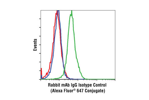
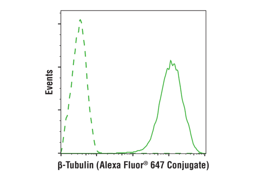
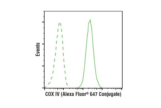
Monoclonal antibodies are produced by immunizing animals with a synthetic peptide corresponding to residues near the amino terminus of human β-actin protein, residues surrounding Lys29 of human COX IV protein, the sequence of human PDI protein, or the amino terminus of human β-tubulin protein.

Background

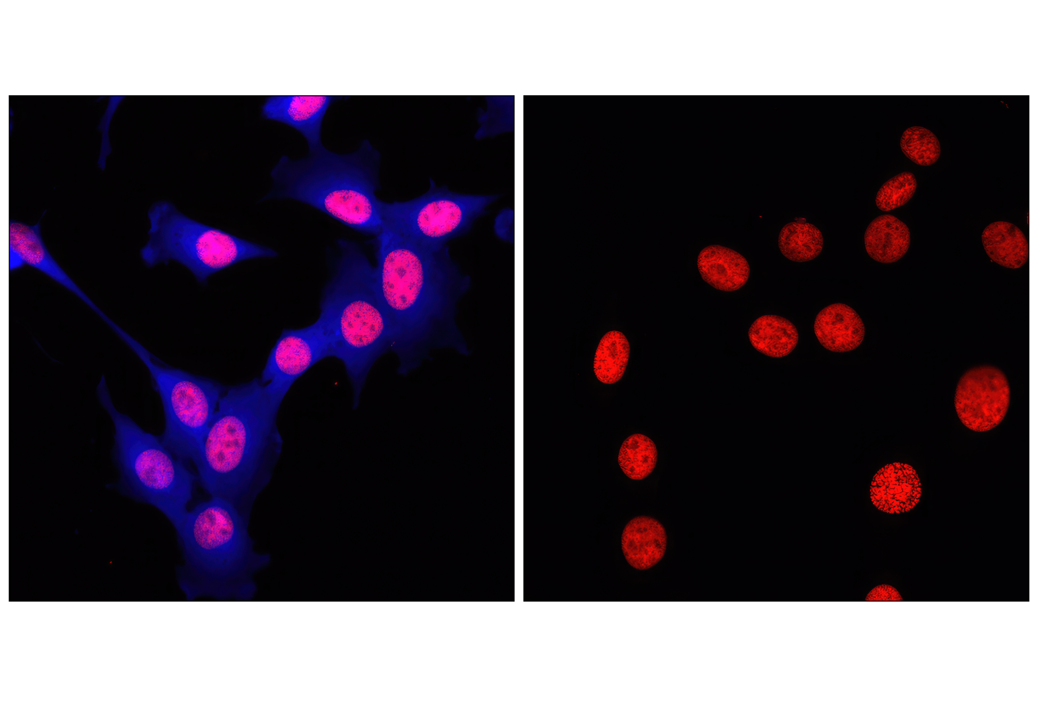
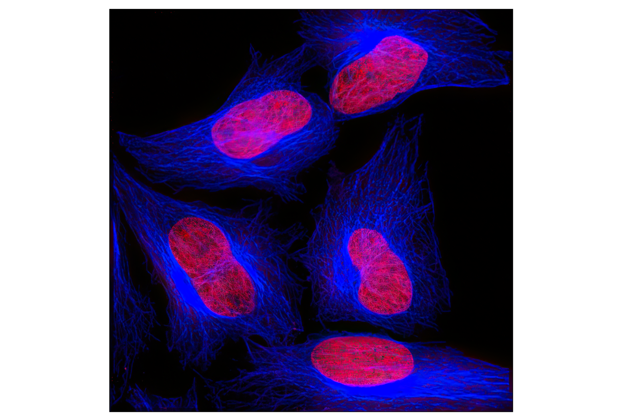
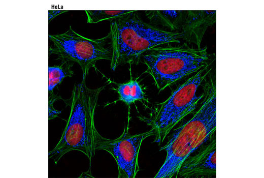
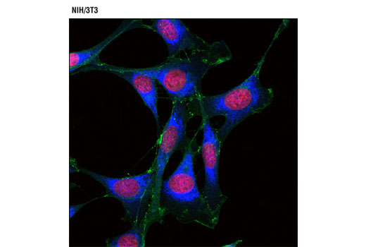
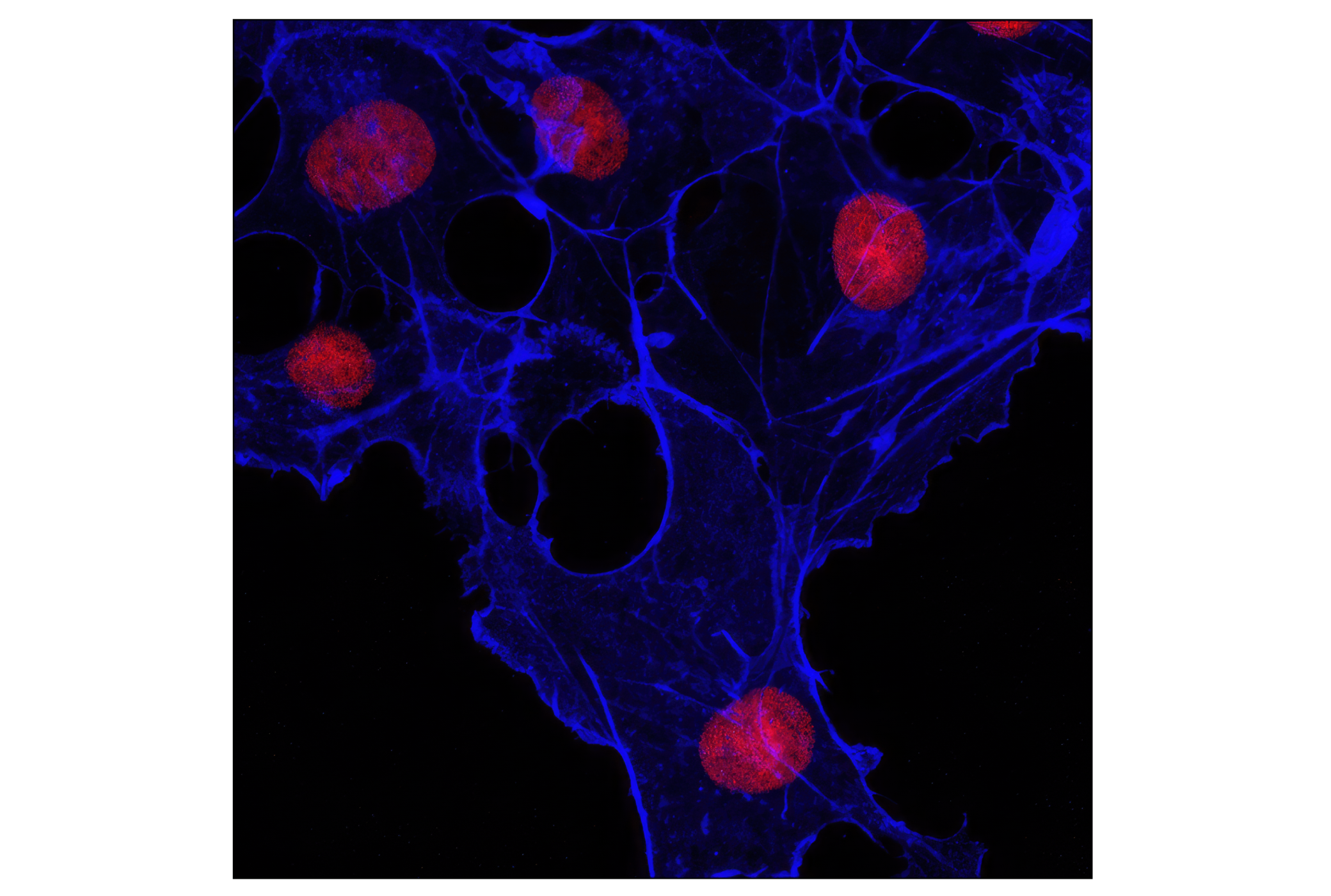
Knowledge of the subcellular location of a protein may reveal the potential role it plays in the cell. The subcellular location of a protein of interest may be confirmed by colocalization with one of the organelle-specific antibodies in this kit.
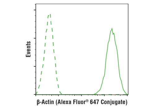
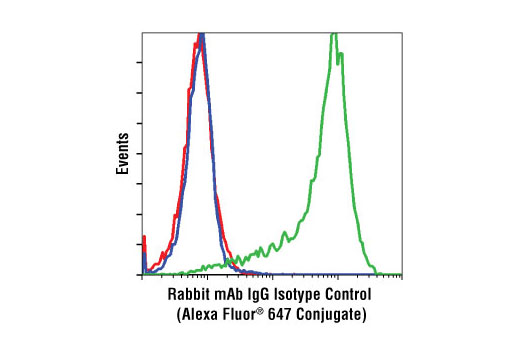
β-Actin and β-tubulin are ubiquitous eukaryotic proteins that form the major component of the cellular cytoskeleton (1,2). Cytochrome c oxidase (COX) IV is part of the hetero-oligomeric enzyme found in the inner mitochondrial membrane (3). Protein disulfide isomerase (PDI) catalyzes the formation and isomerization of disulfide bonds in the endoplasmic reticulum (4).

Limited Uses

Except as otherwise expressly agreed in a writing signed by a legally authorized representative of CST, the following terms apply to Products provided by CST, its affiliates or its distributors. Any Customer's terms and conditions that are in addition to, or different from, those contained herein, unless separately accepted in writing by a legally authorized representative of CST, are rejected and are of no force or effect.

Products are labeled with For Research Use Only or a similar labeling statement and have not been approved, cleared, or licensed by the FDA or other regulatory foreign or domestic entity, for any purpose. Customer shall not use any Product for any diagnostic or therapeutic purpose, or otherwise in any manner that conflicts with its labeling statement. Products sold or licensed by CST are provided for Customer as the end-user and solely for research and development uses. Any use of Product for diagnostic, prophylactic or therapeutic purposes, or any purchase of Product for resale (alone or as a component) or other commercial purpose, requires a separate license from CST. Customer shall (a) not sell, license, loan, donate or otherwise transfer or make available any Product to any third party, whether alone or in combination with other materials, or use the Products to manufacture any commercial products, (b) not copy, modify, reverse engineer, decompile, disassemble or otherwise attempt to discover the underlying structure or technology of the Products, or use the Products for the purpose of developing any products or services that would compete with CST products or services, (c) not alter or remove from the Products any trademarks, trade names, logos, patent or copyright notices or markings, (d) use the Products solely in accordance with CST Product Terms of Sale and any applicable documentation, and (e) comply with any license, terms of service or similar agreement with respect to any third party products or services used by Customer in connection with the Products.

For Research Use Only. Not for Use in Diagnostic Procedures.
Cell Signaling Technology is a trademark of Cell Signaling Technology, Inc.
All other trademarks are the property of their respective owners. Visit our Trademark Information page.