View Featured Offers >>
8815
Active Rac1 Detection Kit
Cellular Assay Kits
Assay Kit

Active Rac1 Detection Kit #8815

Citations (0)
Figure 2. The GTP-bound GTPase pull-down process can be divided into 3 steps as shown. Step 1: Mix sample, binding protein, and glutathione resin in the spin cup and incubate at 4ºC to allow GTP-bound GTPase binding to the glutathione resin through GST-linked binding protein. Step 2: Remove unbound proteins by centrifugation. Step 3: Elute glutathione resin-bound GTPase with SDS buffer. The eluted sample can then be analyzed by western blot.
Figure 1. NIH/3T3 cell lysates (500 µl at 1 mg/ml) were treated in vitro with GTPγS or GDP to activate or inactivate Rac1 (refer to optional step C in protocol). The lysates were then incubated with glutathione resin and GST-PAK1-PBD (lanes 2 and 3). GTPγS-treated lysate was also incubated without GST-PAK1-PBD in the presence of glutathione resin as a negative control (lane 4). Western blot analysis of cell lysate (20 µg, lane 1) or 20 µl of the eluted samples (lanes 2, 3, and 4) was performed using a Rac1 Mouse mAb. Anti-mouse IgG, HRP-linked Antibody #7076 was used as the secondary antibody.
To Purchase # 8815
Cat. # Size Qty. Price
8815S
1 Kit  (30 immunoprecipitations)

Product Includes Quantity (with Count) Storage Temp
GST-Human PAK1-PBD 1 x 600 µg -20°C
Rac1 Mouse Antibody 1 x 50 µl -20°C
GDP 1 x 50 µl -80°C
GTP γS 1 x 50 µl -80°C
Glutathione Resin 1 x 3 ml +4°C
SDS Sample Buffer 1 x 1.5 ml +4°C
Lysis/Binding/Wash Buffer 1 x 100 ml +4°C
Spin Cup and Collection Tubes 1 x 30 ea RT

Storage

GTPγS: Store at -80°C
GDP: Store at -80°C
GST-Human PAK1-PBD: Store at -20°C
Rac1 Mouse mAb: Store at -20°C
Lysis/Binding/Wash Buffer: Store at 4°C
Glutathione Resin: Store at 4°C
SDS Sample Buffer: Store at 4°C
Spin Cup and Collection Tubes: Store at RT

Protocol

PRINT

View >Collapse >

#8815 Active Rac1 Detection Kit Protocol

Additional Materials Required

  • Phenylmethanesulfonyl fluoride (PMSF) #8553
  • Blue Loading Buffer Pack #7722
  • Tris Buffered Saline with Tween® 20 (TBST-10X) #9997
  • Primary antibody dilution buffer: 1X TBST 5% BSA
  • Bovine Serum Albumin (BSA) #9998
  • Nonfat Dry Milk #9999
  • Color-coded Prestained Protein Marker, Broad Range (10-250 kDa) #74124
  • Biotinylated Protein Ladder Detection Pack #7727
  • Anti-mouse IgG, HRP-linked Antibody #7076
  • 20X LumiGLO® Reagent and 20X Peroxide #7003
  • 0.5 M EDTA, pH 8.0 #7011
  • 1 M MgCl2

A. Solutions and Reagents

  • NOTE: Prepare solutions with ddH2O or equivalently purified water.
  • 1X Phosphate Buffered Saline (PBS)
  • 1X Cell Lysis/Binding/Wash Buffer
  • NOTE: Add 1 mM PMSF immediately prior to use.

B. Cell Lysis

  1. To harvest cells under nondenaturing conditions, remove media and rinse cells once with ice-cold PBS. For non-adherent cells, pellet cells at 100xg for 5 min and resuspend cells in 10 ml ice-cold PBS.
  2. Remove PBS and add 0.5 ml ice-cold 1X Lysis/Binding/Wash Buffer plus 1 mM PMSF to each plate (10 cm in diameter), or cell pellets from each 75 cm2 flask (approx 1-2 x 10⁷ cells).
  3. Scrape cells off the plate and transfer to an appropriate tube. For non-adherent cells, just resuspend the pellet.
  4. Votex the tube briefly and incubate on ice for 5 min.
  5. Microcentrifuge at 16,000xg at 4°C for 15 min and transfer the supernatant to a new tube. The supernatant is the cell lysate. Fresh cell lysates are recommended for GTPase assays. Lysate protein concentration can be determined using BCA protein assay.
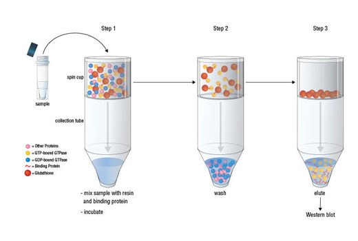
  6. NOTE: We recommend making 1 mg/ml lysate in 1X Lysis/Binding/Wash Buffer for the following steps.

C. In vitro GTPγS or GDP Treatment (Optional)

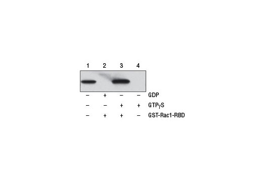
Perform the following treatments, GTPγS (positive control) and GDP (negative control), to ensure the immunoprecipitation procedures are working properly. Use 500 µg of cell lysate for each treatment. For best results, aliquot GTPγS and GDP at first use to minimize freeze/thaw cycles.

  1. For 500 µl lysate, add 10 µl 0.5 M EDTA pH 8.0 (for a final concentration of 10 mM), vortex the sample.
  2. Add 5 µl of 10 mM GTPγS (for a final concentration of 0.1 mM) or 5 µl 100 mM GDP (for a final concentration of 1 mM), vortex the sample.
  3. Incubate the mixture at 30°C for 15 min with constant agitation.
  4. Terminate the reaction by placing the sample on ice and adding 32 µl of 1 M MgCl2 (for a final concentration of 60 mM), vortex the sample.

D. Affinity Precipitation of Activated G protein

  1. Insert a spin cup into a collection tube for each sample.
  2. Swirl the bottle of glutathione resin to thoroughly resuspend the agarose beads. Add 100 µl of the 50% resin slurry to the spin cup with collection tube. Centrifuge the tubes at 6,000xg for 10-30 sec.
  3. Discard the flow-through. Add 400 µl of 1X Lysis/Binding/Wash Buffer to each spin cup with resin. Invert the tubes gently several times. Centrifuge the tubes at 6,000xg for 10-30 sec. Discard the flow-through.
  4. Thaw the GST-PAK1-PBD on ice and immediately make 20 µg aliquots. Store aliquots for later use at -80°C.
  5. Add 20 µg of GST-PAK1-PBD to the spin cup containing the glutathione resin.
  6. Immediately transfer up to 700 µl of the cell lysate (containing at least 500 µg of total protein) to the spin cup, close the cap and vortex the sample.
  7. Seal cap of the collection tube with laboratory film to prevent leakage, which may result from the presence of detergent in the lysate, vortex the sample.
  8. Incubate the reaction mixture at 4°C for 1 hr with gentle rocking.
  9. Centrifuge the spin cup with collection tube at 6,000xg for 10-30 sec.
  10. Remove the laboratory film and transfer the spin cup to a new collection tube.
  11. To wash resin, add 400 µl of 1X Cell Lysis/Binding/Wash Buffer, invert the tube three times, and centrifuge at 6,000xg for 10-30 sec. Discard the buffer. Repeat this wash step two additional times.
  12. Transfer the spin cup to a new collection tube.
  13. Prepare 50 µl of reducing sample buffer for each pull-down reaction by adding dithiothreitol (DTT) to 2X SDS Sample Buffer to a final concentration of 200 mM.
  14. Add 50 µl 2X reducing sample buffer to the resin. Vortex the sample and incubate at room temperature for 2 min.
  15. Centrifuge the tube at 6,000xg for 2 min. Remove and discard the spin cup containing the resin.
  16. Heat the eluted samples for 5 min at 95-100°C. Samples may be electrophoresed on a gel or stored at -20°C for future use.

E. Western blot analysis

    NOTE: Volumes are for 10 cm x 10 cm (100 cm2) of membrane; for different sized membranes, adjust volumes accordingly.

  1. Load 20-25 µl onto SDS-PAGE gel (10 cm x 10 cm).
  2. NOTE: CST recommends loading prestained molecular weight markers (#74124, 10 µl/lane) to verify electrotransfer and biotinylated protein ladder (#7727, 10 µl/lane) to determine molecular weights. 4-20% acrylamide or Tris-glycine gel provides best separation.

  3. Electrotransfer to nitrocellulose membrane.
  4. After transfer, wash nitrocellulose membrane with 25 ml TBST for 5 min at room temperature.
  5. Incubate membrane in 25 ml of blocking buffer (TBS containing 0.1% Tween-20 and 5% BSA) for 1 hr at room temperature.
  6. Wash three times for 5 min each with 15 ml of 1X TBST.
  7. Incubate membrane with Rac1 Mouse mAb (1:1000 dilution) in 10 ml primary antibody dilution buffer with gentle agitation overnight at 4°C.
  8. Wash three times for 5 min each with 15 ml of 1X TBST.
  9. Incubate membrane with Anti-mouse IgG, HRP-linked antibody (1:2000, #7076) and HRP-conjugated anti-biotin antibody (1:1000, #7075) to detect biotinylated protein markers in 10 ml of 1X TBST containing 5% milk with gentle agitation for 1 hour at room temperature.
  10. Wash three times for 5 min each with 15 ml of 1X TBST.
  11. Incubate membrane with 10 ml LumiGLO® (0.5 ml 20X LumiGLO®, 0.5 ml 20X Peroxide, #7003, and 9.0 ml dH20 water) with gentle agitation for 1 min at room temperature.
  12. NOTE: LumiGLO® substrate can be further diluted if signal response is too fast.

  13. Drain membrane of excess developing solution (do not let dry), wrap in plastic wrap and expose to light film. An initial 10-sec exposure should indicate the proper exposure time.
  14. NOTE: Due to the kinetics of the detection reaction, signal is most intense immediately following LumiGLO® incubation and declines over the following 2 hr.

LumiGLO® is a registered trademark of Kirkegaard & Perry Laboratories.

posted June 2020

Protocol Id: 2045

Product Description

The Active Rac1 Detection Kit provides all reagents necessary for measuring activation of Rac1 GTPase in the cell. GST-PAK1-PBD fusion protein is used to bind the activated form of GTP-bound Rac1, which can then be immunoprecipitated with glutathione resin. Rac1 activation levels are then determined by western blot using a Rac1 Mouse mAb.

Specificity / Sensitivity

Active Rac1 Detection Kit detects endogenous levels of GTP-bound (active) Rac1 as shown in Figure 1. This kit detects proteins from the indicated species, as determined through in-house testing, but may also detect homologous proteins from other species.

Species Reactivity:

Human, Mouse

Background

The Ras superfamily of small GTP-binding proteins (G proteins) comprise a large class of proteins (over 150 members) that can be classified into at least five families based on their sequence and functional similarities: Ras, Rho, Rab, Arf, and Ran (1-3). These small G proteins have both GDP/GTP-binding and GTPase activities and function as binary switches in diverse cellular and developmental events that include cell cycle progression, cell survival, actin cytoskeletal organization, cell polarity and movement, and vesicular and nuclear transport (1). An upstream signal stimulates the dissociation of GDP from the GDP-bound form (inactive), which leads to the binding of GTP and formation of the GTP-bound form (active). The activated G protein then goes through a conformational change in its downstream effector-binding region, leading to the binding and regulation of downstream effectors. This activation can be switched off by the intrinsic GTPase activity, which hydrolyzes GTP to GDP and releases the downstream effectors. These intrinsic guanine nucleotide exchange and GTP hydrolysis activities of Ras superfamily proteins are also regulated by guanine nucleotide exchange factors (GEFs) that promote formation of the active GTP-bound form and GTPase activating proteins (GAPs) that return the GTPase to its GDP-bound inactive form (4).
Rac and Cdc42 are members of the Rho-GTPase family. In mammals, Rac exists as three isoforms, Rac1, Rac2, and Rac3, which are highly similar in sequence. Rac1 and Cdc42, the most widely studied of this group, are ubiquitously expressed. Rac2 is expressed in cells of hematopoietic origin, and Rac3, while highly expressed in brain, is also found in many other tissues. Rac and Cdc42 play key signaling roles in cytoskeletal reorganization, membrane trafficking, transcriptional regulation, cell growth, and development (5). GTP binding stimulates the activity of Rac/Cdc42, and the hydrolysis of GTP to GDP through the protein's intrinsic GTPase activity, rendering it inactive. GTP hydrolysis is aided by GTPase activating proteins (GAPs), while exchange of GDP for GTP is facilitated by guanine nucleotide exchange factors (GEFs). Another level of regulation is achieved through the binding of RhoGDI, a guanine nucleotide dissociation inhibitor, which retains Rho family GTPases, including Rac and Cdc42, in their inactive GDP-bound state (6,7).

Pathways

Explore pathways related to this product.

Limited Uses

Except as otherwise expressly agreed in a writing signed by a legally authorized representative of CST, the following terms apply to Products provided by CST, its affiliates or its distributors. Any Customer's terms and conditions that are in addition to, or different from, those contained herein, unless separately accepted in writing by a legally authorized representative of CST, are rejected and are of no force or effect.

Products are labeled with For Research Use Only or a similar labeling statement and have not been approved, cleared, or licensed by the FDA or other regulatory foreign or domestic entity, for any purpose. Customer shall not use any Product for any diagnostic or therapeutic purpose, or otherwise in any manner that conflicts with its labeling statement. Products sold or licensed by CST are provided for Customer as the end-user and solely for research and development uses. Any use of Product for diagnostic, prophylactic or therapeutic purposes, or any purchase of Product for resale (alone or as a component) or other commercial purpose, requires a separate license from CST. Customer shall (a) not sell, license, loan, donate or otherwise transfer or make available any Product to any third party, whether alone or in combination with other materials, or use the Products to manufacture any commercial products, (b) not copy, modify, reverse engineer, decompile, disassemble or otherwise attempt to discover the underlying structure or technology of the Products, or use the Products for the purpose of developing any products or services that would compete with CST products or services, (c) not alter or remove from the Products any trademarks, trade names, logos, patent or copyright notices or markings, (d) use the Products solely in accordance with CST Product Terms of Sale and any applicable documentation, and (e) comply with any license, terms of service or similar agreement with respect to any third party products or services used by Customer in connection with the Products.

For Research Use Only. Not for Use in Diagnostic Procedures.
Cell Signaling Technology is a trademark of Cell Signaling Technology, Inc.
All other trademarks are the property of their respective owners. Visit our Trademark Information page.