| Cat. # | Size | Qty. | Price |
|---|---|---|---|
| 5492S | 1 Kit (500 assays (96 well format)) |
|
| Product Includes | Quantity (with Count) | Solution Color | |||
|---|---|---|---|---|---|
| BrdU | 1 x 150 µl | ||||
| Fixing/denaturing Solution | 2 x 25 ml | ||||
| BrdU Mouse Detection mAb | 1 x 500 µl | Green | |||
| Anti-mouse IgG, HRP-Linked Antibody | 1 x 500 µl | Red | |||
| Detection Antibody Diluent | 1 x 50 ml | Green | |||
| HRP Diluent | 1 x 50 ml | Red | |||
| Luminol/Enhancer Solution | 1 x 25 ml | ||||
| Stable Peroxide Buffer | 1 x 25 ml | ||||
| 20X Wash Buffer | 1 x 50 ml |
Product Information
posted October 2012
Protocol Id: 8
All Species Expected
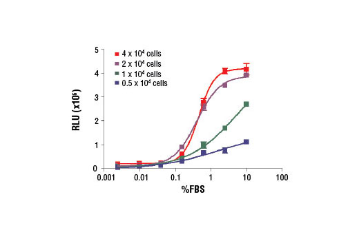
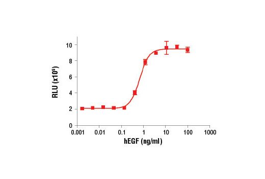
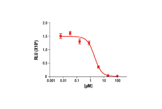
Halogenated nucleotides such as the pyrimidine analog bromodeoxyuridine (BrdU) are useful for labeling nascent DNA in living cells and tissues. BrdU becomes incorporated into replicating DNA in place of thymidine and subsequent immunodetection of BrdU using specific monoclonal antibodies allows labeling of cells in S phase of the cell cycle. After pulse-labeling cells or tissues with bromodeoxyuridine, BrdU (Bu20a) Mouse mAb can be used to detect BrdU incorporated into single stranded DNA. Please see our detailed protocol for information regarding the labeling procedure and denaturation of double stranded DNA for various immunodetection applications (1-4).
Except as otherwise expressly agreed in a writing signed by a legally authorized representative of CST, the following terms apply to Products provided by CST, its affiliates or its distributors. Any Customer's terms and conditions that are in addition to, or different from, those contained herein, unless separately accepted in writing by a legally authorized representative of CST, are rejected and are of no force or effect.
Products are labeled with For Research Use Only or a similar labeling statement and have not been approved, cleared, or licensed by the FDA or other regulatory foreign or domestic entity, for any purpose. Customer shall not use any Product for any diagnostic or therapeutic purpose, or otherwise in any manner that conflicts with its labeling statement. Products sold or licensed by CST are provided for Customer as the end-user and solely for research and development uses. Any use of Product for diagnostic, prophylactic or therapeutic purposes, or any purchase of Product for resale (alone or as a component) or other commercial purpose, requires a separate license from CST. Customer shall (a) not sell, license, loan, donate or otherwise transfer or make available any Product to any third party, whether alone or in combination with other materials, or use the Products to manufacture any commercial products, (b) not copy, modify, reverse engineer, decompile, disassemble or otherwise attempt to discover the underlying structure or technology of the Products, or use the Products for the purpose of developing any products or services that would compete with CST products or services, (c) not alter or remove from the Products any trademarks, trade names, logos, patent or copyright notices or markings, (d) use the Products solely in accordance with CST Product Terms of Sale and any applicable documentation, and (e) comply with any license, terms of service or similar agreement with respect to any third party products or services used by Customer in connection with the Products.