View Featured Offers >>
13859
Cellular Glutathione Detection Assay Kit
Cellular Assay Kits
Assay Kit

Cellular Glutathione Detection Assay Kit #13859

Citations (0)
Figure 1. Reduced Glutathione Standard was diluted in Tris Assay Buffer and samples were assayed to create a standard curve. This standard curve is for demonstration purposes only; in order to accurately determine glutathione levels users should generate a standard curve for each sample set.
Figure 2. Raw 264.7, HeLa, and Jurkat cells were seeded in 96-well plates at various cell densities. Cellular glutathione levels were determined using the Cellular Glutathione Detection Assay Kit using both cell extracts (upper) and live cells (lower).
Figure 3. The relationship between the protein concentration of lysates from HeLa Cells, untreated and treated with Staurosporine #9953 (2.0 μΜ), and RFU as determined by the Cellular Glutathione Detection Assay Kit is shown.
Figure 4. HeLa cells (4x10⁴ cells/well) were treated with terfenadine (indicated concentrations, 4 hr) and reduced glutathione levels were determined using the Cellular Glutathione Detection Assay Kit. Relative fluorescence units (RFU) were measured using a fluorescent plate reader with an excitation wavelength of 380 nm and an emission wavelength of about 460 nm.
Figure 6. Jurkat cells, untreated or terfenadine-treated (200 μΜ, 4 hr), were labeled with MCB (100 μΜ, 30 min) and Propidium Iodide (PI)/RNase Staining Solution #4087 and analyzed under a fluorescent microscope. Live cells (untreated) show high reduced-glutathione level (blue) while dead cells (treated) stained with PI (red) due to their compromised plasma membrane.
To Purchase # 13859
Cat. # Size Qty. Price
13859S
1 Kit  (200 assays, 96 well format)

Product Includes Quantity (with Count) Storage Temp
Reduced Glutathione Standard 1 x 1 ea -20°C
Glutathione-S-Transferase 1 x 1 ea -20°C
Tris Assay Buffer 1 x 25 ml +4°C
Digitonin Lysis Buffer 1 x 11 ml -20°C
Monochlorobimane 1 x 1 ea -20°C

Storage

Upon receipt, Tris Assay Buffer (#13865) should be removed from #13859 and stored at 4°C. And remaining components should be stored at -20ºC.

Protocol

PRINT

View >Collapse >

#13859 Cellular Glutathione Detection Assay Kit Protocol

A. Solutions and Reagents

  1. Reduced Glutathione Standard: Reconstitute lyophilized product in 100 µl of Tris Assay Buffer to create a 100 mM solution.
  2. Glutathione-S-Transferase: Reconstitute lyophilized product with 110 µl of Tris Assay Buffer.
  3. Tris Assay Buffer: Bring to room temperature before use.
  4. Digitonin Lysis Buffer: Thaw and keep on ice while using.
  5. Monochlorobimane: Reconstitute lyophilized product with 44 µl of DMSO.

Reduced Glutathione Working Solution as follows (see Table 1).

Table 1: GSH Working Solution for one 96-well plate (100 assays)

Kit Component Volume (µl)
Monochlorobimane 20
Glutathione-S-Transferase 50
Tris Assay Buffer 4930
Total: 5000

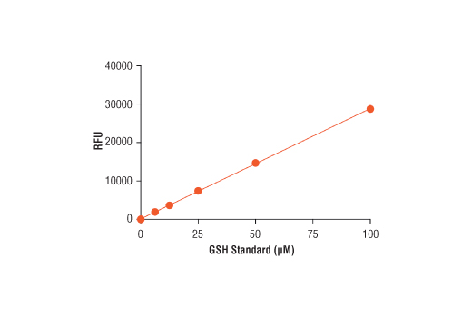
NOTE: To create a GSH standard curve, prepare a 100 µM GSH Standard Solution by diluting the 100 mM Reduced Glutathione Standard 1:1000 in Tris Assay Buffer. Run the standard curve in triplicate using 50 µl of standard per well. Create a five point standard curve by starting at 100 µM and using a 1:2 serial dilution in Tris Assay Buffer. Include a blank control. The GSH Standard Solution is not stable at this concentration and should be used within 2 hours or discarded.

Additional Reagents (not supplied): DMSO ( #12611)

B. Preparing Cell Extracts

  1. Preparation of cell extracts (10 cm dish)
    1. Apply desired treatment protocol and prepare a control sample without treatment.
    2. Following treatment, aspirate media and add 1.0 ml Digitonin Lysis Buffer to each sample, adjusting Digitonin Lysis Buffer volume when using a small surface area vessel.

      NOTE: If using suspended or detached cells, collect and centrifuge the cell suspension for 5 min at 1,200 rpm. Discard the supernatant and add Digitonin Lysis Buffer to the cell pellet.

    3. Incubate the cell lysates on ice for 5-10 min.
    4. Scrape cells off the plate and transfer suspension to a microcentrifuge tube.
    5. Centrifuge cell lysates for 10 min at 14,000 rpm at 4°C.
    6. Collect the supernatant and run assays or store at -80°C for later use.

      NOTE: use a 10 kDa MWCO centrifugal filter and follow the manufacturer’s instructions for cells that require an additional filtration step.

  2. Preparation of cell extracts from 96-well assay plate
    1. Following treatment, centrifuge assay plate for 10 min at 1,200 rpm and carefully remove the supernatant.
    2. Add 50 µl Digitonin Lysis Buffer to each well and incubate at room temperature on shaker for 15 min.
    3. Transfer samples into microcentrifuge tubes and centrifuge samples for 10 min at 14,000 rpm at 4°C.
    4. Collect the supernatants and immediately proceed to GSH assay.

C. Test Procedure

  1. Detection of Reduced Glutathione in Cell Extracts
    1. Mix Tris Assay Buffer with test sample, GSH standard, and Working Solution in a black 96-well plate with a clear bottom to final volume of 100 µl (See table 2).
    2. Table 2: Detection and Standard Curve Assays

      Sample Test Sample (µl) GSH Standard (µl) Working Solution (µl) Assay Buffer (µl) Total Volume (µl)
      Blank - - 50 50 100
      Standard - 50 50 - 100
      Sample 1-50 - 50 50 minus sample volume 100

    3. Incubate the assay plate at room temperature for 60 min, protected from light.
    4. Read plate with a plate reader at an excitation wavelength around 380 nm and emission wavelength of 485 nm.
  2. Detection of Reduced Glutathione (GSH) in Live Cells

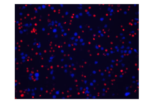
    The following procedure measures reduced glutathione in live cells using a 96-well plate. The same protocol can be modified for fluorescent imaging and flow cytometry detection.

    1. Culture adherent or suspension cells in a 96-well black plate with clear bottom in duplicate for minimum of 16 hr. Determine the background reading by using at least 2 wells containing growth media without cells.

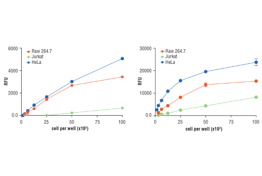
      NOTE: For best results, a cell number titration is recommended to determine the optimal cell seeding density. We recommend not exceeding 1x10⁵ cells per well.

    2. Create a Live Cell Staining Working Solution by diluting the reconstituted Monochlorobimane stock solution 1:50 in Tris Assay Buffer. For example, dilute 20 µl of Monochlorobimane stock solution in 1 ml Tris Assay Buffer to make 1 ml Live Cell Staining Working Solution, which is enough for one 96-well plate. Mix reagents by gently tapping each side of the plate.
    3. Add 10 µl of Live Cell Staining Working Solution to each well containing 100 µl of growth media.
    4. Incubate samples in a CO2 incubator for a minimum of 30 min. For best results, measure the fluorescence intensity every 30 min for up to 3 hr or until the signal has reached a plateau.

posted June 2020

Protocol Id: 2050

Product Description

The Cellular Glutathione Detection Assay Kit employs the cell permeable dye monochlorobimane (MCB) to detect reduced glutathione (GSH) in cellular assays. MCB displays a high affinity for reduced glutathione and exhibits a very low fluorescent yield when free in solution. Upon binding to GSH, the dye exhibits a strong blue fluorescence that can be measured at an excitation wavelength of 380 nm and an emission wavelength of 460 nm. Fluorescent intensity correlates with sample GSH level. This kit can be used to either label cells directly or to detect GSH level in cell extracts. The assay can be easily applied in high throughput plate-format, flow cytometry, or fluorescent imaging.

Specificity / Sensitivity

The Cellular Glutathione Detection Assay Kit is expected to detect the reduced glutathione in cellular assays across all species.

Species Reactivity:

All Species Expected

Species predicted to react based on 100% sequence homology:

All Species Expected

Background

The antioxidant glutathione is found in both reduced and oxidized states in cells. Reduced glutathione can play an important role in preventing cellular damage caused by reactive oxygen species, including free radicals and peroxides. Reduced glutathione (GSH) acts as an electron donor in the presence of free radicals and peroxides to become oxidized (GSSG). GSH also participates in redox signaling through the removal of the cellular second messenger H2O2 (1,2). Diminished glutathione levels are observed during the aging process and in oxidative stress-related diseases. The depletion of GSH is necessary for the progression of apoptosis that is mediated by various signaling pathways (3,4). Intracellular GSH levels can be a very useful indicator for overall cell health, proliferation, and death (2).

Limited Uses

Except as otherwise expressly agreed in a writing signed by a legally authorized representative of CST, the following terms apply to Products provided by CST, its affiliates or its distributors. Any Customer's terms and conditions that are in addition to, or different from, those contained herein, unless separately accepted in writing by a legally authorized representative of CST, are rejected and are of no force or effect.

Products are labeled with For Research Use Only or a similar labeling statement and have not been approved, cleared, or licensed by the FDA or other regulatory foreign or domestic entity, for any purpose. Customer shall not use any Product for any diagnostic or therapeutic purpose, or otherwise in any manner that conflicts with its labeling statement. Products sold or licensed by CST are provided for Customer as the end-user and solely for research and development uses. Any use of Product for diagnostic, prophylactic or therapeutic purposes, or any purchase of Product for resale (alone or as a component) or other commercial purpose, requires a separate license from CST. Customer shall (a) not sell, license, loan, donate or otherwise transfer or make available any Product to any third party, whether alone or in combination with other materials, or use the Products to manufacture any commercial products, (b) not copy, modify, reverse engineer, decompile, disassemble or otherwise attempt to discover the underlying structure or technology of the Products, or use the Products for the purpose of developing any products or services that would compete with CST products or services, (c) not alter or remove from the Products any trademarks, trade names, logos, patent or copyright notices or markings, (d) use the Products solely in accordance with CST Product Terms of Sale and any applicable documentation, and (e) comply with any license, terms of service or similar agreement with respect to any third party products or services used by Customer in connection with the Products.

For Research Use Only. Not for Use in Diagnostic Procedures.
Cell Signaling Technology is a trademark of Cell Signaling Technology, Inc.
All other trademarks are the property of their respective owners. Visit our Trademark Information page.